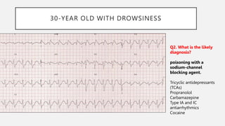
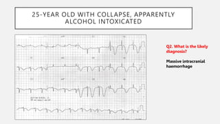
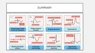
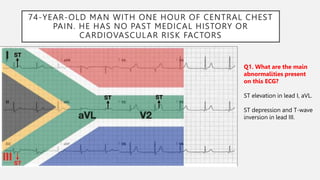
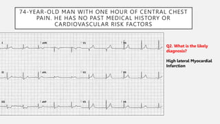
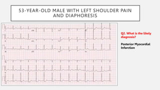
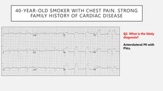
2. 25-YEAR-OLD WITH EXERTIONAL DIZZINESS.
Q1. What are the main
abnormalities present on
this ECG?
Voltage criteria for left
ventricular hypertrophy — S
wave in V1 + R wave in V5 >
35mm.
Deep, narrow Q waves in
lateral leads I, aVL, V5-6.
4. ECG FEATURES IN HCM
• Left ventricular hypertrophy with increased precordial voltages and
non-specific ST segment and T-wave abnormalities
• Deep, narrow (“dagger-like”) Q waves in lateral (I, aVL, V5-6) +/-
inferior (II, III, aVF) leads
5. 17-YEAR-OLD NOTICED “HIGH” HEART RATE ON
SMART WATCH.
Q1. What are the main
abnormalities present
on this ECG?
Very short PR interval (<
120ms)
Broad QRS complexes
with a slurred upstroke,
the delta wave.
6. 17-YEAR-OLD NOTICED “HIGH” HEART RATE ON
SMART WATCH.
Q2. What is the likely
diagnosis?
Wolff-Parkinson-White
Syndrome (WPW)
7. ECG FEATURES IN WPW
• PR interval < 120ms
• Delta wave: slurring slow rise of initial portion of the QRS
• QRS prolongation > 110ms
• ST-segment and T-wave discordant changes – i.e. in the opposite
direction to the major component of the QRS complex
8. 75-YEAR OLD WITH DYSPNEA
Q1. What are the main
abnormalities present
on this ECG?
Sinus tachycardia ~120
bpm.
Low QRS voltages’’.
Electrical alternans.
9. 75-YEAR OLD WITH DYSPNEA
Q1. What are the main
abnormalities present
on this ECG?
Sinus tachycardia ~120
bpm.
Low QRS voltages’’.
Electrical alternans.
10. 75-YEAR OLD WITH DYSPNEA
Q2. What is the likely
diagnosis?
Massive pericardial
effusion.
11. 30-YEAR OLD WITH PALPITATIONS
Q1. What are the main
abnormalities present
on this ECG?
Right axis deviation.
Dominant R wave in V1.
Widespread T-wave
inversion in inferior and
precordial leads.
Epsilon wave.
12. 30-YEAR OLD WITH PALPITATIONS
Q1. What are the main
abnormalities present
on this ECG?
Right axis deviation.
Dominant R wave in V1
Widespread T-wave
inversion in inferior and
precordial leads
Epsilon wave
13. 30-YEAR OLD WITH PALPITATIONS
Q2. What is the likely
diagnosis?
Arrhythmogenic Right
Ventricular Dysplasia.
14. CLINICAL FEATURES OF ARVD
• ARVD causes symptoms due to frequent ventricular ectopic
beats or sustained ventricular tachycardia (with LBBB
morphology) — patients typically present with palpitations,
syncope or cardiac arrest precipitated by exercise
• The first presenting symptom may be sudden cardiac death
15. 60-YEAR OLD WITH SHORTNESS OF BREATH
Q1. What are the main
abnormalities present
on this ECG?
Severe bradycardia.
Flattening, broadening
and near-disappearance of
P waves.
Prolongation of the PR
interval.
Broad QRS complexes.
Symmetrically peaked T
waves in V2-V5.
16. 60-YEAR OLD WITH SHORTNESS OF BREATH
Q2. What is the likely
diagnosis?
Severe hyperkalaemia.
17. ECG FEATURES OF HYPERKALAEMIA
• Peaked, tented T waves
• P wave widening/flattening, PR prolongation
• Bradyarrhythmias: sinus bradycardia, high-grade AV block with slow
junctional and ventricular escape rhythms, slow AF
• Conduction blocks (bundle branch block, fascicular blocks)
• QRS widening with bizarre QRS morpholog
18. 30-YEAR OLD WITH DROWSINESS
Q1. What are the main
abnormalities present
on this ECG?
Sinus tachycardia.
Borderline 1st degree AV
block.
Broad QRS complexes.
Dominant R’ wave in lead
aVR.
19. 30-YEAR OLD WITH DROWSINESS
Q2. What is the likely
diagnosis?
poisoning with a
sodium-channel
blocking agent.
Tricyclic antidepressants
(TCAs)
Propranolol
Carbamazepine
Type IA and IC
antiarrhythmics
Cocaine
20. ECG FEATURES OF SODIUM CHANNEL BLOCKADE
• Intraventricular conduction delay: QRS > 100 ms in lead II
• Terminal R wave > 3 mm in aVR or R/S ratio > 0.7 in aV
21. 25-YEAR OLD WITH COLLAPSE, APPARENTLY
ALCOHOL INTOXICATED
Q1. What are the main
abnormalities present
on this ECG?
Widespread, giant T wave
inversions.
Grossly prolonged QT
interval (~ 600ms)
22. 25-YEAR OLD WITH COLLAPSE, APPARENTLY
ALCOHOL INTOXICATED
Q2. What is the likely
diagnosis?
Massive intracranial
haemorrhage
23. 40-YEAR OLD WITH SYNCOPE
Q1. What are the main
abnormalities present
on this ECG?
RBBB-like pattern with
secondary R’ wave.
ST elevation at the J point
> 2mm with a “coved”
morphology.
Associated T wave
inversion
24. 40-YEAR OLD WITH SYNCOPE
Q1. What are the main
abnormalities present
on this ECG?
RBBB-like pattern with
secondary R’ wave.
ST elevation at the J point
> 2mm with a “coved”
morphology.
Associated T wave
inversion
25. 40-YEAR OLD WITH SYNCOPE
Q2. What is the likely
diagnosis?
Brugada Syndrome.
26. ECG FEATURES OF BRUGADA SYNDROME
• Type 1 = “Coved” ST elevation > 2mm at the J-point, followed by an inverted T
wave
• Type 2 = “Saddleback” ST segments with > 2mm J-point elevation, > 1mm ST
elevation and a positive or biphasic T wave
• Type 3 = Coved or saddleback ST elevation < 1mm
28. 74-YEAR-OLD MAN WITH ONE HOUR OF CENTRAL CHEST
PAIN. HE HAS NO PAST MEDICAL HISTORY OR
CARDIOVASCULAR RISK FACTORS
Q1. What are the main
abnormalities present
on this ECG?
ST elevation in lead I, aVL.
ST depression and T-wave
inversion in lead III.
29. 74-YEAR-OLD MAN WITH ONE HOUR OF CENTRAL CHEST
PAIN. HE HAS NO PAST MEDICAL HISTORY OR
CARDIOVASCULAR RISK FACTORS
Q1. What are the main
abnormalities present
on this ECG?
ST elevation in lead I, aVL.
ST depression and T-wave
inversion in lead III.
30. 74-YEAR-OLD MAN WITH ONE HOUR OF CENTRAL CHEST
PAIN. HE HAS NO PAST MEDICAL HISTORY OR
CARDIOVASCULAR RISK FACTORS
Q2. What is the likely
diagnosis?
High lateral Myocardial
Infarction
31. 53-YEAR-OLD MALE WITH LEFT SHOULDER PAIN
AND DIAPHORESIS
Q1. What are the main
abnormalities present
on this ECG?
ST depression in leads
V1-5 and I.
Trace ST elevation in lead
III
32. 53-YEAR-OLD MALE WITH LEFT SHOULDER PAIN
AND DIAPHORESIS
Q2. What is the likely
diagnosis?
Posterior Myocardial
Infarction
33. 82-YEAR-OLD FEMALE WITH NAUSEA AND
DIZZINESS
Q1. What are the main
abnormalities present
on this ECG?
Up-sloping ST depression
(> 1mm at J point) in
precordial leads V2-6, plus
leads I and II.
De Winter’s T waves.
34. 82-YEAR-OLD FEMALE WITH NAUSEA AND
DIZZINESS
Q1. What are the main
abnormalities present
on this ECG?
Up-sloping ST depression
(> 1mm at J point) in
precordial leads V2-6, plus
leads I and II.
De Winter’s T waves.
35. 82-YEAR-OLD FEMALE WITH NAUSEA AND
DIZZINESS
Q2. What is the likely
diagnosis?
The De Winter T wave
pattern is seen in
approximately 2% of
acute LAD occlusions
36. EARLY 70S FEMALE WITH ATYPICAL CHEST PAIN.
SKIN CLAMMY. STOIC. LOOKS UNWELL
Q1. What are the main
abnormalities present
on this ECG?
T waves in leads III and
aVF appear to tower over
the preceding QRS
complex, and have a wide,
“bulky” appearance. Such
changes are consistent
with hyperacute T waves
(HATW) representative of
hyperacute occlusion and
evolving ST elevation,
There is reciprocal ST
depression in V1-3 and I
37. EARLY 70S FEMALE WITH ATYPICAL CHEST PAIN.
SKIN CLAMMY. STOIC. LOOKS UNWELL
Q2. What is the likely
diagnosis?
Hyperacute inferior MI
38. 60-YEAR OLD WITH LEFT ARM PAIN AND
SHORTNESS OF BREATH. KNOWN LBBB
Q1. What are the main
abnormalities present
on this ECG?
Paced rhythm with left
bundle branch block
(LBBB) morphology.
> 1mm concordant ST
elevation in leads V4-5
and aVL (Sgarbossa
criteria).
ST elevation in leads V2-3
is excessively discordant
(Sgarbossa criteria).
39. 60-YEAR OLD WITH LEFT ARM PAIN AND
SHORTNESS OF BREATH. KNOWN LBBB
Q2. What is the likely
diagnosis?
Anterolateral MI
41. 78-YEAR-OLD WITH RIGHT SHOULDER PAIN AND
DIAPHORESIS. PPM IN SITU
Q1. What are the main
abnormalities present
on this ECG?
Paced rhythm with left
bundle branch block
(LBBB) morphology.
Concordant ST depression
in V2 and V3 (Sgarbossa
criteria).
42. 78-YEAR-OLD WITH RIGHT SHOULDER PAIN AND
DIAPHORESIS. PPM IN SITU
Q2. What is the likely
diagnosis?
Posterior MI
43. 83-YEAR-OLD WITH CHEST PAIN. HISTORY OF 2ND
DEGREE AV BLOCK WITH PPM IN SITU
Q1. What are the main
abnormalities present
on this ECG?
Paced rhythm with left
bundle branch block
(LBBB) morphology.
Degree of ST elevation in
leads V2-5 is > 25% of
depth of the preceding S
wave (Sgarbossa Criteria).
Concordant ST elevation
in leads I and V6
(Sgarbossa criteria).
44. 83-YEAR-OLD WITH CHEST PAIN. HISTORY OF 2ND
DEGREE AV BLOCK WITH PPM IN SITU
Q2. What is the likely
diagnosis?
Anterolateral MI
45. 40-YEAR-OLD SMOKER WITH CHEST PAIN. STRONG
FAMILY HISTORY OF CARDIAC DISEASE
Q1. What are the main
abnormalities present
on this ECG?
ST elevation in V1-6 plus I
and aVL.
Reciprocal ST depression
in III and aVF.
Pathological Q waves in
V1-2, with reduced R wave
height in V3-4.
46. 40-YEAR-OLD SMOKER WITH CHEST PAIN. STRONG
FAMILY HISTORY OF CARDIAC DISEASE
Q2. What is the likely
diagnosis?
Anterolateral MI with
PVcs.
Editor's Notes
Epsilon wave
Localised subtle widening of QRS complexes in V1-3
A small blip following each QRS complex, best seen in V1 and the inferior leads
Epsilon wave
Localised subtle widening of QRS complexes in V1-3
A small blip following each QRS complex, best seen in V1 and the inferior leads
De Winter’s T waves — peaked anterior T waves, with the ascending limb of the T wave commencing below the isoelectric baseline