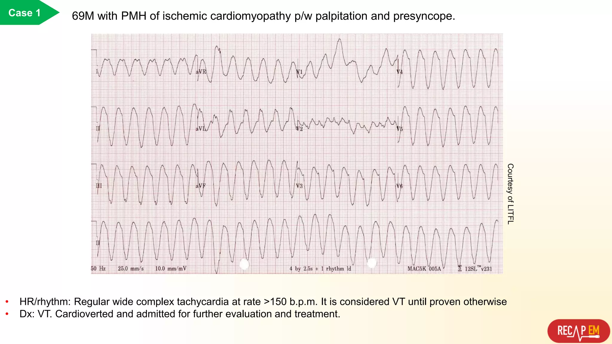
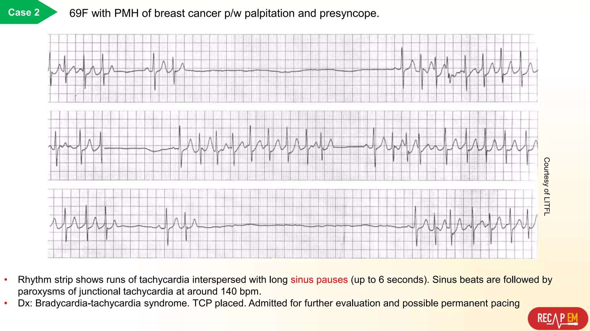
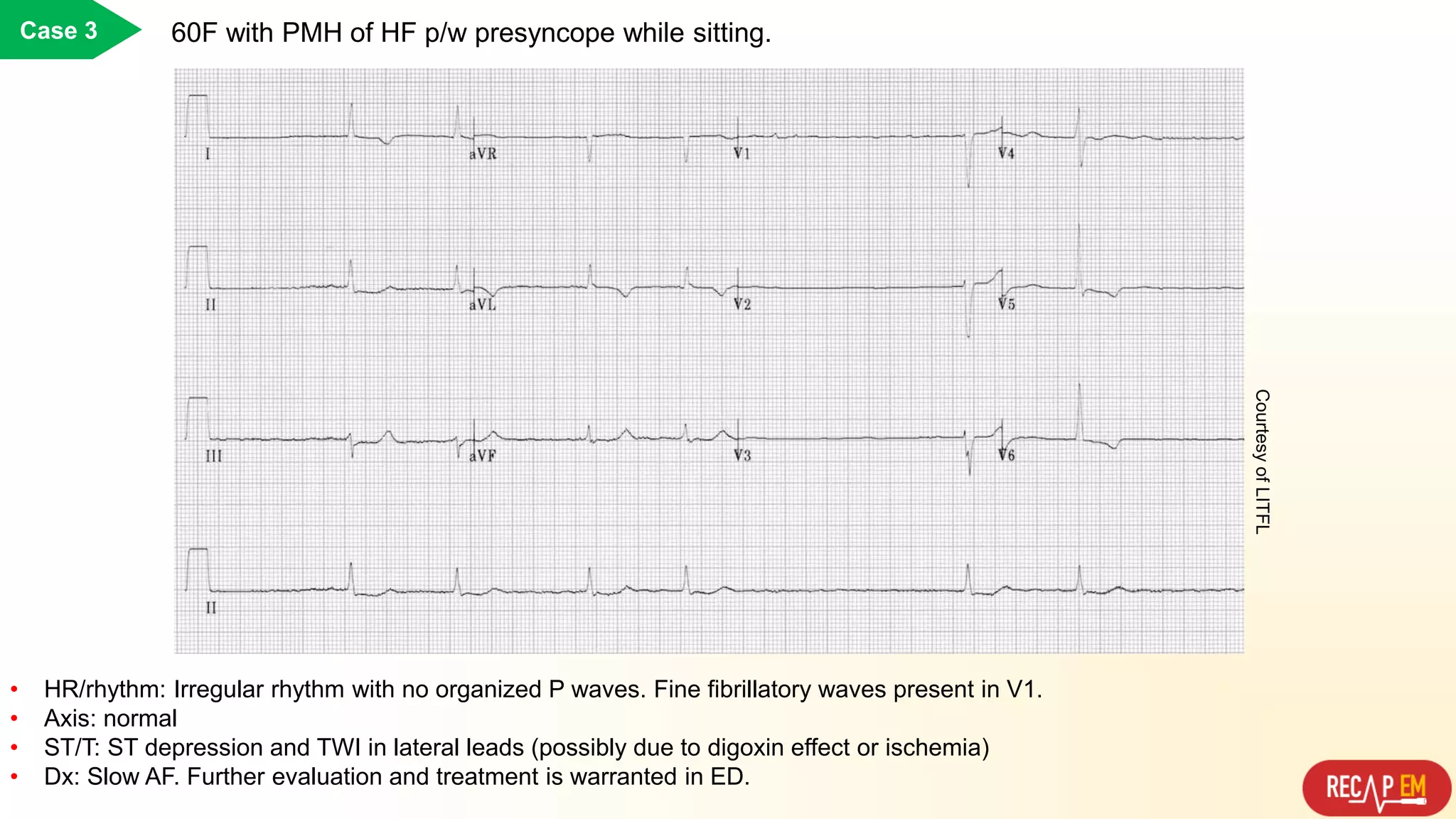
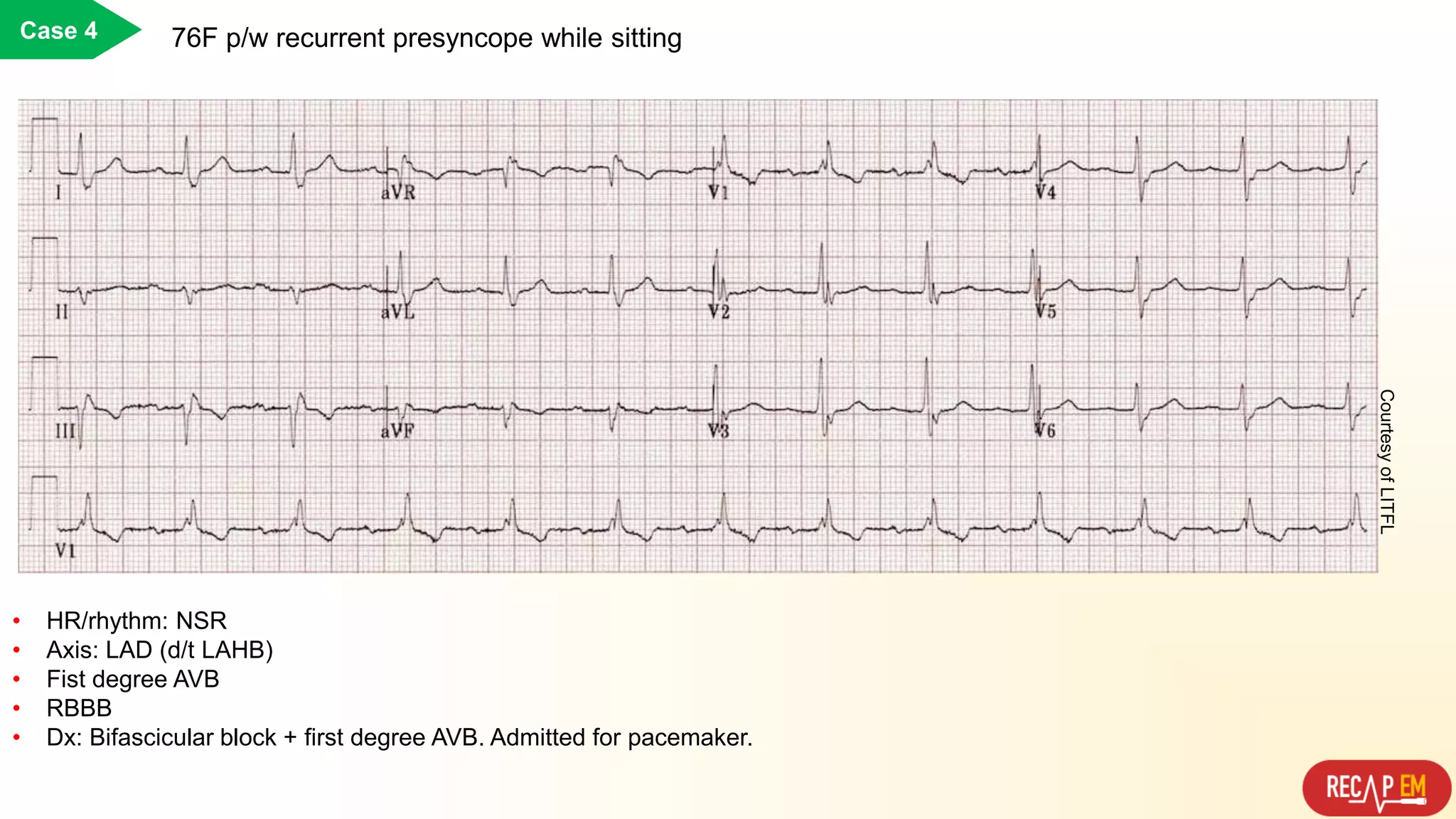
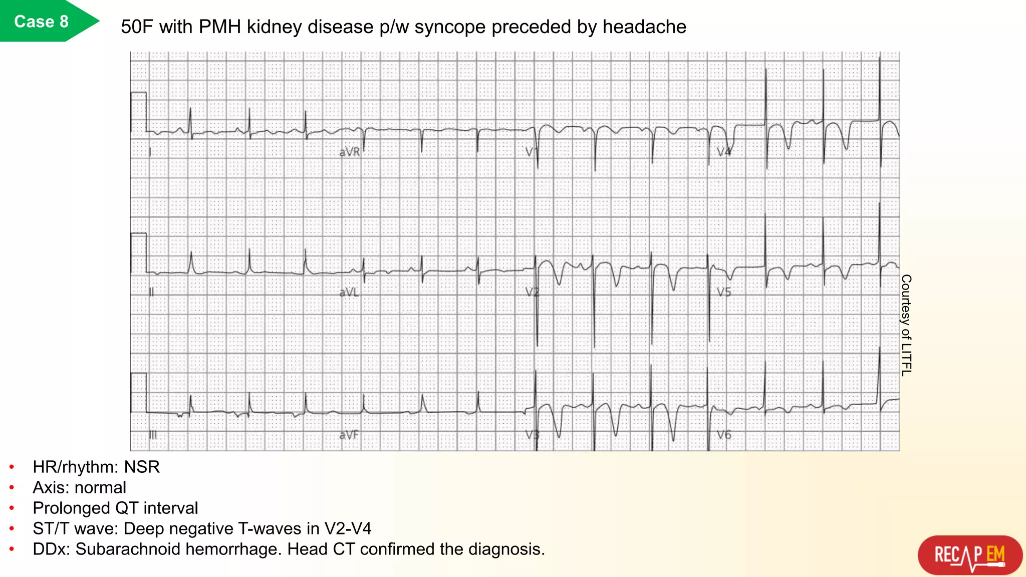
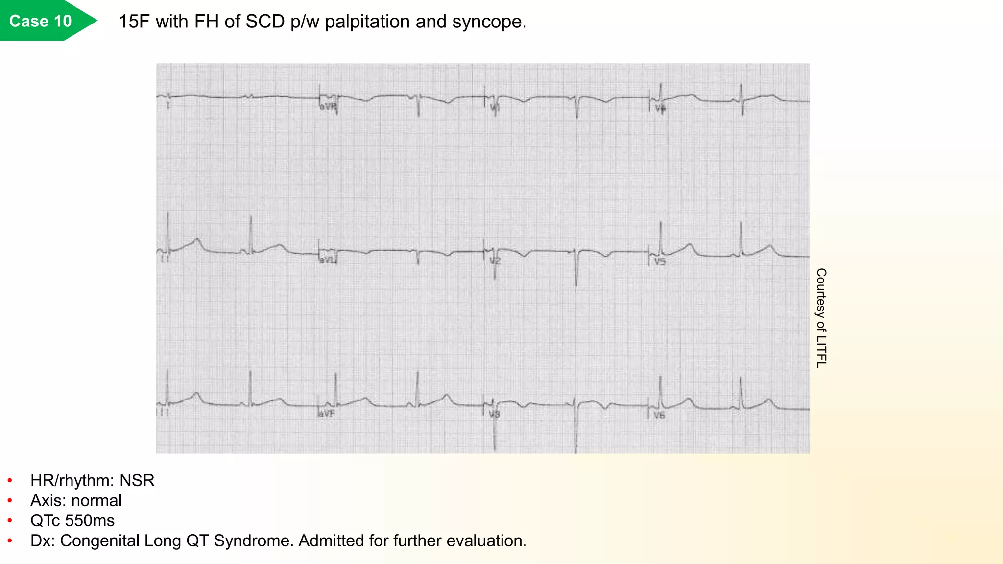
ECG In Syncope
Sh.Lahouti, M.D.
Visit us @https://recapem.com
 Rate?
• Too fast
• Too slow
 Rhythm?
• Sinus rhythm or not?
• Regular vs. irregular?
 Axis?
• Right: PH, PE, LPHB
• Left: LAHB, LBBB
 PR interval?
• Short PRi (WPW)
• Long PRi (AVB)
 QT interval?
• Short (QTc < 350 ms)
• Long (QTc > 450ms. QTc>500ms associates with risk of torsades de pointes)
 QRS complex width and voltage?
• High voltage (and deep narrow Q wave): HCM
• RVH: PH
• Wide QRS: BBB
• Wide QRS + epsilon wave: ARVC
 ST/T changes?
• Brugada, ARVC
• ACS, PE, ICH/SAH
ECG Pearls for Syncope
Case 1Case 1 69M with PMH of ischemic cardiomyopathy p/w palpitation and presyncope.
• HR/rhythm: Regular wide complex tachycardia at rate >150 b.p.m. It is considered VT until proven otherwise
• Dx: VT. Cardioverted and admitted for further evaluation and treatment.
CourtesyofLITFL
• Rhythm strip shows runs of tachycardia interspersed with long sinus pauses (up to 6 seconds). Sinus beats are followed by
paroxysms of junctional tachycardia at around 140 bpm.
• Dx: Bradycardia-tachycardia syndrome. TCP placed. Admitted for further evaluation and possible permanent pacing
Case 2 69F with PMH of breast cancer p/w palpitation and presyncope.
CourtesyofLITFL
Case 3 60F with PMH of HF p/w presyncope while sitting.
• HR/rhythm: Irregular rhythm with no organized P waves. Fine fibrillatory waves present in V1.
• Axis: normal
• ST/T: ST depression and TWI in lateral leads (possibly due to digoxin effect or ischemia)
• Dx: Slow AF. Further evaluation and treatment is warranted in ED.
CourtesyofLITFL
Case 4 76F p/w recurrent presyncope while sitting
• HR/rhythm: NSR
• Axis: LAD (d/t LAHB)
• Fist degree AVB
• RBBB
• Dx: Bifascicular block + first degree AVB. Admitted for pacemaker.
CourtesyofLITFL
Case 5 22M p/w presyncope during exertion
• HR/rhythm: NSR
• Axis: normal
• Voltage criteria for LVH
• Deep, narrow Q wave (< 40ms wide) in the lateral (V5-6, I, AVL) and inferior (II, III, AVF) leads
• Dx: HCM? → Performed POCUS…..
CourtesyofLITFL
PLAX view. A, 2D image shows severe septal hypertrophy (thickness 30 mm) and the anterior and superior motion of the anterior
mitral leaflet during systole (white arrow) and contact of the leaflet with the massive septum. B, Corresponding M-mode image at the
level of the mitral valve leaflet tips demonstrating severe systolic anterior motion with prolonged anterior leaflet–septal contact (white
arrowheads).
• Patient was admitted to the hospital.
Case 5
cont.
Case 6 19M with FH of SCD p/w syncope at bathroom.
• HR/rhythm: NSR
• Axis: RAD
• Presence of epsilon wave in V1.
• TWI in V1-3. Prolonged S-wave upstroke in V1-3 with localized QRS widening
• Dx: ARVC. Admitted for further assessment and possible ICD placement.
CourtesyofLITFL
Case 7 28M p/w syncope while watching TV!
• HR/rhythm: NSR
• Axis: normal
• ST/T wave: V1-2 coved ST elevation
• Dx: Brugada. Patient admitted for further evaluation and possibly ICD placement.
CourtesyofLITFL
Case 8 50F with PMH kidney disease p/w syncope preceded by headache
• HR/rhythm: NSR
• Axis: normal
• Prolonged QT interval
• ST/T wave: Deep negative T-waves in V2-V4
• DDx: Subarachnoid hemorrhage. Head CT confirmed the diagnosis.
CourtesyofLITFL
Case 9 25F with p/w syncope preceded by palpitation.
• HR/rhythm: NSR
• Axis: RAD
• short PR interval (< 120ms), presence of delta wave (broad QRS complex with slurred upstroke) in all precordial leads
• Tall R wave and TWI in V1-V3 mimicking RVH
• Dx: WPW. Referred for ablation.
CourtesyofLITFL
15F with FH of SCD p/w palpitation and syncope.Case 10
• HR/rhythm: NSR
• Axis: normal
• QTc 550ms
• Dx: Congenital Long QT Syndrome. Admitted for further evaluation.
CourtesyofLITFL

Ecg in syncope

  • 1.
    ECG In Syncope Sh.Lahouti,M.D. Visit us @https://recapem.com
  • 2.
     Rate? • Toofast • Too slow  Rhythm? • Sinus rhythm or not? • Regular vs. irregular?  Axis? • Right: PH, PE, LPHB • Left: LAHB, LBBB  PR interval? • Short PRi (WPW) • Long PRi (AVB)  QT interval? • Short (QTc < 350 ms) • Long (QTc > 450ms. QTc>500ms associates with risk of torsades de pointes)  QRS complex width and voltage? • High voltage (and deep narrow Q wave): HCM • RVH: PH • Wide QRS: BBB • Wide QRS + epsilon wave: ARVC  ST/T changes? • Brugada, ARVC • ACS, PE, ICH/SAH ECG Pearls for Syncope
  • 3.
    Case 1Case 169M with PMH of ischemic cardiomyopathy p/w palpitation and presyncope. • HR/rhythm: Regular wide complex tachycardia at rate >150 b.p.m. It is considered VT until proven otherwise • Dx: VT. Cardioverted and admitted for further evaluation and treatment. CourtesyofLITFL
  • 4.
    • Rhythm stripshows runs of tachycardia interspersed with long sinus pauses (up to 6 seconds). Sinus beats are followed by paroxysms of junctional tachycardia at around 140 bpm. • Dx: Bradycardia-tachycardia syndrome. TCP placed. Admitted for further evaluation and possible permanent pacing Case 2 69F with PMH of breast cancer p/w palpitation and presyncope. CourtesyofLITFL
  • 5.
    Case 3 60Fwith PMH of HF p/w presyncope while sitting. • HR/rhythm: Irregular rhythm with no organized P waves. Fine fibrillatory waves present in V1. • Axis: normal • ST/T: ST depression and TWI in lateral leads (possibly due to digoxin effect or ischemia) • Dx: Slow AF. Further evaluation and treatment is warranted in ED. CourtesyofLITFL
  • 6.
    Case 4 76Fp/w recurrent presyncope while sitting • HR/rhythm: NSR • Axis: LAD (d/t LAHB) • Fist degree AVB • RBBB • Dx: Bifascicular block + first degree AVB. Admitted for pacemaker. CourtesyofLITFL
  • 7.
    Case 5 22Mp/w presyncope during exertion • HR/rhythm: NSR • Axis: normal • Voltage criteria for LVH • Deep, narrow Q wave (< 40ms wide) in the lateral (V5-6, I, AVL) and inferior (II, III, AVF) leads • Dx: HCM? → Performed POCUS….. CourtesyofLITFL
  • 8.
    PLAX view. A,2D image shows severe septal hypertrophy (thickness 30 mm) and the anterior and superior motion of the anterior mitral leaflet during systole (white arrow) and contact of the leaflet with the massive septum. B, Corresponding M-mode image at the level of the mitral valve leaflet tips demonstrating severe systolic anterior motion with prolonged anterior leaflet–septal contact (white arrowheads). • Patient was admitted to the hospital. Case 5 cont.
  • 9.
    Case 6 19Mwith FH of SCD p/w syncope at bathroom. • HR/rhythm: NSR • Axis: RAD • Presence of epsilon wave in V1. • TWI in V1-3. Prolonged S-wave upstroke in V1-3 with localized QRS widening • Dx: ARVC. Admitted for further assessment and possible ICD placement. CourtesyofLITFL
  • 10.
    Case 7 28Mp/w syncope while watching TV! • HR/rhythm: NSR • Axis: normal • ST/T wave: V1-2 coved ST elevation • Dx: Brugada. Patient admitted for further evaluation and possibly ICD placement. CourtesyofLITFL
  • 11.
    Case 8 50Fwith PMH kidney disease p/w syncope preceded by headache • HR/rhythm: NSR • Axis: normal • Prolonged QT interval • ST/T wave: Deep negative T-waves in V2-V4 • DDx: Subarachnoid hemorrhage. Head CT confirmed the diagnosis. CourtesyofLITFL
  • 12.
    Case 9 25Fwith p/w syncope preceded by palpitation. • HR/rhythm: NSR • Axis: RAD • short PR interval (< 120ms), presence of delta wave (broad QRS complex with slurred upstroke) in all precordial leads • Tall R wave and TWI in V1-V3 mimicking RVH • Dx: WPW. Referred for ablation. CourtesyofLITFL
  • 13.
    15F with FHof SCD p/w palpitation and syncope.Case 10 • HR/rhythm: NSR • Axis: normal • QTc 550ms • Dx: Congenital Long QT Syndrome. Admitted for further evaluation. CourtesyofLITFL