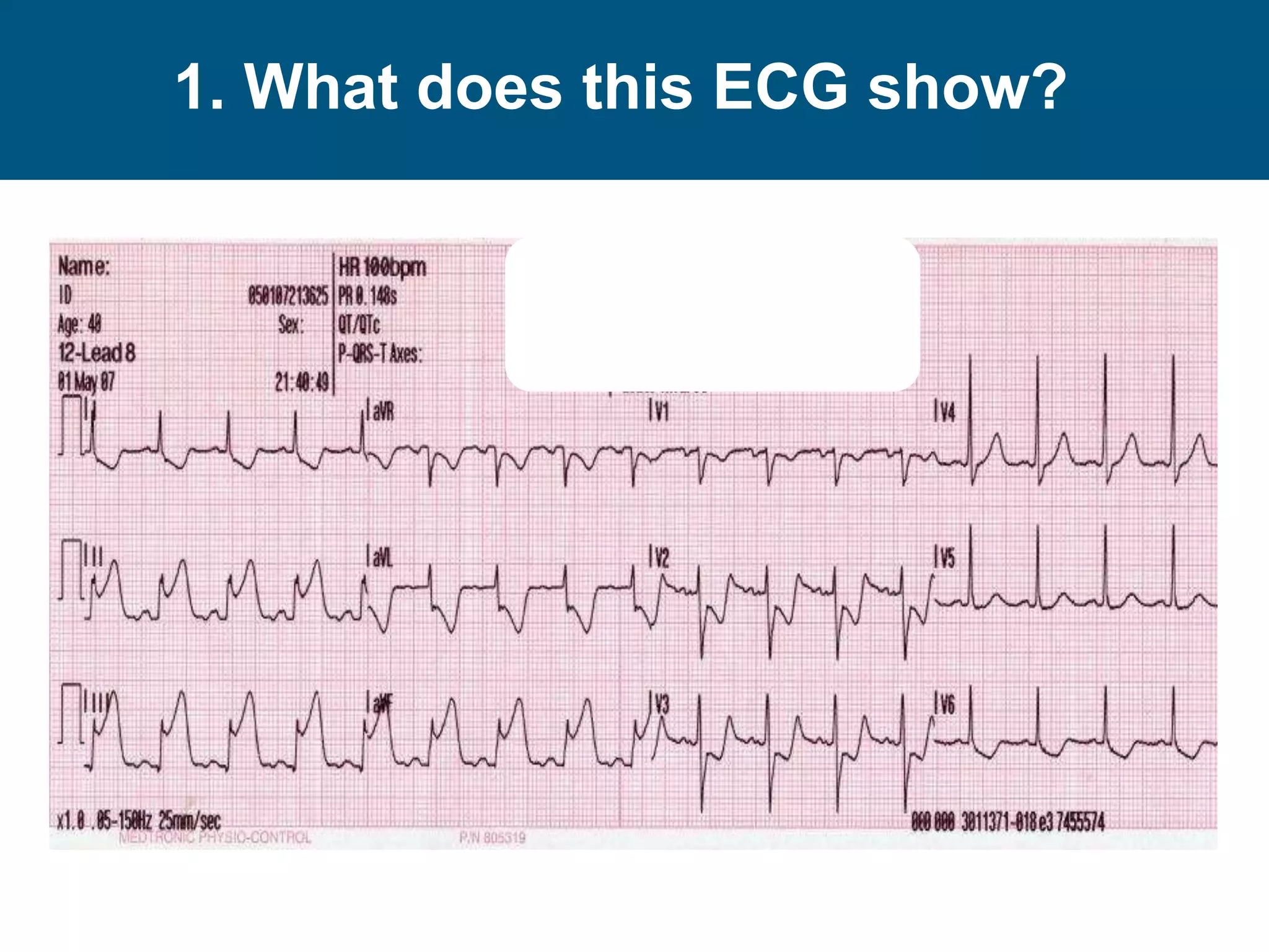
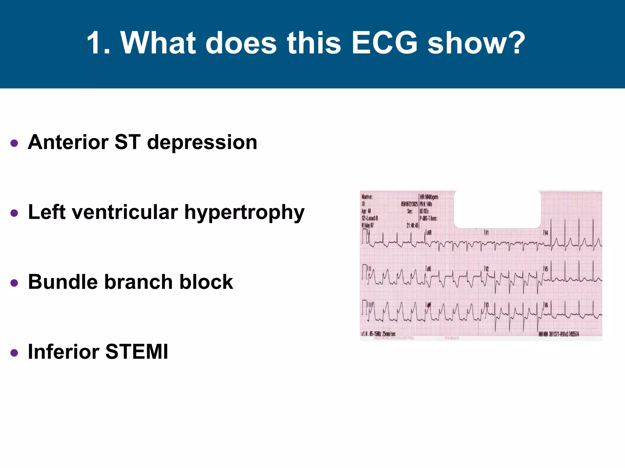
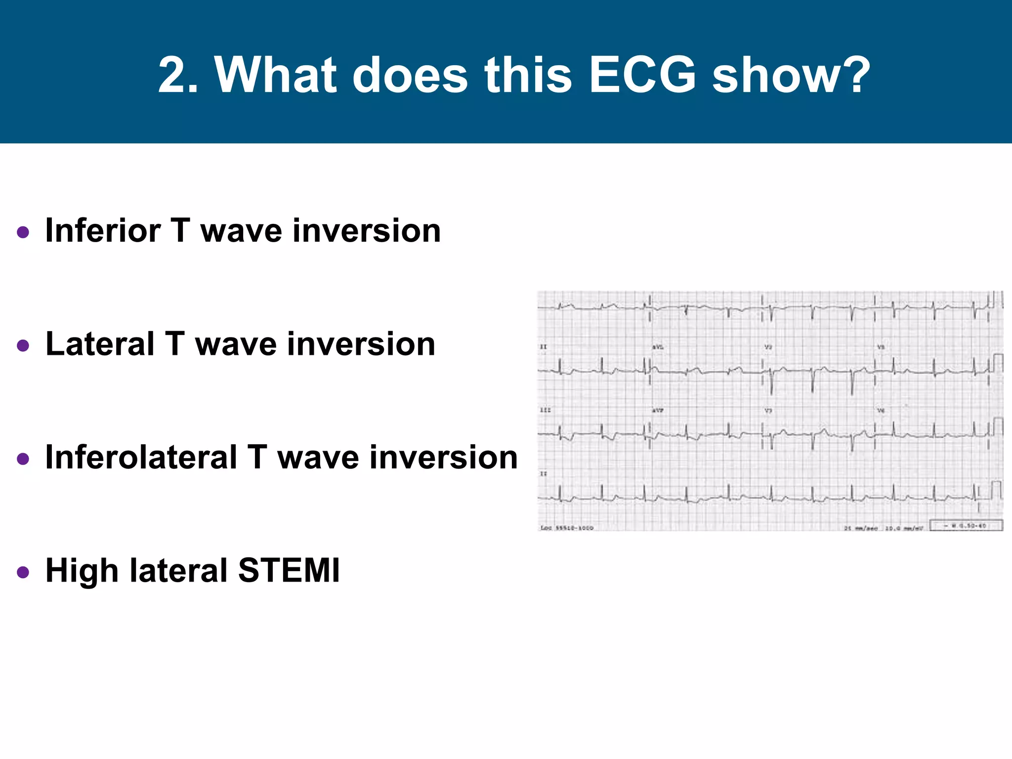
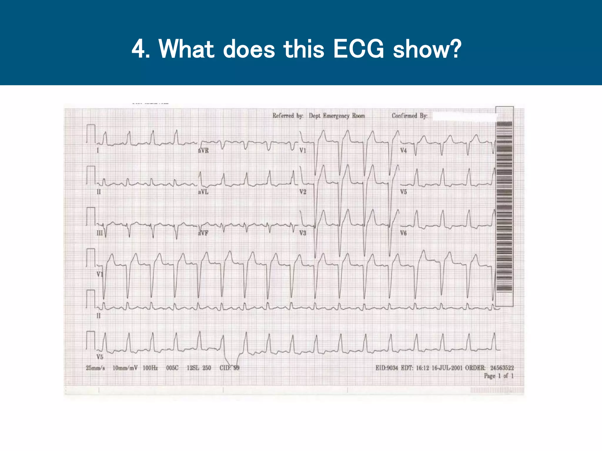
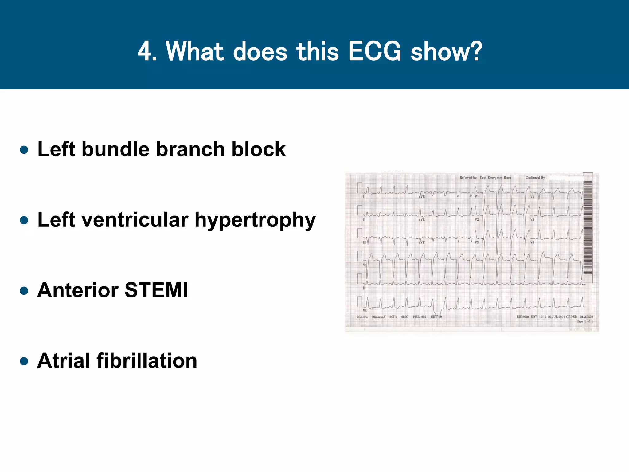
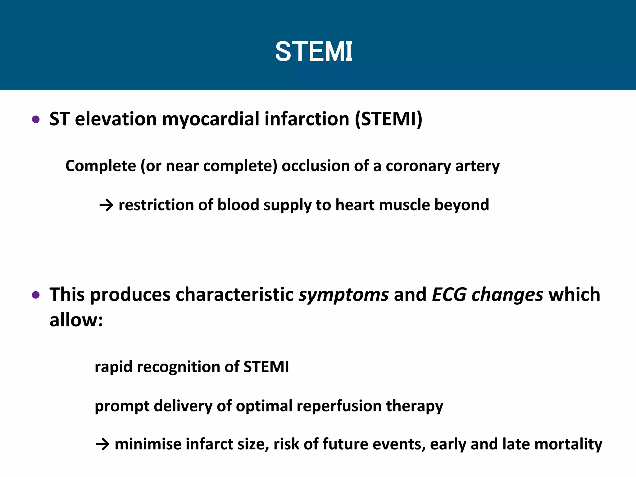
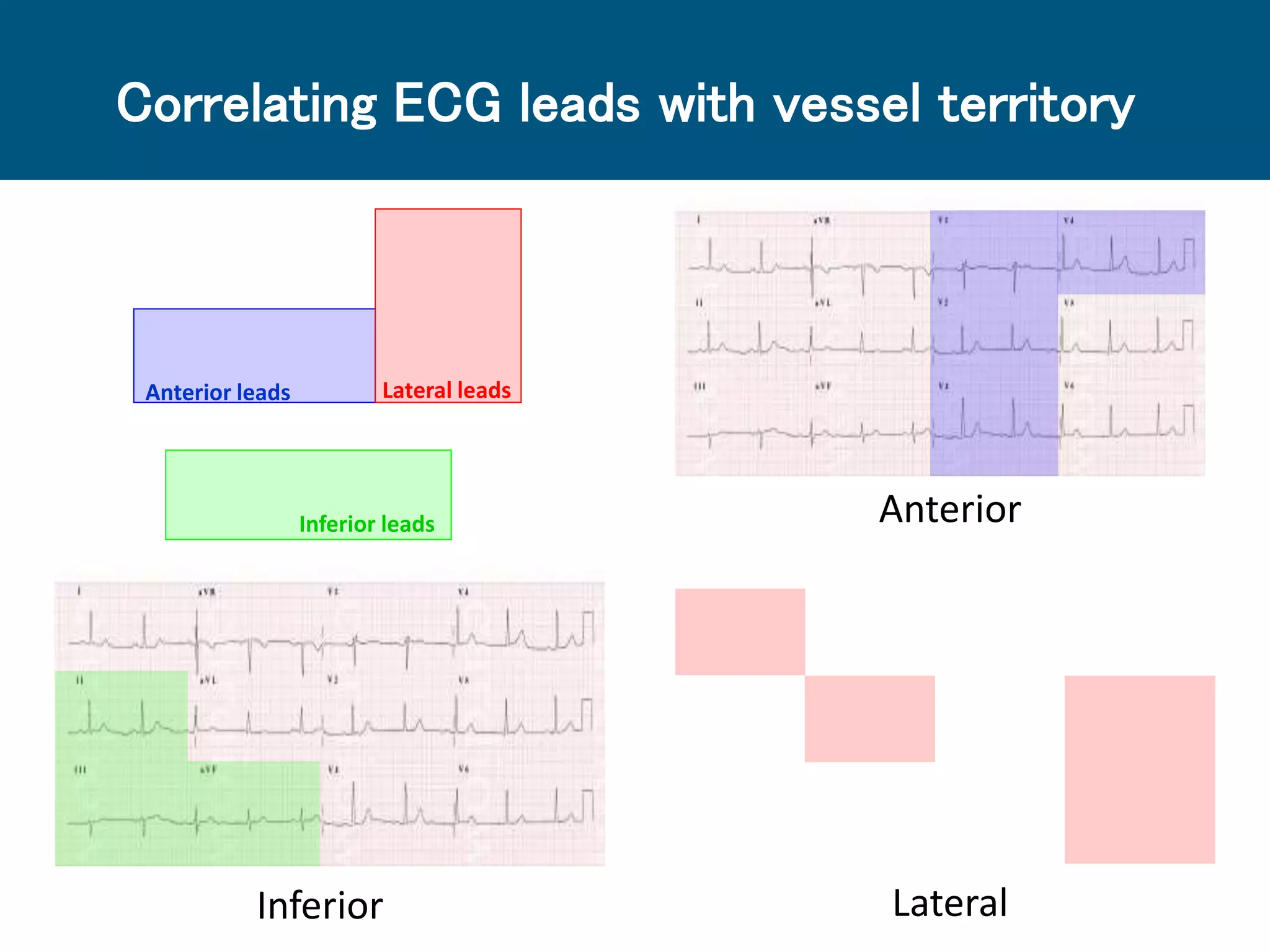
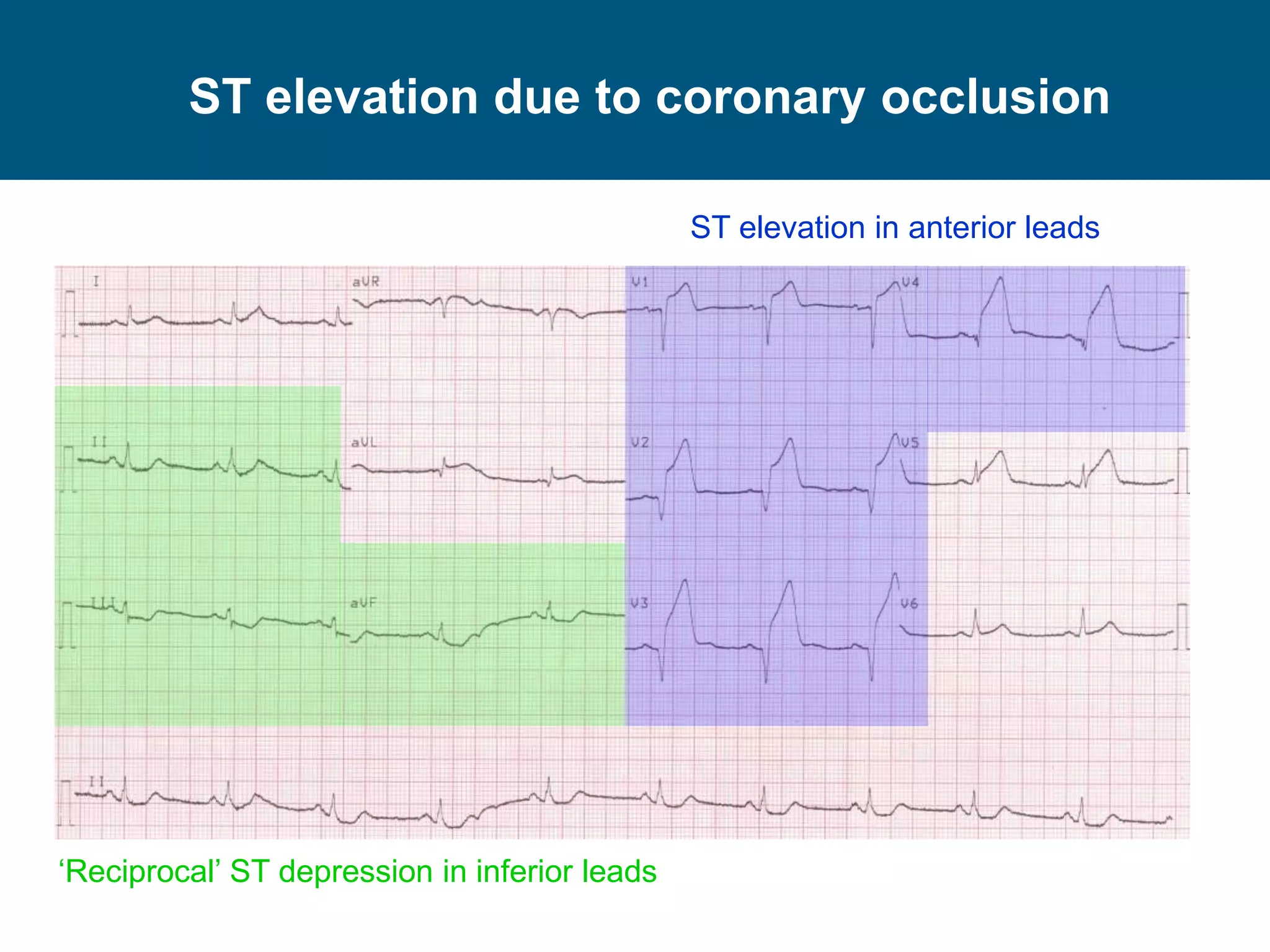
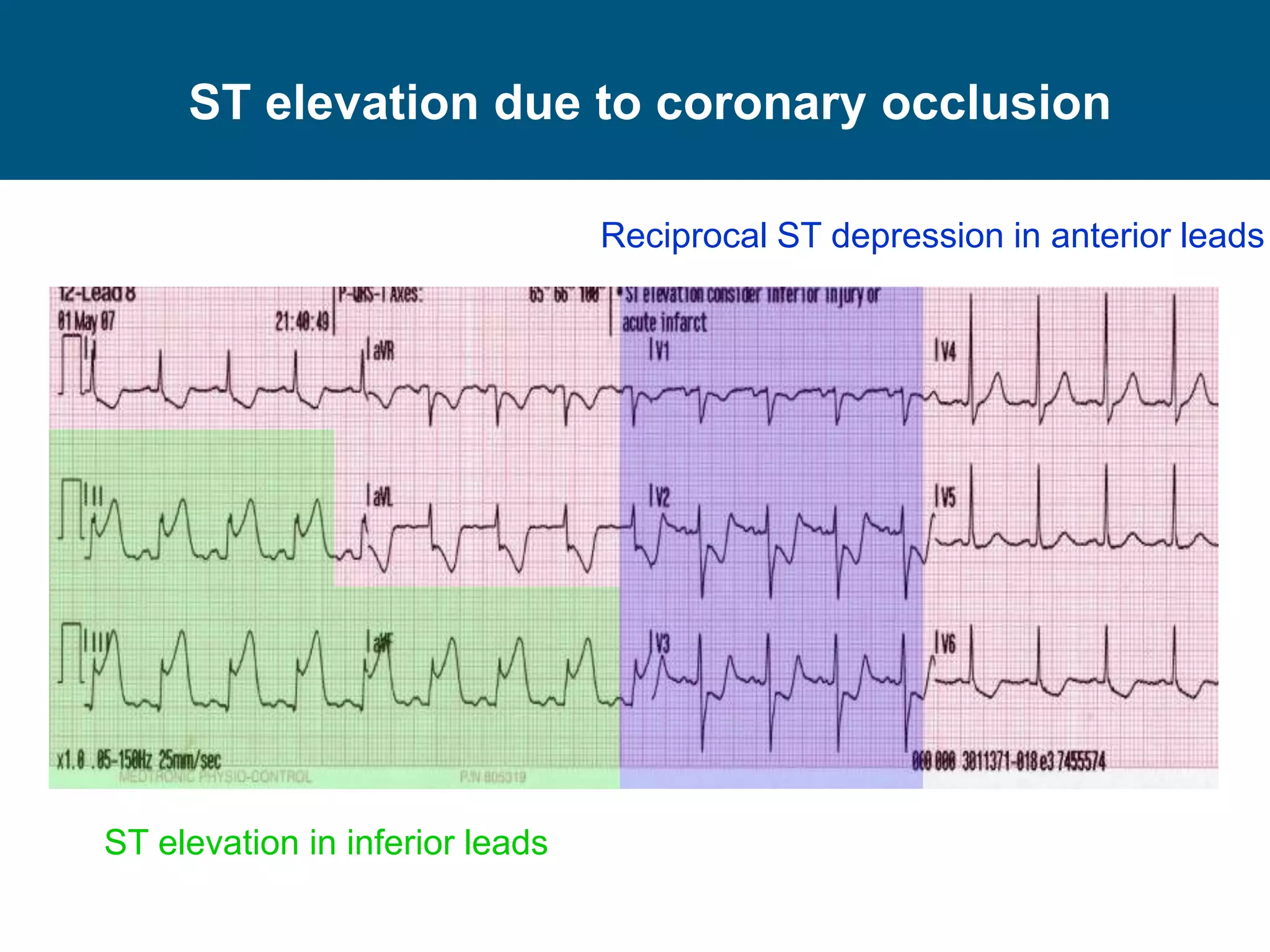
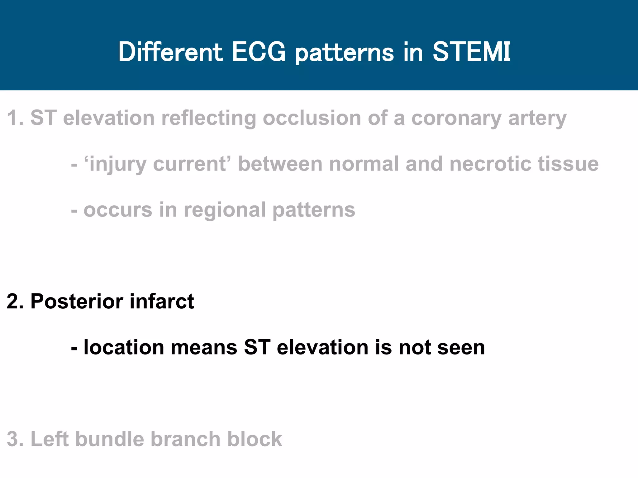
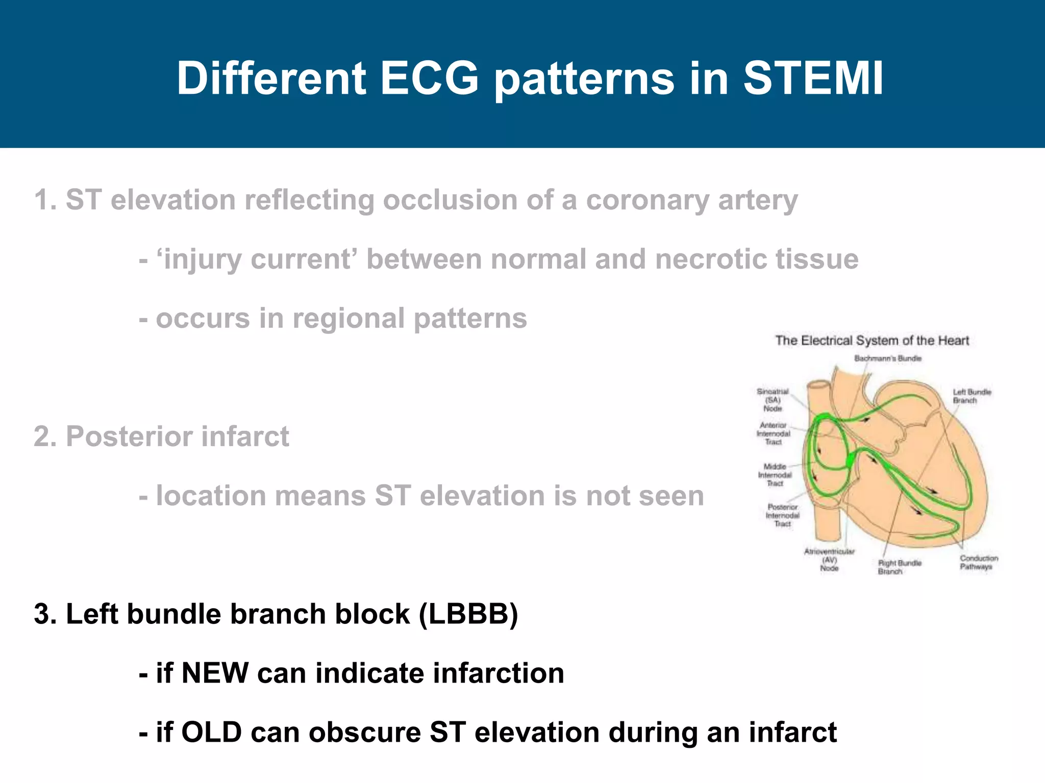
ECG interpretation: STEMI
Primary PCI and direct admission of high
risk NSTEMI
Joanne Simpson
Golden Jubilee National Hospital
Wednesday 17th February 2016
Aims
 Revise the ECG patterns which occur in STEMI
 Focus on those which are more difficult to
recognise, but not uncommon
 Remember:
Some ECG patterns are not clear cut
Minimising missed STEMIs is the priority
1. What does this ECG show?
 Anterior ST depression
 Left ventricular hypertrophy
 Bundle branch block
 Inferior STEMI
2. What does this ECG show?
 Inferior T wave inversion
 Lateral T wave inversion
 Inferolateral T wave inversion
 High lateral STEMI
3. What does this ECG show?
 Left bundle branch block
 Anterior T wave inversion
 Posterior STEMI
 Atrial fibrillation
4. What does this ECG show?
 Left bundle branch block
 Left ventricular hypertrophy
 Anterior STEMI
 Atrial fibrillation
STEMI
 ST elevation myocardial infarction (STEMI)
Complete (or near complete) occlusion of a coronary artery
→ restriction of blood supply to heart muscle beyond
 This produces characteristic symptoms and ECG changes which
allow:
rapid recognition of STEMI
prompt delivery of optimal reperfusion therapy
→ minimise infarct size, risk of future events, early and late mortality
Clinical presentation
Different ECG patterns in STEMI
1. ST elevation reflecting occlusion of a coronary artery
2. Posterior infarct
3. Left bundle branch block
1. ST elevation reflecting occlusion of a coronary artery
- ‘injury current’ between normal and necrotic Tissue
- occurs in regional patterns
2. Posterior infarct
3. Left bundle branch block
Different ECG patterns in STEMI
ST elevation due to coronary occlusion
ECG leads over the territory of the occluded artery will show ST
elevation as the injury current travels toward them
ST elevation correlates to the territory of the occluded artery:
Inferior = RCA / LCx
Posterior = RCA / LCx
Lateral = LCx
Anteroseptal = LAD
Circumflex
artery (LCx)
Left
anterior
descending
artery (LAD)
Right coronary
artery (RCA)
How do ECG leads look at the heart?
Chest leadsFrontal leads
Inferior leads
Anterior leads Lateral leads
How do ECG leads look at the heart?
Correlating ECG leads with vessel territory
Inferior Lateral
AnteriorInferior leads
Anterior leads Lateral leads
ST elevation due to coronary occlusion
ST elevation in anterior leads
‘Reciprocal’ ST depression in inferior leads
ST elevation in inferior leads
Reciprocal ST depression in anterior leads
ST elevation due to coronary occlusion
ST elevation in high lateral leads
‘Reciprocal’ ST depression in inferior leads
Different ECG patterns in STEMI
1. ST elevation reflecting occlusion of a coronary artery
- ‘injury current’ between normal and necrotic tissue
- occurs in regional patterns
2. Posterior infarct
- location means ST elevation is not seen
3. Left bundle branch block
Posterior wall infarction
Posterior infarct may be caused by RCA or LCx occlusion
- often associated with inferior or lateral ST elevation
Circumflex
artery (LCx)
Left
anterior
descending
artery (LAD)
Right coronary
artery (RCA)
Posterior wall infarction
 No ECG leads ‘look’ directly at the posterior wall of the heart
 Anterior leads are directly opposite and will see the opposite
of any current generated at the posterior wall
 posterior ST elevation= anterior ST depression
Posterior wall infarction
ST depression in anterior leads
Subtle ST elevation in inferior leads
Different ECG patterns in STEMI
1. ST elevation reflecting occlusion of a coronary artery
- ‘injury current’ between normal and necrotic tissue
- occurs in regional patterns
2. Posterior infarct
- location means ST elevation is not seen
3. Left bundle branch block (LBBB)
- if NEW can indicate infarction
- if OLD can obscure ST elevation during an infarct
Left bundle branch block
QRS duration > 3 small squares
Negative QRS across anterior leads with poor R wave progression
Notched R wave in V6
V1
V6
Summary
 The clinical presentation is paramount
 Check each ECG territory in turn for ST elevation
 Look for ST depression
- is this reciprocal change?
- if anterior, could it be a posterior infarct?
 Check the QRS duration
- if > 3 small squares, is there LBBB?

ECG interpretation in NSTEMI

  • 1.
    ECG interpretation: STEMI PrimaryPCI and direct admission of high risk NSTEMI Joanne Simpson Golden Jubilee National Hospital Wednesday 17th February 2016
  • 2.
    Aims  Revise theECG patterns which occur in STEMI  Focus on those which are more difficult to recognise, but not uncommon  Remember: Some ECG patterns are not clear cut Minimising missed STEMIs is the priority
  • 3.
    1. What doesthis ECG show?
  • 4.
    1. What doesthis ECG show?  Anterior ST depression  Left ventricular hypertrophy  Bundle branch block  Inferior STEMI
  • 5.
    2. What doesthis ECG show?
  • 6.
    2. What doesthis ECG show?  Inferior T wave inversion  Lateral T wave inversion  Inferolateral T wave inversion  High lateral STEMI
  • 7.
    3. What doesthis ECG show?
  • 8.
    3. What doesthis ECG show?  Left bundle branch block  Anterior T wave inversion  Posterior STEMI  Atrial fibrillation
  • 9.
    4. What doesthis ECG show?
  • 10.
    4. What doesthis ECG show?  Left bundle branch block  Left ventricular hypertrophy  Anterior STEMI  Atrial fibrillation
  • 11.
    STEMI  ST elevationmyocardial infarction (STEMI) Complete (or near complete) occlusion of a coronary artery → restriction of blood supply to heart muscle beyond  This produces characteristic symptoms and ECG changes which allow: rapid recognition of STEMI prompt delivery of optimal reperfusion therapy → minimise infarct size, risk of future events, early and late mortality
  • 12.
    STEMI  ST elevationmyocardial infarction (STEMI) Complete (or near complete) occlusion of a coronary artery → restriction of blood supply to heart muscle beyond  This produces characteristic symptoms and ECG changes which allow: rapid recognition of STEMI prompt delivery of optimal reperfusion therapy → minimise infarct size, risk of future events, early and late mortality
  • 13.
  • 14.
    Different ECG patternsin STEMI 1. ST elevation reflecting occlusion of a coronary artery 2. Posterior infarct 3. Left bundle branch block
  • 15.
    1. ST elevationreflecting occlusion of a coronary artery - ‘injury current’ between normal and necrotic Tissue - occurs in regional patterns 2. Posterior infarct 3. Left bundle branch block Different ECG patterns in STEMI
  • 16.
    ST elevation dueto coronary occlusion ECG leads over the territory of the occluded artery will show ST elevation as the injury current travels toward them ST elevation correlates to the territory of the occluded artery: Inferior = RCA / LCx Posterior = RCA / LCx Lateral = LCx Anteroseptal = LAD Circumflex artery (LCx) Left anterior descending artery (LAD) Right coronary artery (RCA)
  • 17.
    How do ECGleads look at the heart?
  • 18.
    How do ECGleads look at the heart? Chest leadsFrontal leads
  • 19.
    Inferior leads Anterior leadsLateral leads How do ECG leads look at the heart?
  • 20.
    Correlating ECG leadswith vessel territory Inferior Lateral AnteriorInferior leads Anterior leads Lateral leads
  • 21.
    ST elevation dueto coronary occlusion ST elevation in anterior leads ‘Reciprocal’ ST depression in inferior leads
  • 22.
    ST elevation ininferior leads Reciprocal ST depression in anterior leads ST elevation due to coronary occlusion
  • 23.
    ST elevation dueto coronary occlusion ST elevation in high lateral leads ‘Reciprocal’ ST depression in inferior leads
  • 24.
    Different ECG patternsin STEMI 1. ST elevation reflecting occlusion of a coronary artery - ‘injury current’ between normal and necrotic tissue - occurs in regional patterns 2. Posterior infarct - location means ST elevation is not seen 3. Left bundle branch block
  • 25.
    Posterior wall infarction Posteriorinfarct may be caused by RCA or LCx occlusion - often associated with inferior or lateral ST elevation Circumflex artery (LCx) Left anterior descending artery (LAD) Right coronary artery (RCA)
  • 26.
    Posterior wall infarction No ECG leads ‘look’ directly at the posterior wall of the heart  Anterior leads are directly opposite and will see the opposite of any current generated at the posterior wall  posterior ST elevation= anterior ST depression
  • 27.
    Posterior wall infarction STdepression in anterior leads Subtle ST elevation in inferior leads
  • 28.
    Different ECG patternsin STEMI 1. ST elevation reflecting occlusion of a coronary artery - ‘injury current’ between normal and necrotic tissue - occurs in regional patterns 2. Posterior infarct - location means ST elevation is not seen 3. Left bundle branch block (LBBB) - if NEW can indicate infarction - if OLD can obscure ST elevation during an infarct
  • 29.
  • 30.
    Left bundle branchblock QRS duration > 3 small squares Negative QRS across anterior leads with poor R wave progression Notched R wave in V6 V1 V6
  • 31.
    Summary  The clinicalpresentation is paramount  Check each ECG territory in turn for ST elevation  Look for ST depression - is this reciprocal change? - if anterior, could it be a posterior infarct?  Check the QRS duration - if > 3 small squares, is there LBBB?