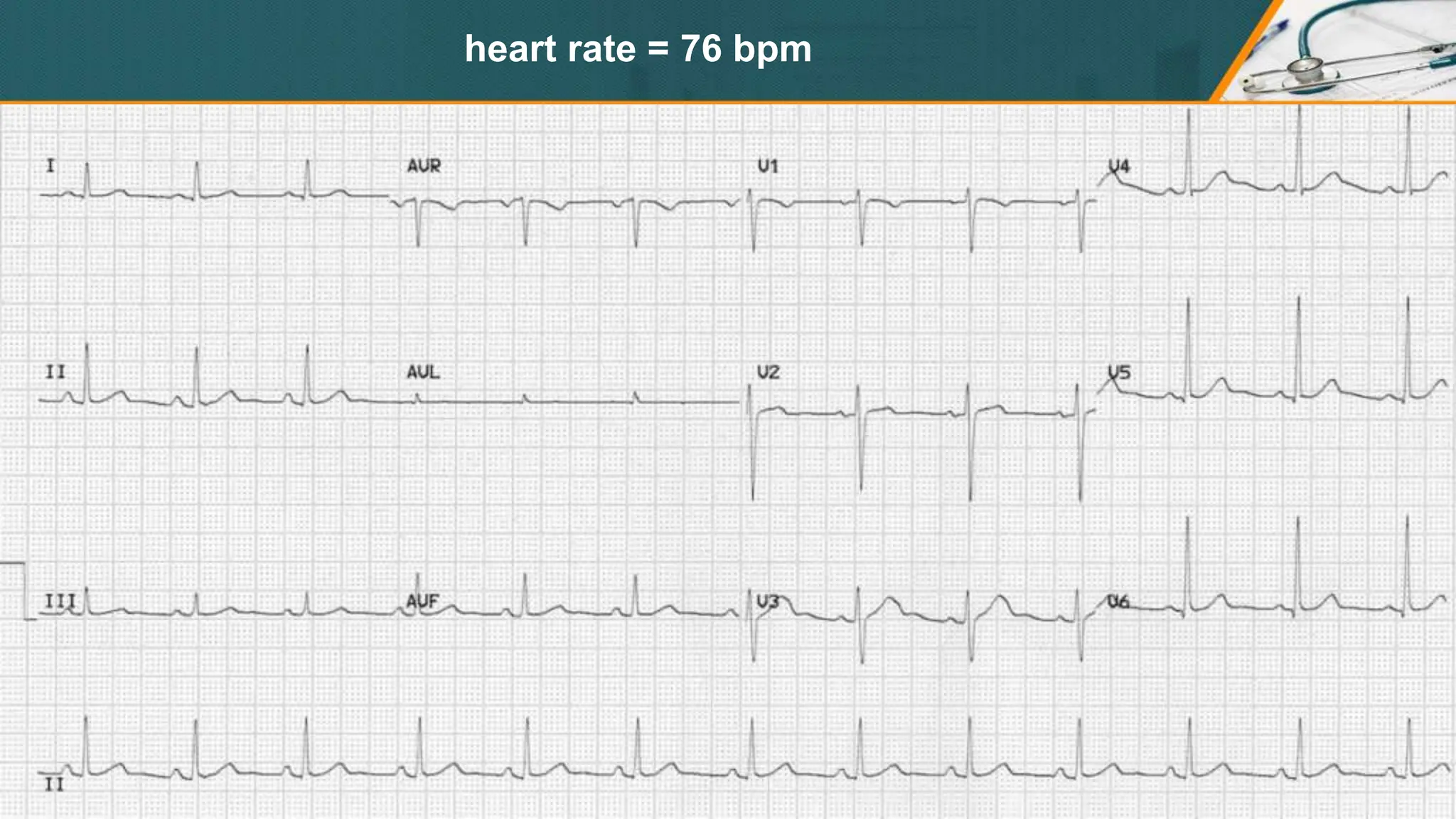
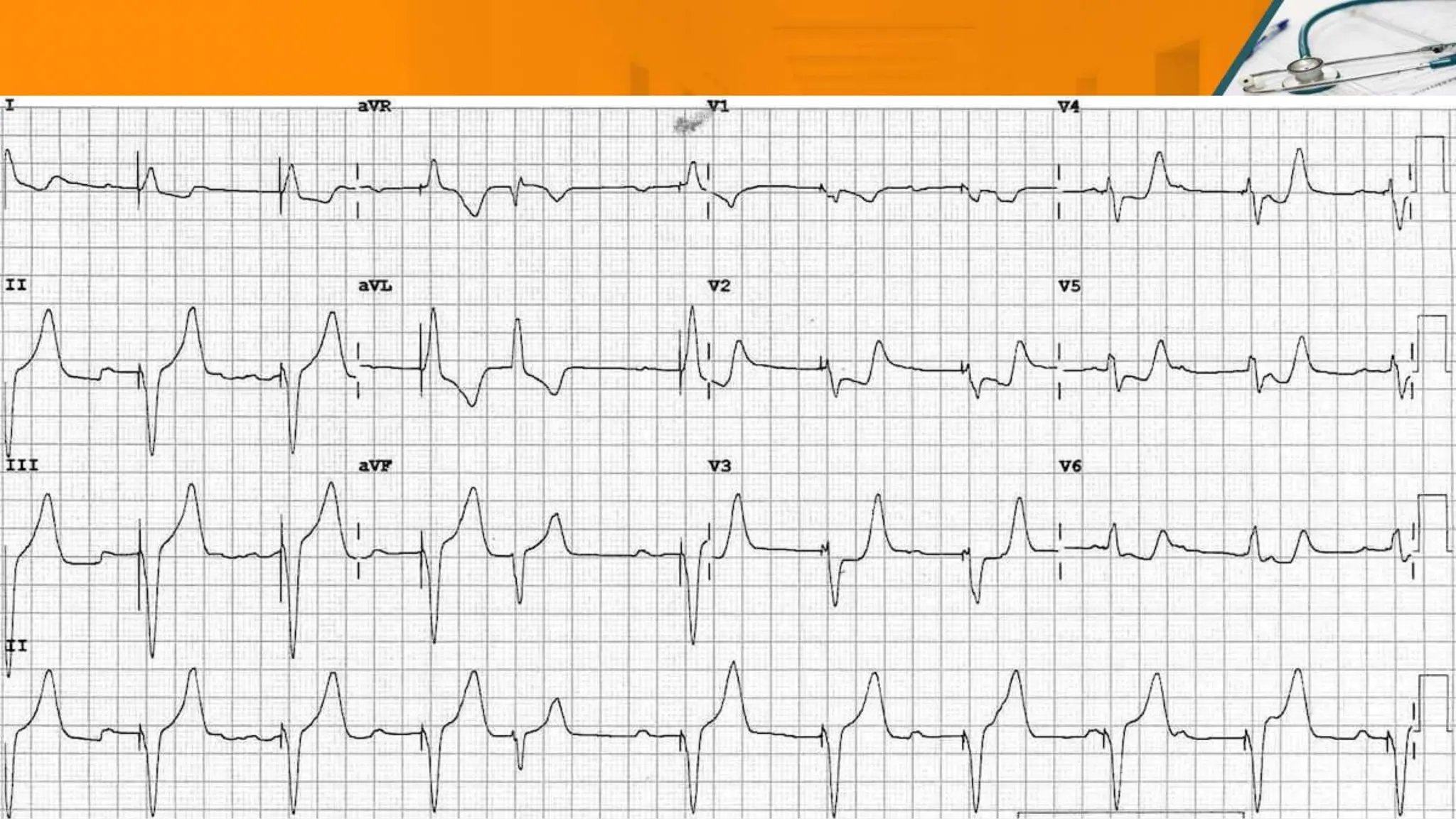
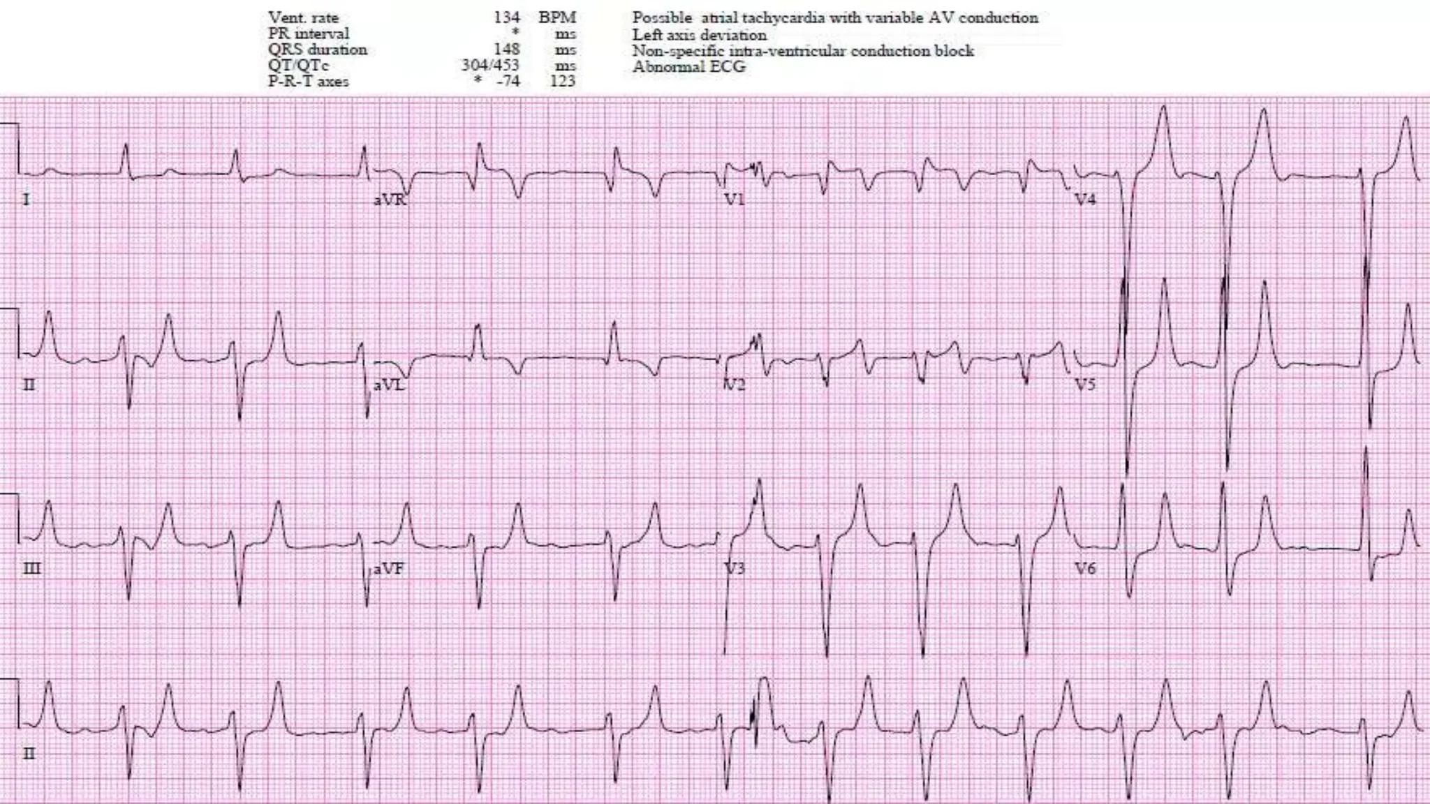
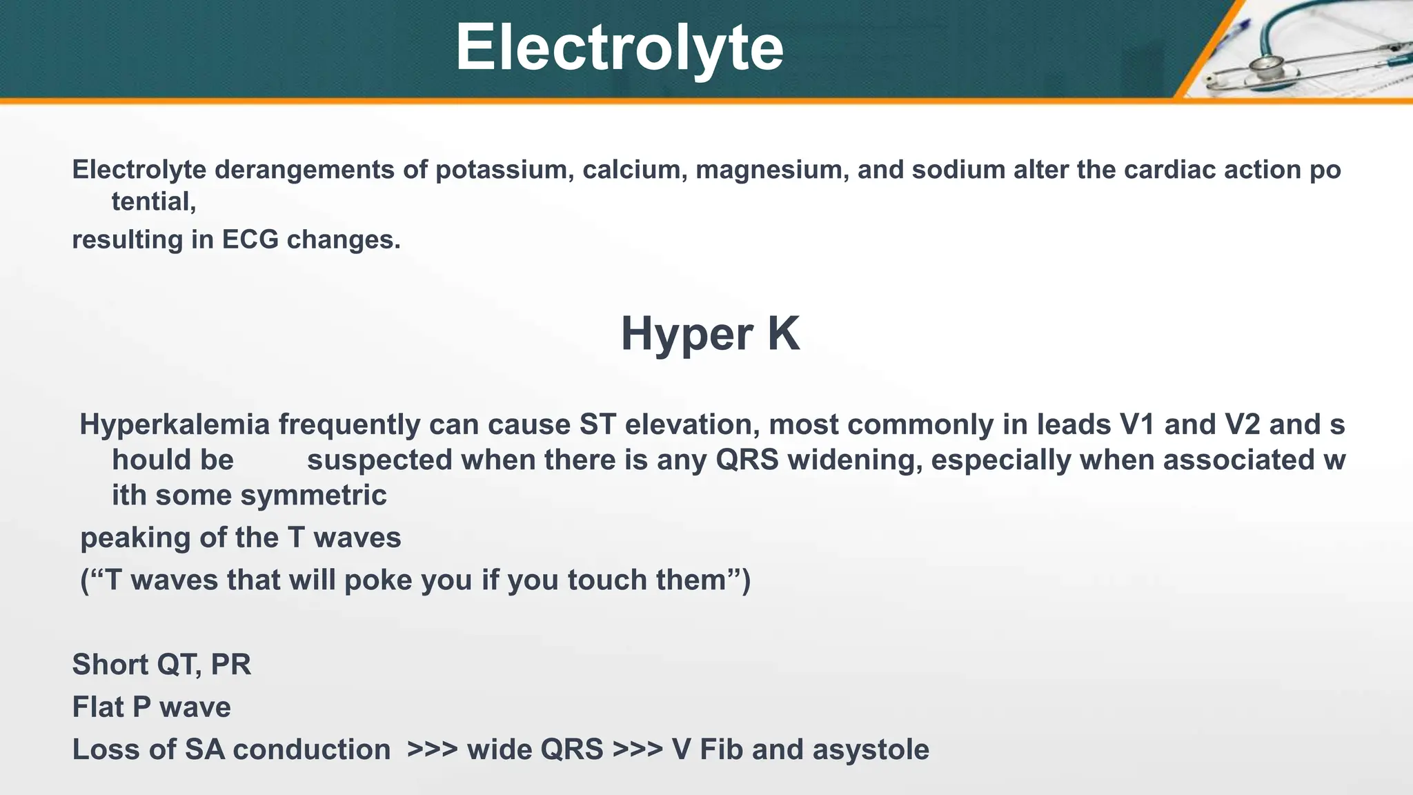
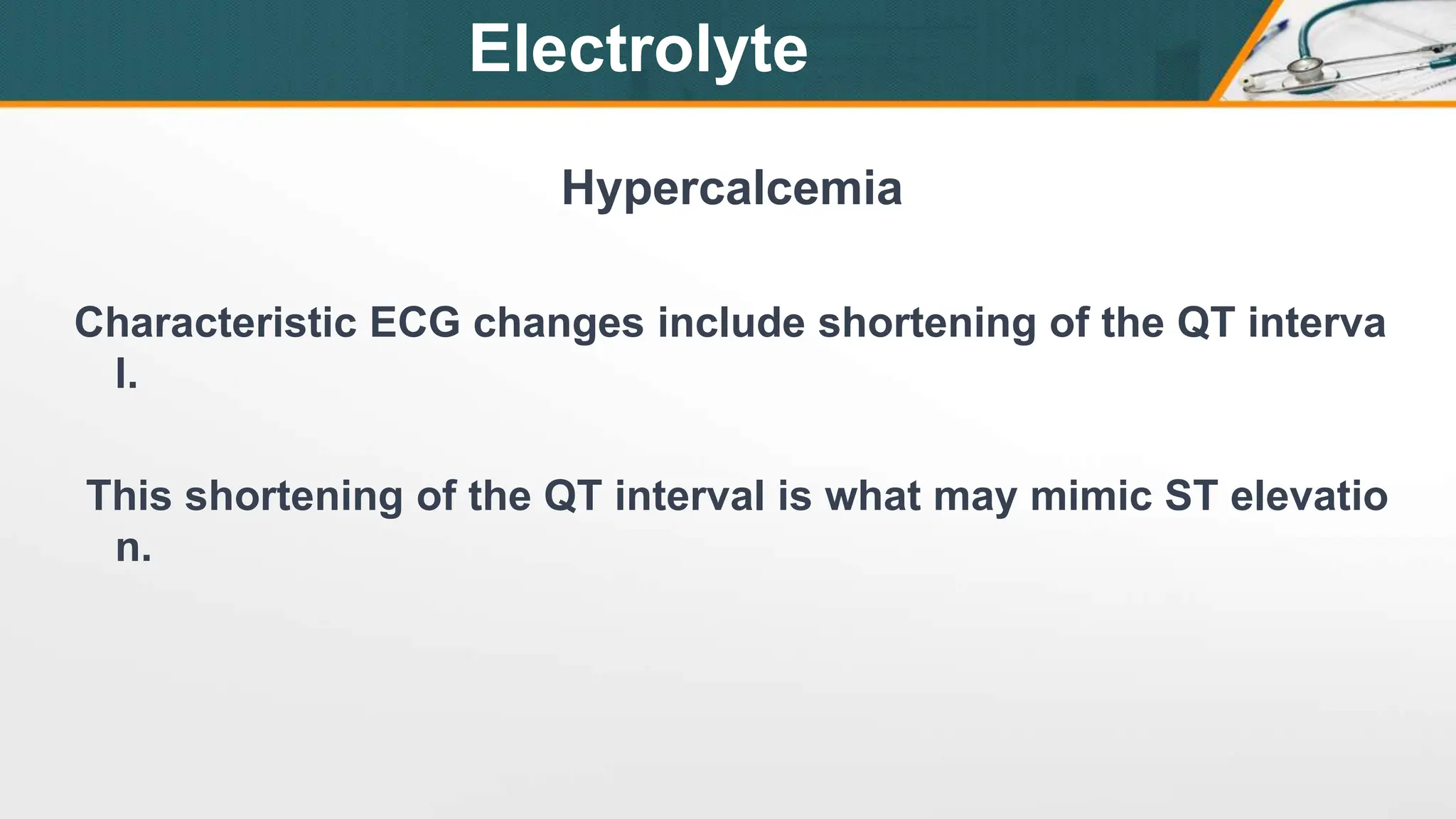
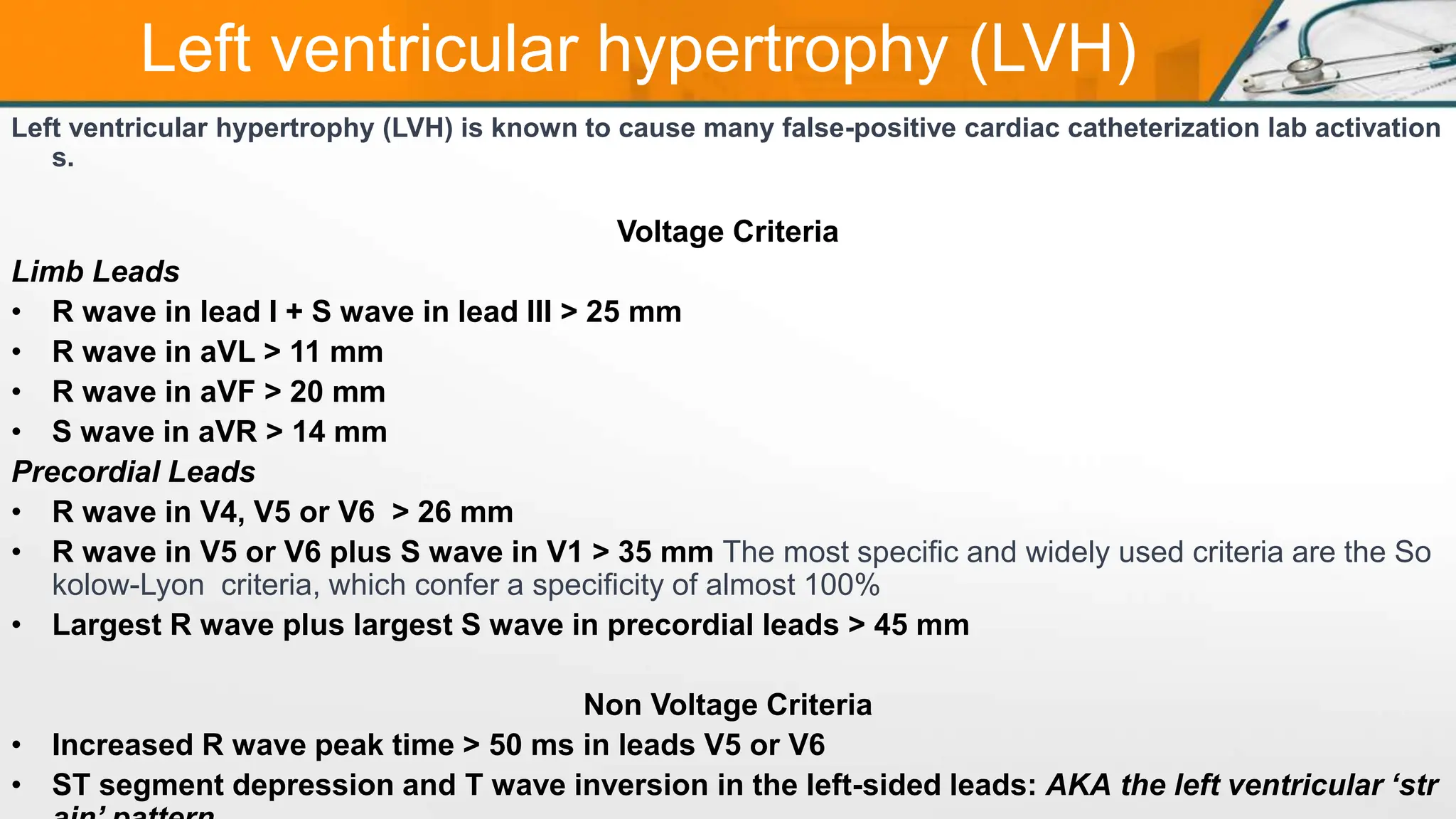
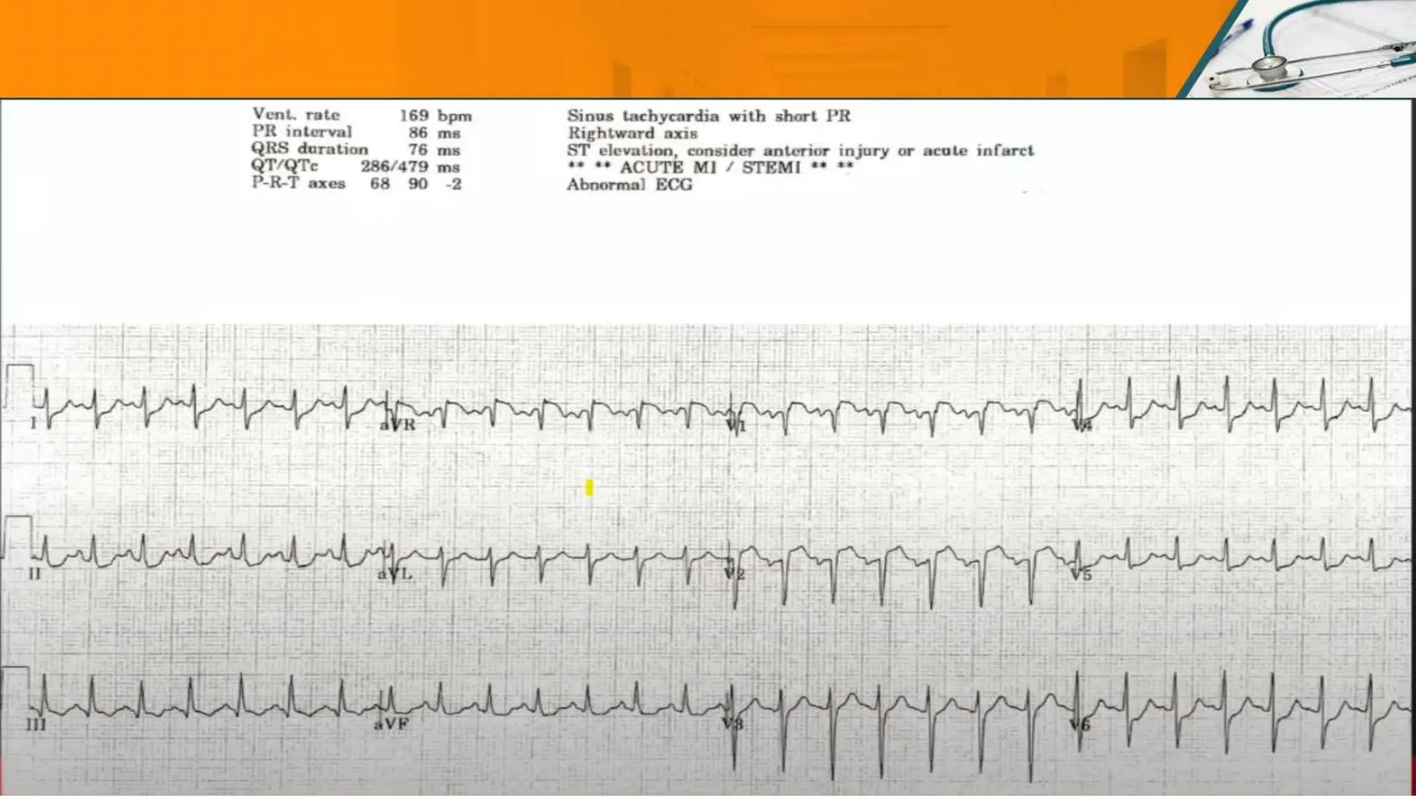
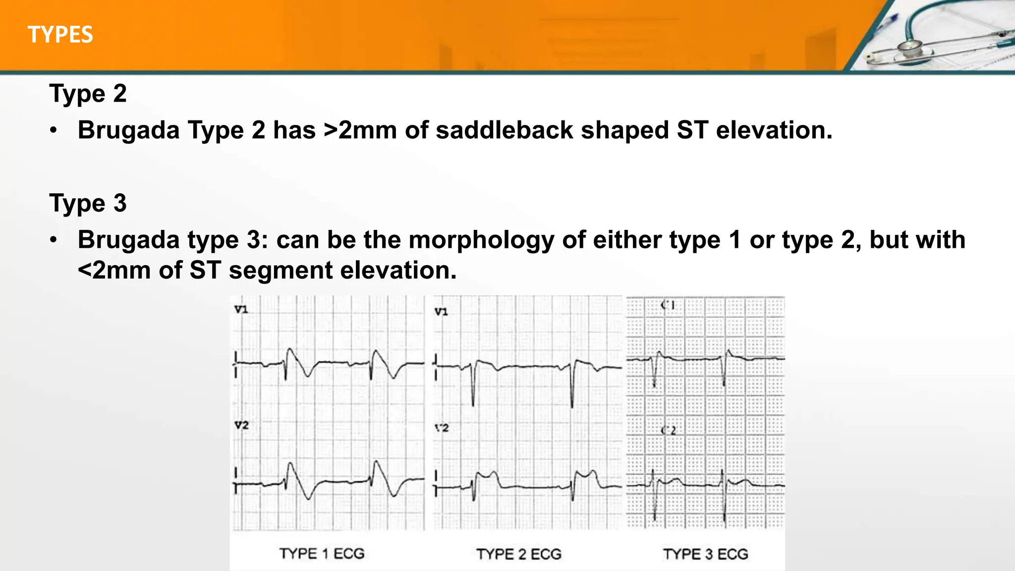
SlideShare a Scribd company logo
STEMI MIMICS
Dr. HAITHAM S HABTAR
SBEM, PGY-4
WHY IS THIS IMPORTEANT ?
Chest pain is a common cause of ED visits.
In patients with ECG findings consistent with a STEMI, the primary goal is t
o salvage myocardium through rapid reperfusion.
However, before activating the cardiac catheterization lab for ECG changes
,
it is important to consider non-ischemic causes of ST-segment elevation
Early repolarization was initially associated with young healthy athletes,
but increasingly is found in a wider variety of individuals.
The dramatic appearance of ST segment in multiple contiguous leads results in early repolarization being cited as the most
common cause of false-positive catheterization laboratory activations in patients without elevated cardiac biomarkers.
Over the years, different authors have used different criteria for the diagnosis of early repolarization. To provide consistency, in
2015, Hancock et al proposed three criteria that are required for the diagnosis of benign early repolarization:
• The QRS slur or notch (termed a J wave) must be on the downslope of the R wave and be above the isoelectric
line.
• The peak of the J point must be elevated ≥ 0.1 mV in two or more contiguous leads, except V1-V3.
• The QRS duration must be < 120 ms.
Early Repolarization
ST segment / T wave morphology
The ST segment-T wave complex in ER has a characteristic appearance:
• There is elevation of the J point
• The T wave is peaked and slightly asymmetrical
• The ST segment and the ascending limb of the T wave form an upward concavity (smiley
-shaped )
• The descending limb of the T wave is straighter and slightly steeper than the ascending li
mb
J-point morphology
One characteristic feature of ER is the presence of a notched or irregular J-point so-called
“fish hook” pattern.
This is often best seen in lead V4.
a healthy 17-year old female who was admitted to hospital following a benz
odiazepine overdose.
She had no chest pain, and cardiac biomarkers were normal.
heart rate = 76 bpm
heart rate = 54 bpm
Left Bundle Branch Block
ECG Diagnostic criteria of LBBB:
• QRS duration ≥ 120ms
• Dominant S wave in V1
• Broad monophasic R wave in lateral leads (I, aVL, V5-6)
• Absence of Q waves in lateral leads
• Prolonged R wave peak time > 60ms in leads V5-6
In hemodynamically stable patients with a presumed new LBBB, evaluation of their sympt
oms requires both measurement of cardiac biomarkers and observation.
In patients with hemodynamic compromise (including acute heart failure), revascularizatio
n should be emergently considered.
Sgarbossa criteria for LBBB
The Sgarbossa criteria can help guide the decision for emergent catheterization and coronary intervention in the
presence of both new and old LBBBs.
• Concordant ST elevation > 1mm in leads
with a positive QRS complex (score 5)
• Concordant ST depression > 1 mm in V1-V3 (score 3)
• Excessively discordant ST elevation > 5 mm in leads
with a -ve QRS complex (score 2)
These criteria are specific, but not sensitive (36%)
for myocardial infarction
. A total score of ≥ 3 is reported to have a specificity of 90%
for diagnosing myocardial infarction.
Smith-Modified Sgarbossa Criteria
Electrolyte derangements of potassium, calcium, magnesium, and sodium alter the cardiac action po
tential,
resulting in ECG changes.
Hyper K
Hyperkalemia frequently can cause ST elevation, most commonly in leads V1 and V2 and s
hould be suspected when there is any QRS widening, especially when associated w
ith some symmetric
peaking of the T waves
(“T waves that will poke you if you touch them”)
Short QT, PR
Flat P wave
Loss of SA conduction <<< wide QRS <<< V Fib and asystole
Electrolyte
Hypercalcemia
Characteristic ECG changes include shortening of the QT interva
l.
This shortening of the QT interval is what may mimic ST elevatio
n.
Electrolyte
Sodium channel blocker (SCB) toxicity
Sodium channel blocker (SCB) toxicity may manifest as ST elevation,
particularly in lead aVR.
SCB TOXICITY Like ??
Electrolyte
Left ventricular hypertrophy (LVH)
Left ventricular hypertrophy (LVH) is known to cause many false-positive cardiac catheterization lab activation
s.
Voltage Criteria
Limb Leads
• R wave in lead I + S wave in lead III > 25 mm
• R wave in aVL > 11 mm
• R wave in aVF > 20 mm
• S wave in aVR > 14 mm
Precordial Leads
• R wave in V4, V5 or V6 > 26 mm
• R wave in V5 or V6 plus S wave in V1 > 35 mm The most specific and widely used criteria are the So
kolow-Lyon criteria, which confer a specificity of almost 100%
• Largest R wave plus largest S wave in precordial leads > 45 mm
Non Voltage Criteria
• Increased R wave peak time > 50 ms in leads V5 or V6
• ST segment depression and T wave inversion in the left-sided leads: AKA the left ventricular ‘str
Acute Pulmonary Embolism
ECG findings compared to Acute Coronary Syndrome
• ACS is rarely associated with tachycardia
• Rt Axis deviation rarely seen in STEMI
• Bedside echo may be useful in differentiating the two, demonstrating features of RV dilatation and
pulmonary
arterial hypertension
• Kosuge et al have shown that simultaneous inversion in III and V1 are diagnostically significant:
Negative T waves in leads III and V1 were observed in only 1% of patients with ACS compared with 88% of patient
s with Acute PE (p less than 0.001). The sensitivity, specificity, positive predictive value, and negative predictive
value of this finding for the diagnosis of PE were 88%, 99%, 97%, and 95%, respectively. In conclusion, the presen
ce of negative T waves in both leads III and V1 allows PE to be differentiated simply but accurately from ACS in p
atients with negative T waves in the
precordial leads.
Acute Pulmonary Embolism
When a transmural infarct is not aborted by therapeutic intervention and the AMI completes itself, the
myocardium is replaced by a thin, fibrous layer, which is called an LV aneurysm.
On the ECG, a left ventricular aneurysm may manifest as persistent ST elevation in the territory of a p
rior infarct,
commonly concomitant with Q waves.
The rule states that when the differential diagnosis is acute LAD occlusion vs. anterior LV aneurysm,
if any of
leads V1-V4 has a T wave amplitude to QRS amplitude ratio of > 0.36, then STEMI is likely.
In general, aneurysm is favored by prominent Q waves in leads V1-V4 with corresponding diminishe
d T wave
amplitudes.
• T-wave/QRS ratio < 0.36 in all precordial leads favours LV aneurysm
• T-wave/QRS ratio > 0.36 in any precordial lead favours anterior STEMI
Aneurysm
Brugada syndrome
Brugada Syndrome is an ECG abnormality with a high incidence of sudden death in patient
s with structurally normal hearts.
Brugada syndrome is due to a mutation in the cardiac sodium channel gene.
• Diagnosis depends on a characteristic ECG finding AND clinical criteria.
• Further risk stratification is controversial.
• Definitive treatment = ICD.
• Brugada sign in isolation is of questionable significance.
Diagnostic Criteria
This ECG abnormality must be associated with one of the following clinical criteria to make
the diagnosis:
• Documented ventricular fibrillation (VF) or polymorphic ventricular tachycardia (VT).
• Family history of sudden cardiac death at <45 years old .
• Coved-type ECGs in family members.
• Inducibility of VT with programmed electrical stimulation .
• Syncope.
• Nocturnal agonal respiration.
TYPES
Type 1
• Coved ST segment elevation >2mm in >1 of V1-V3 followed by a negative
T wave.
• This is the only ECG abnormality that is potentially diagnostic.
• It is often referred to as Brugada sign.
TYPES
Type 2
• Brugada Type 2 has >2mm of saddleback shaped ST elevation.
Type 3
• Brugada type 3: can be the morphology of either type 1 or type 2, but with
<2mm of ST segment elevation.
Look for features of STEMI first:
• Search for ST depression in leads other than aVR and V1
• Look for ST elevation in lead III > II
• Search for horizontal or convex upward ST elevation
• New Q wave
pericarditis
Temp: 32 c
Hypothermia
Hypothermia is defined as a core body temperature of < 35 °C
The Osborn wave (J wave) is a positive deflection
at the J point (negative in aVR and V1).
• It is usually most prominent in the precordial leads.
• The height of the Osborn wave is roughly proportional
to the degree of hypothermia
RESOURSE
THANK YOU

More Related Content

Similar to STEMI Mimic WHAT IS IT AND HOW TO IDENTIFY IT ?

Stemi equivalents
Stemi equivalentsStemi equivalents
Stemi equivalents
Mohamed Hamoda
 
Presentation the electrocardiogram in the acs patient
Presentation the electrocardiogram in the acs patientPresentation the electrocardiogram in the acs patient
Presentation the electrocardiogram in the acs patient
drwaque
 
RK ECG.pptx
RK ECG.pptxRK ECG.pptx
RK ECG.pptx
RigilKent7
 
Ekg Cases 7 15 09 Level 2 Part 1
Ekg Cases 7 15 09 Level 2 Part 1Ekg Cases 7 15 09 Level 2 Part 1
Ekg Cases 7 15 09 Level 2 Part 1Michael LaCombe
 
ECG life in the fastlane.pptx
ECG life in the fastlane.pptxECG life in the fastlane.pptx
ECG life in the fastlane.pptx
abdogamala2
 
Ekg Cases 7 8 09 Level 1 Part2
Ekg Cases 7 8 09 Level 1 Part2Ekg Cases 7 8 09 Level 1 Part2
Ekg Cases 7 8 09 Level 1 Part2Michael LaCombe
 
A vr case presentation +kfhh c shock
A vr case presentation +kfhh c shockA vr case presentation +kfhh c shock
A vr case presentation +kfhh c shock
asadsoomro1960
 
How to read ECG systematically with practice strips
How to read ECG systematically with practice strips How to read ECG systematically with practice strips
How to read ECG systematically with practice strips
Khaled AlKhodari
 
INTERESTING ECGS -- PART II
INTERESTING ECGS -- PART IIINTERESTING ECGS -- PART II
INTERESTING ECGS -- PART II
Praveen Nagula
 
Lethal ECG pattern reading and diagnosis.pptx
Lethal ECG pattern reading and diagnosis.pptxLethal ECG pattern reading and diagnosis.pptx
Lethal ECG pattern reading and diagnosis.pptx
Hussein Alwais
 
Basic ecg
Basic ecgBasic ecg
Mimicks Masks of Myocardial Infarction.pptx
Mimicks Masks of Myocardial Infarction.pptxMimicks Masks of Myocardial Infarction.pptx
Mimicks Masks of Myocardial Infarction.pptx
RajeshPonnada3
 
Systematic approach to wide qrs tachycardia
Systematic approach to wide qrs tachycardiaSystematic approach to wide qrs tachycardia
Systematic approach to wide qrs tachycardiasalah_atta
 
Complete EKG Interpretation Course
Complete EKG Interpretation Course Complete EKG Interpretation Course
Complete EKG Interpretation Course
Kerolus Shehata
 
Ecg in athletes and young age
Ecg in athletes and young ageEcg in athletes and young age
Ecg in athletes and young age
Magdy Khames Aly
 
EKG Dasar medis michael johandadasdas.ppt
EKG Dasar medis michael johandadasdas.pptEKG Dasar medis michael johandadasdas.ppt
EKG Dasar medis michael johandadasdas.ppt
michaeljohan1211
 
ECG Basics
ECG BasicsECG Basics

Similar to STEMI Mimic WHAT IS IT AND HOW TO IDENTIFY IT ? (20)

Stemi equivalents
Stemi equivalentsStemi equivalents
Stemi equivalents
 
Presentation the electrocardiogram in the acs patient
Presentation the electrocardiogram in the acs patientPresentation the electrocardiogram in the acs patient
Presentation the electrocardiogram in the acs patient
 
RK ECG.pptx
RK ECG.pptxRK ECG.pptx
RK ECG.pptx
 
Ekg Cases 7 15 09 Level 2 Part 1
Ekg Cases 7 15 09 Level 2 Part 1Ekg Cases 7 15 09 Level 2 Part 1
Ekg Cases 7 15 09 Level 2 Part 1
 
ECG life in the fastlane.pptx
ECG life in the fastlane.pptxECG life in the fastlane.pptx
ECG life in the fastlane.pptx
 
Ekg Cases 7 8 09 Level 1 Part2
Ekg Cases 7 8 09 Level 1 Part2Ekg Cases 7 8 09 Level 1 Part2
Ekg Cases 7 8 09 Level 1 Part2
 
A vr case presentation +kfhh c shock
A vr case presentation +kfhh c shockA vr case presentation +kfhh c shock
A vr case presentation +kfhh c shock
 
How to read ECG systematically with practice strips
How to read ECG systematically with practice strips How to read ECG systematically with practice strips
How to read ECG systematically with practice strips
 
44 E C G
44 E C G44 E C G
44 E C G
 
44 E C G
44 E C G44 E C G
44 E C G
 
INTERESTING ECGS -- PART II
INTERESTING ECGS -- PART IIINTERESTING ECGS -- PART II
INTERESTING ECGS -- PART II
 
Lethal ECG pattern reading and diagnosis.pptx
Lethal ECG pattern reading and diagnosis.pptxLethal ECG pattern reading and diagnosis.pptx
Lethal ECG pattern reading and diagnosis.pptx
 
Basic ecg
Basic ecgBasic ecg
Basic ecg
 
Mimicks Masks of Myocardial Infarction.pptx
Mimicks Masks of Myocardial Infarction.pptxMimicks Masks of Myocardial Infarction.pptx
Mimicks Masks of Myocardial Infarction.pptx
 
Systematic approach to wide qrs tachycardia
Systematic approach to wide qrs tachycardiaSystematic approach to wide qrs tachycardia
Systematic approach to wide qrs tachycardia
 
Complete EKG Interpretation Course
Complete EKG Interpretation Course Complete EKG Interpretation Course
Complete EKG Interpretation Course
 
Ecg in athletes and young age
Ecg in athletes and young ageEcg in athletes and young age
Ecg in athletes and young age
 
EKG Dasar medis michael johandadasdas.ppt
EKG Dasar medis michael johandadasdas.pptEKG Dasar medis michael johandadasdas.ppt
EKG Dasar medis michael johandadasdas.ppt
 
Mi in lbbb i.tammi raju
Mi in lbbb i.tammi rajuMi in lbbb i.tammi raju
Mi in lbbb i.tammi raju
 
ECG Basics
ECG BasicsECG Basics
ECG Basics
 

More from Haitham Habtar

Massive Transfusion Protocol ( MTP ) HOW AND WHY ?
Massive Transfusion Protocol ( MTP ) HOW AND WHY ?Massive Transfusion Protocol ( MTP ) HOW AND WHY ?
Massive Transfusion Protocol ( MTP ) HOW AND WHY ?
Haitham Habtar
 
Acute Myopericardial Syndromes
Acute Myopericardial SyndromesAcute Myopericardial Syndromes
Acute Myopericardial Syndromes
Haitham Habtar
 
NECK TRAUMA
NECK TRAUMANECK TRAUMA
NECK TRAUMA
Haitham Habtar
 
Diabetic Ketoacidosis and Hyperglycemic Hyperosmolar State
Diabetic Ketoacidosis and Hyperglycemic Hyperosmolar StateDiabetic Ketoacidosis and Hyperglycemic Hyperosmolar State
Diabetic Ketoacidosis and Hyperglycemic Hyperosmolar State
Haitham Habtar
 
peripheral and CNS infection
peripheral and CNS infectionperipheral and CNS infection
peripheral and CNS infection
Haitham Habtar
 
ELEVATED SERUM AMINOTRANSFERASES
ELEVATED SERUM AMINOTRANSFERASESELEVATED SERUM AMINOTRANSFERASES
ELEVATED SERUM AMINOTRANSFERASES
Haitham Habtar
 
Approach to patient with chronic complications of diabetes
Approach to patient with chronic complications of diabetesApproach to patient with chronic complications of diabetes
Approach to patient with chronic complications of diabetes
Haitham Habtar
 

More from Haitham Habtar (7)

Massive Transfusion Protocol ( MTP ) HOW AND WHY ?
Massive Transfusion Protocol ( MTP ) HOW AND WHY ?Massive Transfusion Protocol ( MTP ) HOW AND WHY ?
Massive Transfusion Protocol ( MTP ) HOW AND WHY ?
 
Acute Myopericardial Syndromes
Acute Myopericardial SyndromesAcute Myopericardial Syndromes
Acute Myopericardial Syndromes
 
NECK TRAUMA
NECK TRAUMANECK TRAUMA
NECK TRAUMA
 
Diabetic Ketoacidosis and Hyperglycemic Hyperosmolar State
Diabetic Ketoacidosis and Hyperglycemic Hyperosmolar StateDiabetic Ketoacidosis and Hyperglycemic Hyperosmolar State
Diabetic Ketoacidosis and Hyperglycemic Hyperosmolar State
 
peripheral and CNS infection
peripheral and CNS infectionperipheral and CNS infection
peripheral and CNS infection
 
ELEVATED SERUM AMINOTRANSFERASES
ELEVATED SERUM AMINOTRANSFERASESELEVATED SERUM AMINOTRANSFERASES
ELEVATED SERUM AMINOTRANSFERASES
 
Approach to patient with chronic complications of diabetes
Approach to patient with chronic complications of diabetesApproach to patient with chronic complications of diabetes
Approach to patient with chronic complications of diabetes
 

Recently uploaded

Effective-Soaps-for-Fungal-Skin-Infections.pptx
Effective-Soaps-for-Fungal-Skin-Infections.pptxEffective-Soaps-for-Fungal-Skin-Infections.pptx
Effective-Soaps-for-Fungal-Skin-Infections.pptx
SwisschemDerma
 
Cervical & Brachial Plexus By Dr. RIG.pptx
Cervical & Brachial Plexus By Dr. RIG.pptxCervical & Brachial Plexus By Dr. RIG.pptx
Cervical & Brachial Plexus By Dr. RIG.pptx
Dr. Rabia Inam Gandapore
 
Top-Vitamin-Supplement-Brands-in-India.pptx
Top-Vitamin-Supplement-Brands-in-India.pptxTop-Vitamin-Supplement-Brands-in-India.pptx
Top-Vitamin-Supplement-Brands-in-India.pptx
SwisschemDerma
 
Light House Retreats: Plant Medicine Retreat Europe
Light House Retreats: Plant Medicine Retreat EuropeLight House Retreats: Plant Medicine Retreat Europe
Light House Retreats: Plant Medicine Retreat Europe
Lighthouse Retreat
 
Tom Selleck Health: A Comprehensive Look at the Iconic Actor’s Wellness Journey
Tom Selleck Health: A Comprehensive Look at the Iconic Actor’s Wellness JourneyTom Selleck Health: A Comprehensive Look at the Iconic Actor’s Wellness Journey
Tom Selleck Health: A Comprehensive Look at the Iconic Actor’s Wellness Journey
greendigital
 
Colonic and anorectal physiology with surgical implications
Colonic and anorectal physiology with surgical implicationsColonic and anorectal physiology with surgical implications
Colonic and anorectal physiology with surgical implications
Dr Maria Tamanna
 
Ophthalmology Clinical Tests for OSCE exam
Ophthalmology Clinical Tests for OSCE examOphthalmology Clinical Tests for OSCE exam
Ophthalmology Clinical Tests for OSCE exam
KafrELShiekh University
 
Aortic Association CBL Pilot April 19 – 20 Bern
Aortic Association CBL Pilot April 19 – 20 BernAortic Association CBL Pilot April 19 – 20 Bern
Aortic Association CBL Pilot April 19 – 20 Bern
suvadeepdas911
 
Non-respiratory Functions of the Lungs.pdf
Non-respiratory Functions of the Lungs.pdfNon-respiratory Functions of the Lungs.pdf
Non-respiratory Functions of the Lungs.pdf
MedicoseAcademics
 
Adv. biopharm. APPLICATION OF PHARMACOKINETICS : TARGETED DRUG DELIVERY SYSTEMS
Adv. biopharm. APPLICATION OF PHARMACOKINETICS : TARGETED DRUG DELIVERY SYSTEMSAdv. biopharm. APPLICATION OF PHARMACOKINETICS : TARGETED DRUG DELIVERY SYSTEMS
Adv. biopharm. APPLICATION OF PHARMACOKINETICS : TARGETED DRUG DELIVERY SYSTEMS
AkankshaAshtankar
 
CDSCO and Phamacovigilance {Regulatory body in India}
CDSCO and Phamacovigilance {Regulatory body in India}CDSCO and Phamacovigilance {Regulatory body in India}
CDSCO and Phamacovigilance {Regulatory body in India}
NEHA GUPTA
 
BRACHYTHERAPY OVERVIEW AND APPLICATORS
BRACHYTHERAPY OVERVIEW  AND  APPLICATORSBRACHYTHERAPY OVERVIEW  AND  APPLICATORS
BRACHYTHERAPY OVERVIEW AND APPLICATORS
Krishan Murari
 
Maxilla, Mandible & Hyoid Bone & Clinical Correlations by Dr. RIG.pptx
Maxilla, Mandible & Hyoid Bone & Clinical Correlations by Dr. RIG.pptxMaxilla, Mandible & Hyoid Bone & Clinical Correlations by Dr. RIG.pptx
Maxilla, Mandible & Hyoid Bone & Clinical Correlations by Dr. RIG.pptx
Dr. Rabia Inam Gandapore
 
Pharynx and Clinical Correlations BY Dr.Rabia Inam Gandapore.pptx
Pharynx and Clinical Correlations BY Dr.Rabia Inam Gandapore.pptxPharynx and Clinical Correlations BY Dr.Rabia Inam Gandapore.pptx
Pharynx and Clinical Correlations BY Dr.Rabia Inam Gandapore.pptx
Dr. Rabia Inam Gandapore
 
263778731218 Abortion Clinic /Pills In Harare ,
263778731218 Abortion Clinic /Pills In Harare ,263778731218 Abortion Clinic /Pills In Harare ,
263778731218 Abortion Clinic /Pills In Harare ,
sisternakatoto
 
Hemodialysis: Chapter 3, Dialysis Water Unit - Dr.Gawad
Hemodialysis: Chapter 3, Dialysis Water Unit - Dr.GawadHemodialysis: Chapter 3, Dialysis Water Unit - Dr.Gawad
Hemodialysis: Chapter 3, Dialysis Water Unit - Dr.Gawad
NephroTube - Dr.Gawad
 
SURGICAL ANATOMY OF THE RETROPERITONEUM, ADRENALS, KIDNEYS AND URETERS.pptx
SURGICAL ANATOMY OF THE RETROPERITONEUM, ADRENALS, KIDNEYS AND URETERS.pptxSURGICAL ANATOMY OF THE RETROPERITONEUM, ADRENALS, KIDNEYS AND URETERS.pptx
SURGICAL ANATOMY OF THE RETROPERITONEUM, ADRENALS, KIDNEYS AND URETERS.pptx
Bright Chipili
 
Role of Mukta Pishti in the Management of Hyperthyroidism
Role of Mukta Pishti in the Management of HyperthyroidismRole of Mukta Pishti in the Management of Hyperthyroidism
Role of Mukta Pishti in the Management of Hyperthyroidism
Dr. Jyothirmai Paindla
 
NVBDCP.pptx Nation vector borne disease control program
NVBDCP.pptx Nation vector borne disease control programNVBDCP.pptx Nation vector borne disease control program
NVBDCP.pptx Nation vector borne disease control program
Sapna Thakur
 
Gram Stain introduction, principle, Procedure
Gram Stain introduction, principle, ProcedureGram Stain introduction, principle, Procedure
Gram Stain introduction, principle, Procedure
Suraj Goswami
 

Recently uploaded (20)

Effective-Soaps-for-Fungal-Skin-Infections.pptx
Effective-Soaps-for-Fungal-Skin-Infections.pptxEffective-Soaps-for-Fungal-Skin-Infections.pptx
Effective-Soaps-for-Fungal-Skin-Infections.pptx
 
Cervical & Brachial Plexus By Dr. RIG.pptx
Cervical & Brachial Plexus By Dr. RIG.pptxCervical & Brachial Plexus By Dr. RIG.pptx
Cervical & Brachial Plexus By Dr. RIG.pptx
 
Top-Vitamin-Supplement-Brands-in-India.pptx
Top-Vitamin-Supplement-Brands-in-India.pptxTop-Vitamin-Supplement-Brands-in-India.pptx
Top-Vitamin-Supplement-Brands-in-India.pptx
 
Light House Retreats: Plant Medicine Retreat Europe
Light House Retreats: Plant Medicine Retreat EuropeLight House Retreats: Plant Medicine Retreat Europe
Light House Retreats: Plant Medicine Retreat Europe
 
Tom Selleck Health: A Comprehensive Look at the Iconic Actor’s Wellness Journey
Tom Selleck Health: A Comprehensive Look at the Iconic Actor’s Wellness JourneyTom Selleck Health: A Comprehensive Look at the Iconic Actor’s Wellness Journey
Tom Selleck Health: A Comprehensive Look at the Iconic Actor’s Wellness Journey
 
Colonic and anorectal physiology with surgical implications
Colonic and anorectal physiology with surgical implicationsColonic and anorectal physiology with surgical implications
Colonic and anorectal physiology with surgical implications
 
Ophthalmology Clinical Tests for OSCE exam
Ophthalmology Clinical Tests for OSCE examOphthalmology Clinical Tests for OSCE exam
Ophthalmology Clinical Tests for OSCE exam
 
Aortic Association CBL Pilot April 19 – 20 Bern
Aortic Association CBL Pilot April 19 – 20 BernAortic Association CBL Pilot April 19 – 20 Bern
Aortic Association CBL Pilot April 19 – 20 Bern
 
Non-respiratory Functions of the Lungs.pdf
Non-respiratory Functions of the Lungs.pdfNon-respiratory Functions of the Lungs.pdf
Non-respiratory Functions of the Lungs.pdf
 
Adv. biopharm. APPLICATION OF PHARMACOKINETICS : TARGETED DRUG DELIVERY SYSTEMS
Adv. biopharm. APPLICATION OF PHARMACOKINETICS : TARGETED DRUG DELIVERY SYSTEMSAdv. biopharm. APPLICATION OF PHARMACOKINETICS : TARGETED DRUG DELIVERY SYSTEMS
Adv. biopharm. APPLICATION OF PHARMACOKINETICS : TARGETED DRUG DELIVERY SYSTEMS
 
CDSCO and Phamacovigilance {Regulatory body in India}
CDSCO and Phamacovigilance {Regulatory body in India}CDSCO and Phamacovigilance {Regulatory body in India}
CDSCO and Phamacovigilance {Regulatory body in India}
 
BRACHYTHERAPY OVERVIEW AND APPLICATORS
BRACHYTHERAPY OVERVIEW  AND  APPLICATORSBRACHYTHERAPY OVERVIEW  AND  APPLICATORS
BRACHYTHERAPY OVERVIEW AND APPLICATORS
 
Maxilla, Mandible & Hyoid Bone & Clinical Correlations by Dr. RIG.pptx
Maxilla, Mandible & Hyoid Bone & Clinical Correlations by Dr. RIG.pptxMaxilla, Mandible & Hyoid Bone & Clinical Correlations by Dr. RIG.pptx
Maxilla, Mandible & Hyoid Bone & Clinical Correlations by Dr. RIG.pptx
 
Pharynx and Clinical Correlations BY Dr.Rabia Inam Gandapore.pptx
Pharynx and Clinical Correlations BY Dr.Rabia Inam Gandapore.pptxPharynx and Clinical Correlations BY Dr.Rabia Inam Gandapore.pptx
Pharynx and Clinical Correlations BY Dr.Rabia Inam Gandapore.pptx
 
263778731218 Abortion Clinic /Pills In Harare ,
263778731218 Abortion Clinic /Pills In Harare ,263778731218 Abortion Clinic /Pills In Harare ,
263778731218 Abortion Clinic /Pills In Harare ,
 
Hemodialysis: Chapter 3, Dialysis Water Unit - Dr.Gawad
Hemodialysis: Chapter 3, Dialysis Water Unit - Dr.GawadHemodialysis: Chapter 3, Dialysis Water Unit - Dr.Gawad
Hemodialysis: Chapter 3, Dialysis Water Unit - Dr.Gawad
 
SURGICAL ANATOMY OF THE RETROPERITONEUM, ADRENALS, KIDNEYS AND URETERS.pptx
SURGICAL ANATOMY OF THE RETROPERITONEUM, ADRENALS, KIDNEYS AND URETERS.pptxSURGICAL ANATOMY OF THE RETROPERITONEUM, ADRENALS, KIDNEYS AND URETERS.pptx
SURGICAL ANATOMY OF THE RETROPERITONEUM, ADRENALS, KIDNEYS AND URETERS.pptx
 
Role of Mukta Pishti in the Management of Hyperthyroidism
Role of Mukta Pishti in the Management of HyperthyroidismRole of Mukta Pishti in the Management of Hyperthyroidism
Role of Mukta Pishti in the Management of Hyperthyroidism
 
NVBDCP.pptx Nation vector borne disease control program
NVBDCP.pptx Nation vector borne disease control programNVBDCP.pptx Nation vector borne disease control program
NVBDCP.pptx Nation vector borne disease control program
 
Gram Stain introduction, principle, Procedure
Gram Stain introduction, principle, ProcedureGram Stain introduction, principle, Procedure
Gram Stain introduction, principle, Procedure
 

STEMI Mimic WHAT IS IT AND HOW TO IDENTIFY IT ?

  • 1. STEMI MIMICS Dr. HAITHAM S HABTAR SBEM, PGY-4
  • 2. WHY IS THIS IMPORTEANT ? Chest pain is a common cause of ED visits. In patients with ECG findings consistent with a STEMI, the primary goal is t o salvage myocardium through rapid reperfusion. However, before activating the cardiac catheterization lab for ECG changes , it is important to consider non-ischemic causes of ST-segment elevation
  • 3.
  • 4.
  • 5. Early repolarization was initially associated with young healthy athletes, but increasingly is found in a wider variety of individuals. The dramatic appearance of ST segment in multiple contiguous leads results in early repolarization being cited as the most common cause of false-positive catheterization laboratory activations in patients without elevated cardiac biomarkers. Over the years, different authors have used different criteria for the diagnosis of early repolarization. To provide consistency, in 2015, Hancock et al proposed three criteria that are required for the diagnosis of benign early repolarization: • The QRS slur or notch (termed a J wave) must be on the downslope of the R wave and be above the isoelectric line. • The peak of the J point must be elevated ≥ 0.1 mV in two or more contiguous leads, except V1-V3. • The QRS duration must be < 120 ms. Early Repolarization
  • 6. ST segment / T wave morphology The ST segment-T wave complex in ER has a characteristic appearance: • There is elevation of the J point • The T wave is peaked and slightly asymmetrical • The ST segment and the ascending limb of the T wave form an upward concavity (smiley -shaped ) • The descending limb of the T wave is straighter and slightly steeper than the ascending li mb
  • 7. J-point morphology One characteristic feature of ER is the presence of a notched or irregular J-point so-called “fish hook” pattern. This is often best seen in lead V4.
  • 8. a healthy 17-year old female who was admitted to hospital following a benz odiazepine overdose. She had no chest pain, and cardiac biomarkers were normal.
  • 9. heart rate = 76 bpm
  • 10. heart rate = 54 bpm
  • 11.
  • 12. Left Bundle Branch Block ECG Diagnostic criteria of LBBB: • QRS duration ≥ 120ms • Dominant S wave in V1 • Broad monophasic R wave in lateral leads (I, aVL, V5-6) • Absence of Q waves in lateral leads • Prolonged R wave peak time > 60ms in leads V5-6 In hemodynamically stable patients with a presumed new LBBB, evaluation of their sympt oms requires both measurement of cardiac biomarkers and observation. In patients with hemodynamic compromise (including acute heart failure), revascularizatio n should be emergently considered.
  • 13.
  • 14. Sgarbossa criteria for LBBB The Sgarbossa criteria can help guide the decision for emergent catheterization and coronary intervention in the presence of both new and old LBBBs. • Concordant ST elevation > 1mm in leads with a positive QRS complex (score 5) • Concordant ST depression > 1 mm in V1-V3 (score 3) • Excessively discordant ST elevation > 5 mm in leads with a -ve QRS complex (score 2) These criteria are specific, but not sensitive (36%) for myocardial infarction . A total score of ≥ 3 is reported to have a specificity of 90% for diagnosing myocardial infarction.
  • 16.
  • 17.
  • 18. Electrolyte derangements of potassium, calcium, magnesium, and sodium alter the cardiac action po tential, resulting in ECG changes. Hyper K Hyperkalemia frequently can cause ST elevation, most commonly in leads V1 and V2 and s hould be suspected when there is any QRS widening, especially when associated w ith some symmetric peaking of the T waves (“T waves that will poke you if you touch them”) Short QT, PR Flat P wave Loss of SA conduction <<< wide QRS <<< V Fib and asystole Electrolyte
  • 19.
  • 20. Hypercalcemia Characteristic ECG changes include shortening of the QT interva l. This shortening of the QT interval is what may mimic ST elevatio n. Electrolyte
  • 21.
  • 22. Sodium channel blocker (SCB) toxicity Sodium channel blocker (SCB) toxicity may manifest as ST elevation, particularly in lead aVR. SCB TOXICITY Like ?? Electrolyte
  • 23.
  • 24. Left ventricular hypertrophy (LVH) Left ventricular hypertrophy (LVH) is known to cause many false-positive cardiac catheterization lab activation s. Voltage Criteria Limb Leads • R wave in lead I + S wave in lead III > 25 mm • R wave in aVL > 11 mm • R wave in aVF > 20 mm • S wave in aVR > 14 mm Precordial Leads • R wave in V4, V5 or V6 > 26 mm • R wave in V5 or V6 plus S wave in V1 > 35 mm The most specific and widely used criteria are the So kolow-Lyon criteria, which confer a specificity of almost 100% • Largest R wave plus largest S wave in precordial leads > 45 mm Non Voltage Criteria • Increased R wave peak time > 50 ms in leads V5 or V6 • ST segment depression and T wave inversion in the left-sided leads: AKA the left ventricular ‘str
  • 25.
  • 26.
  • 27.
  • 28. Acute Pulmonary Embolism ECG findings compared to Acute Coronary Syndrome • ACS is rarely associated with tachycardia • Rt Axis deviation rarely seen in STEMI • Bedside echo may be useful in differentiating the two, demonstrating features of RV dilatation and pulmonary arterial hypertension • Kosuge et al have shown that simultaneous inversion in III and V1 are diagnostically significant: Negative T waves in leads III and V1 were observed in only 1% of patients with ACS compared with 88% of patient s with Acute PE (p less than 0.001). The sensitivity, specificity, positive predictive value, and negative predictive value of this finding for the diagnosis of PE were 88%, 99%, 97%, and 95%, respectively. In conclusion, the presen ce of negative T waves in both leads III and V1 allows PE to be differentiated simply but accurately from ACS in p atients with negative T waves in the precordial leads.
  • 30.
  • 31. When a transmural infarct is not aborted by therapeutic intervention and the AMI completes itself, the myocardium is replaced by a thin, fibrous layer, which is called an LV aneurysm. On the ECG, a left ventricular aneurysm may manifest as persistent ST elevation in the territory of a p rior infarct, commonly concomitant with Q waves. The rule states that when the differential diagnosis is acute LAD occlusion vs. anterior LV aneurysm, if any of leads V1-V4 has a T wave amplitude to QRS amplitude ratio of > 0.36, then STEMI is likely. In general, aneurysm is favored by prominent Q waves in leads V1-V4 with corresponding diminishe d T wave amplitudes. • T-wave/QRS ratio < 0.36 in all precordial leads favours LV aneurysm • T-wave/QRS ratio > 0.36 in any precordial lead favours anterior STEMI Aneurysm
  • 32.
  • 33.
  • 34. Brugada syndrome Brugada Syndrome is an ECG abnormality with a high incidence of sudden death in patient s with structurally normal hearts. Brugada syndrome is due to a mutation in the cardiac sodium channel gene. • Diagnosis depends on a characteristic ECG finding AND clinical criteria. • Further risk stratification is controversial. • Definitive treatment = ICD. • Brugada sign in isolation is of questionable significance.
  • 35. Diagnostic Criteria This ECG abnormality must be associated with one of the following clinical criteria to make the diagnosis: • Documented ventricular fibrillation (VF) or polymorphic ventricular tachycardia (VT). • Family history of sudden cardiac death at <45 years old . • Coved-type ECGs in family members. • Inducibility of VT with programmed electrical stimulation . • Syncope. • Nocturnal agonal respiration.
  • 36. TYPES Type 1 • Coved ST segment elevation >2mm in >1 of V1-V3 followed by a negative T wave. • This is the only ECG abnormality that is potentially diagnostic. • It is often referred to as Brugada sign.
  • 37. TYPES Type 2 • Brugada Type 2 has >2mm of saddleback shaped ST elevation. Type 3 • Brugada type 3: can be the morphology of either type 1 or type 2, but with <2mm of ST segment elevation.
  • 38.
  • 39. Look for features of STEMI first: • Search for ST depression in leads other than aVR and V1 • Look for ST elevation in lead III > II • Search for horizontal or convex upward ST elevation • New Q wave pericarditis
  • 40.
  • 42. Hypothermia Hypothermia is defined as a core body temperature of < 35 °C The Osborn wave (J wave) is a positive deflection at the J point (negative in aVR and V1). • It is usually most prominent in the precordial leads. • The height of the Osborn wave is roughly proportional to the degree of hypothermia

Editor's Notes

  1. Early repolarization showing J-point elevation in multiple contiguous leads and slurring and/or notching on the downstroke of the R wave in leads II, III, aVF, V5, and V6.
  2. ST elevation and J-point notching are more prominent at a slower heart rate.
  3. There is concordant ST depression in V2-5 (= Sgarbossa positive). The morphology in V2-5 is reminiscent of posterior STEMI, with horizontal ST depression and prominent upright T waves. This patient had a confirmed posterior infarction, requiring PCI to a completely occluded posterolateral branch of the RCA.
  4. STE V1 +AVR + RAD = PE
  5. STE AVR + RAD = PE
  6. STE V1 +AVR + RAD = PE
  7. Old q in lead ll ,ll wave this is AP