Downloaded 14 times
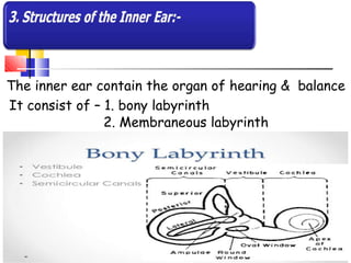
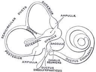
![MEMBRANEOUS LABYRINTH
It contains endolymph.
Vestibule:– contains utricle and saccule
Cochlea:- cross section of cochlea have 3
compartments.
1. Scala vastibuli : originate at oval window
2. Sacala tympani : ends at round window.
[two compartments are continous with
each other]
3. Scala media (cochlear duct)](https://image.slidesharecdn.com/earanatomybypari-200211100804/85/Ear-Anatomy-Physiology-24-320.jpg)

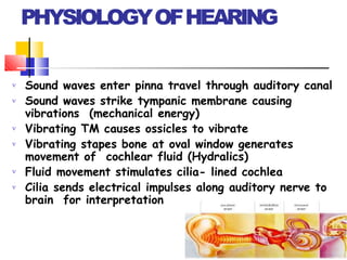
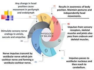
![Sound waves & vibration
from air travels [332mtr/sec]
enters in pinna
O1
Through auditory canal it
goes and cause vibration in
Tympanic membrane.
O2
Tympanic membrane
vibration transmitted and
amplify middle ear by
ossicle movements
O3
Stapes bone moves to
and fro in oval window.
O4
Fluid waves transmitted:
perilymph & endolymph
into cochlear duct
O5
It stiimulate auditory
receptors O6
Nerve impulses generate &
pass to brain by 8th cranial
nerve and reaches to hearing
areaof cerebrum and sound
preserved.
O7](https://image.slidesharecdn.com/earanatomybypari-200211100804/85/Ear-Anatomy-Physiology-30-320.jpg)


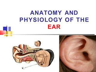
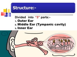
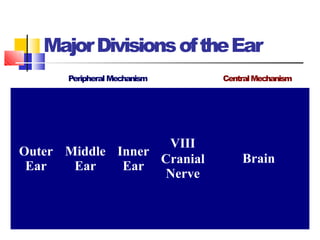
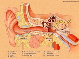
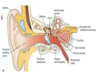
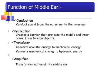
The document discusses the anatomy and physiology of the human ear. It describes the ear as having three main parts: the outer, middle, and inner ear. The outer ear collects sounds and directs them through the external auditory canal to the tympanic membrane. The middle ear contains three small bones called ossicles that transmit vibrations from the tympanic membrane to the inner ear. The inner ear converts these vibrations into neural signals for hearing and maintains balance. The cochlea and vestibular system within the inner ear work together to provide hearing and equilibrium.


