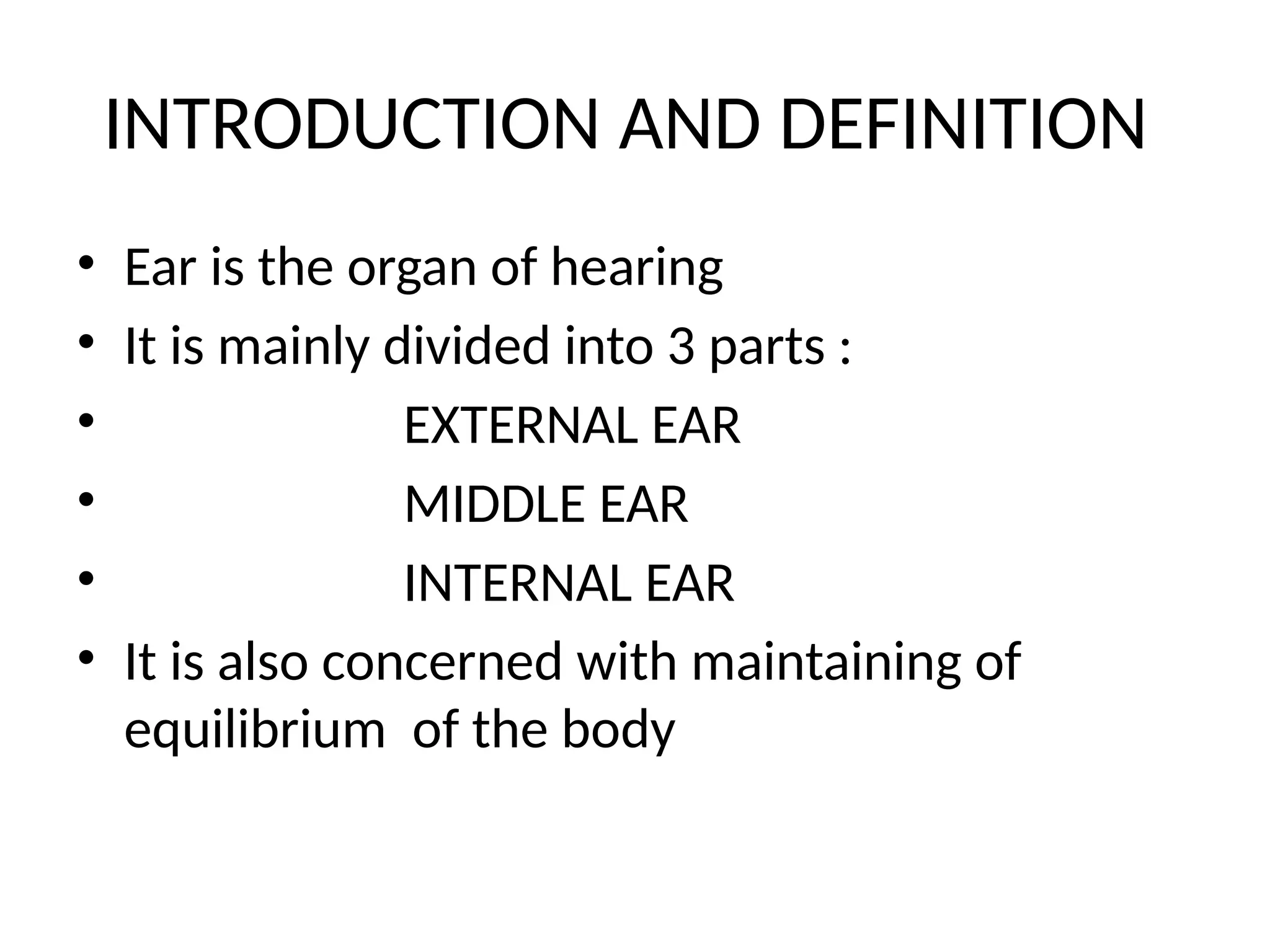
ANATOMY AND PHYSIOLOGY OF
EAR
SAGAR MADIVAL
4TH
YEAR BAMS
NKJAMC
Karna
• Definition :
• Indriya which is not seen with naked eye along with shashkuli
( hole / coiled ) is called karna.
• Synonym- shotra
• Origin : karn is originated from aakash mahabhuta.
• Karna sharira:
• Charka stated that karnendriya adhishthana exhibits 2 parts
i.e, 2 karnashashkuli ,
2karnaputraka.
• Chakrapani stated that karnashashkuli is karnagata avarta and
karnaputraka is bahyakarna.
• Karnashashkuli is that part which resembles avarta.
• Sushruta included bahya karna in 9 bahirmukha shrotas.
Contents
• Asthi :1 asthi.
• 1 taruna asthi.
• Sandhi : 1 sandhi.
• Peshi :2 peshi present in ear
• Sira : 10 siras
• 4 vatavaha sira. 2 pittavaha sira
• 2 kaphavaha sira. 2 raktavaha sira.
• Dhamani : 2 dhamani that convey shabda.
• Marma : 2 marma
• Vidhura marma
• Shringataka marma.
INTRODUCTION AND DEFINITION
• Ear is the organ of hearing
• It is mainly divided into 3 parts :
• EXTERNAL EAR
• MIDDLE EAR
• INTERNAL EAR
• It is also concerned with maintaining of
equilibrium of the body
ANATOMY OF EXTERNAL EAR
• It contains the following
The auricle or pinna
The external acoustic meatus
Auricle
• It is a flap of elastic cartilage
• It is covered by skin
• It is the opening of the external auditory canal
External auditory canal
• It is the passage and it extends up to the ear
drum
• It is 2.5 cm in length
• At the anterior end It contains glands and
hairs
• Gland –ceruminous gland which secrete the
ear wax
BLOOD SUPPLY OF EXTERNAL EAR
Nerve and lymphatic
• NERVE SUPPLY –Auriculotemporal nerve
• LYMPHATICS – PREAURICULAR
• POST AURICULAR
• CERVICAL LYMPH NODES
ANATOMY OF MIDDLE EAR
• It is a small air filled cavity in the temporal bone
• It is also lined by the epithelium
• It is separated by the inner ear by the oval
window
• Contains: ossicles
• oval window
• Eustachian tube
• tympanic membrane
AUDITORY OSSICLES
• THE MALLEUS
• THE INCUS
• THE STAPES
• Above mentioned bones are the smallest
bones in the body
• These are connected by the synovial joints
OSSICLES
MALLEUS
• In latin malleus is called as hammer so
because if it the name is given as malleus
• It resembles as the shape of hammer
• It is the largest among the ossicles
• It articulates with the body of the incus
Incus
• It looks like the anvil which are used by the
blacksmith
• It also resembles like the molar teeth .
• It has ;
• Body
• Long process
• Short process
• Lentiform process
Stapes
• It is shaped as stirrup
• It is the smallest of the ear ossicles
• It contains ;
• Two limbs :anterior and posterior
• Head
• Neck
• Base
Oval window
• It is the intersection between the middle ear
and the inner ear
• It is a membrane cover that leads to vestibule
part of inner ear
• When the sounds reach the oval window the
sound that is travelled is amplified by 10times
Tympanic membrane
• It is thin translucent partition between the
external and middle ear
• It is oval in shape
• Dimensions =9x10mm
• It is placed obliquely at an angle of 55 degree
with the floor
• It is held inward pull by the muscle tensor
tympani which is further inserted into the
malleus
.
• It is covered by the simple cuboidal epithelium
Eustachian tube
• It is the air filled cavity
• It consist of bony and hyaline cartilage
• It connects middle ear to the nasopharnyx
• It is normally closed at the pharyngeal end
• It maintains the pressure on each side
• If there is disturbed pressure then it affects
with the hearing
Oval window and eustachian tube
Blood supply
• Maxillary artery
• Auricular artery
• Meningial artery
• Pharyngeal artery
• Internal carotid artery
LYMPHATIC SUPPLY :
PREAURICULAR AND RETROPHARYNGEAL
NERVE SUPPLY :Glossopharyngeal nerve
caroticotympanic nerve
Inner ear
Inner ear
• It is also called as labyrinth
• It has two mains division
• outer bony labyrinth
• inner membranous labyrinth
REAL IMAGE
OUTER BONY LABYRINTH
• It is a series of cavity in the temporal bone
• It is divided into three regions
• Cochlea
• Vestibule
• 3 Semicircular canals
Bony labyrinth is lined by the periosteum and
contains the perilymph which is similar to the CSF
Bony labyrinth
cochlea
INNER MEMBRANOUS LABYRINTH
• It is a series of sacs and tubes inside the bony
labyrinth
• It is lined by the epithelium
• It contains the endolymph
• Here the pottasium level is high because it will
generate the auditory signals
• It consists of :
• Utricle
• Saccule
Membranous labyrinth
.
• Blood supply :labyrinthine branch of basilar
artery
• Nerve supply :vestibulocochlear nerve
Clinical anatomy
• Acoustic neuroma is the tumor of schwan cells
of 8 th nerve
• Why do we feel the clogged sensation in the
ear when we are in the plane or underwater
or specially if there is altitude change ?
Answer
• It happens because the difference between the
pressure on the outside and inside we know
that when we are in the higher or the lower
altitude there will be the pressure difference
• It can be managed by maintaining the pressure
which can be done by stimulating the
eustachian tube
• Keep making the mouth movements by eating
and drinking
Physiology of ear
• External ear :collecting and conveying the
sound waves
• To protect against the pathogens by producing
the ear wax
• To filter out the dust and unwanted substance
entering the ear
Ear canal
• * Once the sound waves have passed the
pinna, they move two to three
centimeters into the auditory canal
before hitting the eardrum, also known
as tympanic membrane. The function of
ear canal is to transmit sound from pinna
to the eardrum.
Ear drum
• ➤ Ear drum is the end point of the external
ear.
• ➤The eardrum is extremely sensitive and
pressure from sound wave makes the ear
drum vibrate.
• ➤The auditory canal is slightly curved in order
to protect the eardrum and also it
automatically amplifies low and less
penetrating sound of human voice.
Physiology of middle ear
• Amplifying the sound waves by the bones of
middle ear
• Conveying the sounds energy to the internal
ear through the bones
• Maintaining the pressure on both the sides
via eustachian tube which connects the
middle ear to the pharynx
Middle ear
• Ossicles will amplify the sounds
• THE OVAL WINDOW :
• ➤The main function of the oval window is to
amplify the sound before they move to inner ear.
• THE ROUND WINDOW:
• ➤It allows fluid in the cochlea to move
EUSTACHIAN TUBE:
• ➤The function of Eustachian tube is to equalize
the air pressure inside and outside the ear.
Inner ear
• To maintain the balance or the posture of the
body it helps in walking dancing and all other
activities
• To convert the sound which is reached to
inner ear by through middle ear into
electrical signals
• To send the electrical signal to the auditory
area in the brain via nerves
Inner ear
• COCHLEA: The cochlea resembles a snail which is
filled with fluid. When the fluid moves inside the
cochlea, thousands of microscopic hair fibers comes
in motion. The hair fibers send auditory nerve which
is connected to the auditory center of the brain.
• In cochlea sound waves are transformed into
electrical impulses which are sent to brains The
brain then translates the impulses into sound that
we know and understand
Bony labyrinth
cochlea
Organ of corti
Scala media
Endolymph
Mechanism
The pathway of hearing
• Auditory Pathway Flowchart
• Action potential generated in the cochlea
• ↓
• Mid Pons (superior olive)
• ↓
• Pons-midbrain junction (lateral lemniscus)
• ↓
• Caudal midbrain (inferior colliculus)
• ↓
• Rostral midbrain (medial geniculate complex of the
thalamus)
• ↓
• Primary auditory cortex
Mechanism of hearing
Balance
Semicircular organ
Otholith organ
Mechanism
Semicircular canals one for the up down and
one for side ways and other for the right and
left movements
The otolith organ which has the saccule and
utricle are involved in the head positioning
Both the organs have stereo cilia which move
according to the position of head and sends
the signal to the brain
Semicircular canals
Nodding the head up and down
Side ways movements
Left and right movements
Utricle and saccule
• Utricle that is sensitive to the horizontal
motion we can see that in the car motion
• Saccule is sensitive to the vertical motion we
can observe it in elevator
• Some actions are dealt by combination of
both the Saccule and utricle
THANK YOU

ANATOMY AND PHYSIOLOGY OF EAR sagar-2.pptx

  • 1.
    ANATOMY AND PHYSIOLOGYOF EAR SAGAR MADIVAL 4TH YEAR BAMS NKJAMC
  • 2.
    Karna • Definition : •Indriya which is not seen with naked eye along with shashkuli ( hole / coiled ) is called karna. • Synonym- shotra • Origin : karn is originated from aakash mahabhuta. • Karna sharira: • Charka stated that karnendriya adhishthana exhibits 2 parts i.e, 2 karnashashkuli , 2karnaputraka. • Chakrapani stated that karnashashkuli is karnagata avarta and karnaputraka is bahyakarna. • Karnashashkuli is that part which resembles avarta. • Sushruta included bahya karna in 9 bahirmukha shrotas.
  • 3.
    Contents • Asthi :1asthi. • 1 taruna asthi. • Sandhi : 1 sandhi. • Peshi :2 peshi present in ear • Sira : 10 siras • 4 vatavaha sira. 2 pittavaha sira • 2 kaphavaha sira. 2 raktavaha sira. • Dhamani : 2 dhamani that convey shabda. • Marma : 2 marma • Vidhura marma • Shringataka marma.
  • 4.
    INTRODUCTION AND DEFINITION •Ear is the organ of hearing • It is mainly divided into 3 parts : • EXTERNAL EAR • MIDDLE EAR • INTERNAL EAR • It is also concerned with maintaining of equilibrium of the body
  • 5.
    ANATOMY OF EXTERNALEAR • It contains the following The auricle or pinna The external acoustic meatus
  • 6.
    Auricle • It isa flap of elastic cartilage • It is covered by skin • It is the opening of the external auditory canal
  • 7.
    External auditory canal •It is the passage and it extends up to the ear drum • It is 2.5 cm in length • At the anterior end It contains glands and hairs • Gland –ceruminous gland which secrete the ear wax
  • 8.
    BLOOD SUPPLY OFEXTERNAL EAR
  • 9.
    Nerve and lymphatic •NERVE SUPPLY –Auriculotemporal nerve • LYMPHATICS – PREAURICULAR • POST AURICULAR • CERVICAL LYMPH NODES
  • 10.
    ANATOMY OF MIDDLEEAR • It is a small air filled cavity in the temporal bone • It is also lined by the epithelium • It is separated by the inner ear by the oval window • Contains: ossicles • oval window • Eustachian tube • tympanic membrane
  • 11.
    AUDITORY OSSICLES • THEMALLEUS • THE INCUS • THE STAPES • Above mentioned bones are the smallest bones in the body • These are connected by the synovial joints
  • 12.
  • 13.
    MALLEUS • In latinmalleus is called as hammer so because if it the name is given as malleus • It resembles as the shape of hammer • It is the largest among the ossicles • It articulates with the body of the incus
  • 14.
    Incus • It lookslike the anvil which are used by the blacksmith • It also resembles like the molar teeth . • It has ; • Body • Long process • Short process • Lentiform process
  • 15.
    Stapes • It isshaped as stirrup • It is the smallest of the ear ossicles • It contains ; • Two limbs :anterior and posterior • Head • Neck • Base
  • 17.
    Oval window • Itis the intersection between the middle ear and the inner ear • It is a membrane cover that leads to vestibule part of inner ear • When the sounds reach the oval window the sound that is travelled is amplified by 10times
  • 18.
    Tympanic membrane • Itis thin translucent partition between the external and middle ear • It is oval in shape • Dimensions =9x10mm • It is placed obliquely at an angle of 55 degree with the floor • It is held inward pull by the muscle tensor tympani which is further inserted into the malleus
  • 19.
    . • It iscovered by the simple cuboidal epithelium
  • 20.
    Eustachian tube • Itis the air filled cavity • It consist of bony and hyaline cartilage • It connects middle ear to the nasopharnyx • It is normally closed at the pharyngeal end • It maintains the pressure on each side • If there is disturbed pressure then it affects with the hearing
  • 21.
    Oval window andeustachian tube
  • 22.
    Blood supply • Maxillaryartery • Auricular artery • Meningial artery • Pharyngeal artery • Internal carotid artery
  • 23.
    LYMPHATIC SUPPLY : PREAURICULARAND RETROPHARYNGEAL NERVE SUPPLY :Glossopharyngeal nerve caroticotympanic nerve
  • 24.
  • 25.
    Inner ear • Itis also called as labyrinth • It has two mains division • outer bony labyrinth • inner membranous labyrinth REAL IMAGE
  • 26.
    OUTER BONY LABYRINTH •It is a series of cavity in the temporal bone • It is divided into three regions • Cochlea • Vestibule • 3 Semicircular canals Bony labyrinth is lined by the periosteum and contains the perilymph which is similar to the CSF
  • 27.
  • 28.
    INNER MEMBRANOUS LABYRINTH •It is a series of sacs and tubes inside the bony labyrinth • It is lined by the epithelium • It contains the endolymph • Here the pottasium level is high because it will generate the auditory signals • It consists of : • Utricle • Saccule
  • 29.
  • 30.
    . • Blood supply:labyrinthine branch of basilar artery • Nerve supply :vestibulocochlear nerve
  • 31.
    Clinical anatomy • Acousticneuroma is the tumor of schwan cells of 8 th nerve • Why do we feel the clogged sensation in the ear when we are in the plane or underwater or specially if there is altitude change ?
  • 32.
    Answer • It happensbecause the difference between the pressure on the outside and inside we know that when we are in the higher or the lower altitude there will be the pressure difference • It can be managed by maintaining the pressure which can be done by stimulating the eustachian tube • Keep making the mouth movements by eating and drinking
  • 33.
    Physiology of ear •External ear :collecting and conveying the sound waves • To protect against the pathogens by producing the ear wax • To filter out the dust and unwanted substance entering the ear
  • 34.
    Ear canal • *Once the sound waves have passed the pinna, they move two to three centimeters into the auditory canal before hitting the eardrum, also known as tympanic membrane. The function of ear canal is to transmit sound from pinna to the eardrum.
  • 35.
    Ear drum • ➤Ear drum is the end point of the external ear. • ➤The eardrum is extremely sensitive and pressure from sound wave makes the ear drum vibrate. • ➤The auditory canal is slightly curved in order to protect the eardrum and also it automatically amplifies low and less penetrating sound of human voice.
  • 36.
    Physiology of middleear • Amplifying the sound waves by the bones of middle ear • Conveying the sounds energy to the internal ear through the bones • Maintaining the pressure on both the sides via eustachian tube which connects the middle ear to the pharynx
  • 37.
    Middle ear • Ossicleswill amplify the sounds • THE OVAL WINDOW : • ➤The main function of the oval window is to amplify the sound before they move to inner ear. • THE ROUND WINDOW: • ➤It allows fluid in the cochlea to move EUSTACHIAN TUBE: • ➤The function of Eustachian tube is to equalize the air pressure inside and outside the ear.
  • 38.
    Inner ear • Tomaintain the balance or the posture of the body it helps in walking dancing and all other activities • To convert the sound which is reached to inner ear by through middle ear into electrical signals • To send the electrical signal to the auditory area in the brain via nerves
  • 39.
    Inner ear • COCHLEA:The cochlea resembles a snail which is filled with fluid. When the fluid moves inside the cochlea, thousands of microscopic hair fibers comes in motion. The hair fibers send auditory nerve which is connected to the auditory center of the brain. • In cochlea sound waves are transformed into electrical impulses which are sent to brains The brain then translates the impulses into sound that we know and understand
  • 40.
  • 41.
    Organ of corti Scalamedia Endolymph
  • 42.
  • 43.
    The pathway ofhearing • Auditory Pathway Flowchart • Action potential generated in the cochlea • ↓ • Mid Pons (superior olive) • ↓ • Pons-midbrain junction (lateral lemniscus) • ↓ • Caudal midbrain (inferior colliculus) • ↓ • Rostral midbrain (medial geniculate complex of the thalamus) • ↓ • Primary auditory cortex
  • 44.
  • 45.
  • 46.
    Mechanism Semicircular canals onefor the up down and one for side ways and other for the right and left movements The otolith organ which has the saccule and utricle are involved in the head positioning Both the organs have stereo cilia which move according to the position of head and sends the signal to the brain
  • 47.
  • 48.
  • 49.
    Left and rightmovements
  • 50.
    Utricle and saccule •Utricle that is sensitive to the horizontal motion we can see that in the car motion • Saccule is sensitive to the vertical motion we can observe it in elevator • Some actions are dealt by combination of both the Saccule and utricle
  • 51.