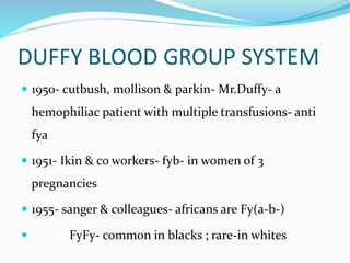
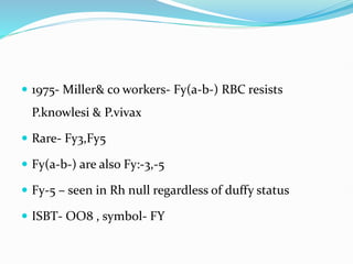
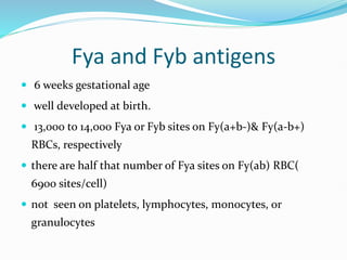
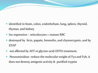
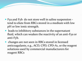
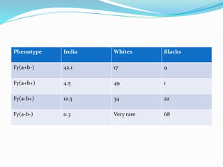
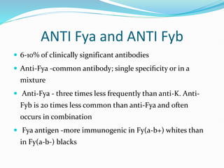
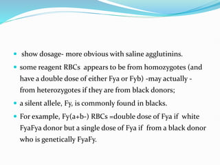
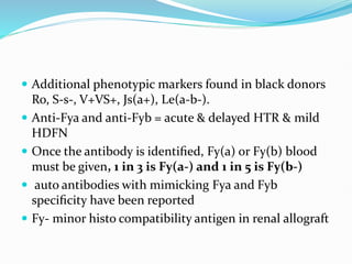
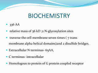
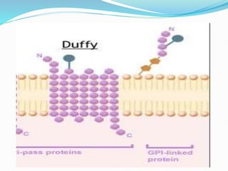
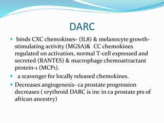
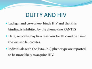
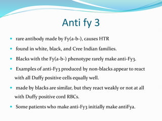
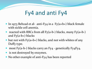
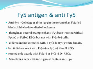
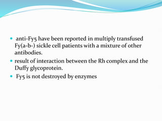
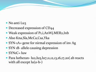
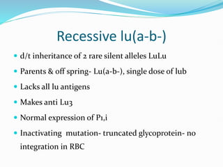
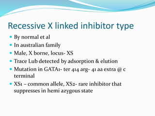
IHBT
DUFFY BLOOD GROUP SYSTEM
 1950- cutbush, mollison & parkin- Mr.Duffy- a
hemophiliac patient with multiple transfusions- anti
fya
 1951- Ikin & co workers- fyb- in women of 3
pregnancies
 1955- sanger & colleagues- africans are Fy(a-b-)
 FyFy- common in blacks ; rare-in whites
 1975- Miller& co workers- Fy(a-b-) RBC resists
P.knowlesi & P.vivax
 Rare- Fy3,Fy5
 Fy(a-b-) are also Fy:-3,-5
 Fy-5 – seen in Rh null regardless of duffy status
 ISBT- OO8 , symbol- FY
Fya and Fyb antigens
 6 weeks gestational age
 well developed at birth.
 13,000 to 14,000 Fya or Fyb sites on Fy(a+b-)& Fy(a-b+)
RBCs, respectively
 there are half that number of Fya sites on Fy(ab) RBC(
6900 sites/cell)
 not seen on platelets, lymphocytes, monocytes, or
granulocytes
 identified in brain, colon, endothelium, lung, spleen, thyroid,
thymus, and kidney
 Inc expression – reticulocytes > mature RBC
 destroyed by ficin, papain, bromelin, and chymotrypsin, and by
ZZAP
 not affected by AET or glycine-acid-EDTA treatment.
 Neuraminidase -reduce the molecular weight of Fya and Fyb, it
does not destroy antigenic activity & purified trypsin
 Fya and Fyb do not store well in saline suspension -
tend to elute from RBCs stored in a medium with low
pH or low ionic strength.
 leads to inhibitory substances in the supernatant
fluid, which can weaken the reactivity of an anti-Fya or
anti-Fyb.
 changes are not seen in RBCs stored in licensed
anticoagulants, e.g., ACD, CPD, CPD-A1, or the reagent
solutions used by commercial manufacturers for
reagent RBCs
Phenotype India Whites Blacks
Fy(a+b-) 42.1 17 9
Fy(a+b+) 4.5 49 1
Fy(a-b+) 12.3 34 22
Fy(a-b-) 0.3 Very rare 68
ANTI Fya and ANTI Fyb
 6-10% of clinically significant antibodies
 Anti-Fya -common antibody; single specificity or in a
mixture
 Anti-Fya - three times less frequently than anti-K. Anti-
Fyb is 20 times less common than anti-Fya and often
occurs in combination
 Fya antigen -more immunogenic in Fy(a-b+) whites than
in Fy(a-b-) blacks
 usually IgG1 - react best at AHG phase.
 50% anti-Fya and anti-Fyb bind complement upto C3
 A few examples are saline agglutinins.
 Antibody activity is enhanced in a low ionic strength medium.
 Because anti-Fya and anti-Fyb do not react with enzyme treated
RBCs, this is a helpful technique when multiple antibodies are
present.
 Naturally occurring - rare
 show dosage- more obvious with saline agglutinins.
 some reagent RBCs appears to be from homozygotes (and
have a double dose of either Fya or Fyb) -may actually -
from heterozygotes if they are from black donors;
 a silent allele, Fy, is commonly found in blacks.
 For example, Fy(a+b-) RBCs =double dose of Fya if white
FyaFya donor but a single dose of Fya if from a black donor
who is genetically FyaFy.
 Additional phenotypic markers found in black donors
Ro, S-s-, V+VS+, Js(a+), Le(a-b-).
 Anti-Fya and anti-Fyb = acute & delayed HTR & mild
HDFN
 Once the antibody is identified, Fy(a) or Fy(b) blood
must be given, 1 in 3 is Fy(a-) and 1 in 5 is Fy(b-)
 auto antibodies with mimicking Fya and Fyb
specificity have been reported
 Fy- minor histo compatibility antigen in renal allograft
BIOCHEMISTRY
 336 AA
 relative mass of 36 kD ;2 N-glycosylation sites
 traverse the cell membrane seven times ( 7 trans
membrane alpha helical domains)and 2 disulfide bridges.
 Extracellular N terminus- 65AA,
 C terminus- intracellular
 Homologous to protein of G protein coupled receptor
 Fya/Fyb- SNP in exon 2- gly42asp on extra cellular N
terminus
 Fy3 - on the third extracellular loop
 Fy6 - amino acids 19 through 25
 a member of the superfamily of chemokine receptors
DARC (Duffy antigen receptor for chemokine)
 Receptor for P.vivax in N terminal domain( sulphation
of Tyr 42- increases affinity of binding by 1000 fold)
DARC
 binds CXC chemokines- (IL8) & melanocyte growth-
stimulating activity (MGSA)& CC chemokines
regulated on activation, normal T-cell expressed and
secreted (RANTES) & macrophage chemoattractant
protein-1 (MCP1).
 a scavenger for locally released chemokines.
 Decreases angiogenesis- ca prostate progression
decreases ( erythroid DARC is inc in ca prostate pts of
african ancestry)
 Present in capillaries & post capillary venules-
 Inhibits cancer metastasis & induction of cellular
senescence
 Facilitates movement of chemokines across
endothelium
 IL-8 binding – residues on EC domain 1 and 4 –
requires –S=S-
 Ligands for parasites- glycophorin A,B,C,D and NANA
 leucocyte recruitment to sites of inflammation
 regulate chemokine bioavailability between the
circulation and extravascular sites during
inflammation
 In Fy(a− b−) phenotype and many patients with sickle
cell disease have the Fy(a− b−) phenotype- elicit
markers of inflammation, & inc chances of allo
immunization
 more susceptible to chronic organ damage
&proteinuria & more susceptible to asthma, worst
outcomes for Acute lung injury
GENETICS
 chromosome 1
 the first human gene to be assigned to a specific
chromosome- DONAHUE
 2 exons- exon1 – encodes first 7 AA
 CO DOMINANT alleles
 Fy locus is syntenic to Rh
 Fya and Fyb allele- Fya and Fyb
 silent allele, Fy, that is the major allele in blacks
Fy allele
 found to be an Fyb variant - change in the promoter
region(GATA1) of the gene
 T-C substitution
 which disrupts the binding site for mRNA
transcription in the RBC
 do not express Fyb on their RBCs but express Fyb in
other tissues,
 no anti-Fyb is made by these individuals.
 in new guinea- GATA mutation seen in Fya allele
 In whites - 14 bp deletion in the Fy gene, resulting in a
reading frame shift and the introduction of a
translation stop codon.
 no Duffy protein on their RBCs or on other tissues and
--form anti-Fyb and anti-Fy3.
 Fy3 developed first, then Fyb, and that Fya arose
during human evolution.
DUFFY AND HIV
 Lachgar and co-worker- binds HIV and that this
binding is inhibited by the chemokine RANTES
 Here, red cells may be a reservoir for HIV and transmit
the virus to leucocytes.
 Individuals with the Fy(a− b−) phenotype are reported
to be more likely to acquire HIV.
Fyx
 1965 by Chown - new allele at the Fy locus.
 inherited weak form of Fyb
 Mis sense mutation- Arg89Cys in 1st EC loop of
cytosolic domain- reduced amount of Duffy
glycoprotein on the surface of RBCs
 reacts with some but not all examples of anti-Fyb.
 in white populations.
 Individuals with Fyx may type Fy(b-), but their RBCs
adsorb and elute anti-Fyb.
 depressed expression of Fy3 & Fy5 antigens.
 no anti-Fyx.
Fy3 antigen & anti Fy3
 In 1971 Albrey et al- anti-Fy3 in the serum of an Fy(a-b-
) white Australian female.
 It reacted with all RBCs tested except Fy(a-b-)
 an inseparable anti-FyaFyb,
 react with an antigenic determinant or precursor
common to both Fya and Fyb and was called Fy3.
 the Fy3 antigen is not destroyed by enzymes.
Anti fy 3
 rare antibody made by Fy(a-b-), causes HTR
 found in white, black, and Cree Indian families.
 Blacks with the Fy(a-b-) phenotype rarely make anti-Fy3.
 Examples of anti-Fy3 produced by non-blacks appear to react
with all Duffy positive cells equally well.
 made by blacks are similar, but they react weakly or not at all
with Duffy positive cord RBCs.
 Some patients who make anti-Fy3 initially make antiFya.
Fy4 and anti Fy4
 In 1973 Behzad et al- anti-Fy4 in a Fy(a+b+) black female
with sickle cell anemia.
 reacted with RBCs from all Fy(a-b-) blacks, many Fy(a+b-)
and Fy(a-b+) blacks
 but not with Fy(a+b+) blacks, and not with whites of any
Duffy type.
 most Fy(a-b-) blacks carry an Fy4 - genetically Fy4Fy4.
 is not destroyed by enzymes.
 No other example of anti-Fy4 has been reported
Fy5 antigen & anti Fy5
 Anti-Fy5 - Colledge et al- in 1973 in the serum of an Fy(a-b-)
black child who later died of leukemia.
 thought as second example of anti-Fy3 bcoz- reacted with all
Fy(a+) or Fy(b+) RBCs but not with Fy(a-b-) cells.
 differed in that it reacted with a Fy(a-b-)Fy:-3 white female,
 but it did not react with Fy(a+) or Fy(b+) Rhnull RBCs
 reacted only weakly with Fy(a+) or Fy(b+) D- RBCs.
 Sometimes, sera with anti-Fy5 also contain anti-Fya.
 anti-Fy5 have been reported in multiply transfused
Fy(a-b-) sickle cell patients with a mixture of other
antibodies.
 result of interaction between the Rh complex and the
Duffy glycoprotein.
 Fy5 is not destroyed by enzymes
Fy6 antigen
 In 1987 Nichols et al -a murine monoclonal ab
 reacted much like anti-Fy3, except that its reactivity was
destroyed by ficin, papain, and chymotrypsin.
 Trypsin enhanced its reactivity.
 The antibody appeared to define the Duffy receptor used by P.
vivax to penetrate RBCs
 Fy6 involves amino acids 19 to 25 on the extracellular domain of
the Duffy glycoprotein.
 No human examples of anti-Fy6 have been reported to date.
LUTHERAN
 1945, anti-Lua was discovered in SLE patient
following the transfusion with LPA- ( also made anti-c,
anti-N, anti-CW, and anti-Levay, now known as Kpc!)
 In 1956 Cutbush & Chanarin - anti-Lub
 1961- Crawford- Lu(a-b-) with dominant inheritance.
 In 1963 Darnborough et al Lu(a-b-)phenotype
inherited as a recessive silent allele.
 22 antigens- Lu 10,LU15 = obsolete
 4 anti thetical ag
 HPA- Lub,
Lu3,lu4,lu5,lu6,lu7,lu8,lu11,lu12,lu13,lu16,lu17,lu20,lu21,
lu22
 Lua-8% in whites& 5 in blacks
 Lu9- 2%
 Lu14-2.4%
 Aua,lu18=80% whites
 Aub,lu19=50% whites
Phenotype Prevalence in most population
Lu(a+b-) 0.15
Lu(a+b+) 7.5
Lu(a-b+) 92.35
Lu(a—b-) Very rare
Basic concepts
 either very HPA/LPA.
 10 to 12 weeks
 poorly developed at birth and do not reach adult
levels until age 15 years.
 not been detected on platelets, lymphocytes,
monocytes, or granulocytes
 seen in brain, lung, pancreas, placenta, skeletal
muscle, & hepatocytes (especially fetal hepatic
epithelial cells
 Lu on placenta- adsorption of maternal Ab to lu Ag-
low risk of HDFN
 Resistant to papain/ficin/glycine acid EDTA
 Destroyed by trypsin,chymotrypsin,pronase
 Do not react with DTT/AET treated RBC
 4 antithetical Ag- lu1/lu2,lu6/lu9(
ser275phe),lu8/lu14,lu18/lu19
Lua and Lub antigens
 Co dominant
 Most- lu(b+)
 8% of whites & 5 % in blacks- lu(a+)
 Lua/lub= his77arg
 expression is variable from one individual to another; one
individual’s RBCs can also vary.
 Lub sites= 1640 -4070 on lu(a-b+)
 850-1820 on lu( a+b+)
 Transient loss of lua- in AITP
 Shows dosage effect
 Lub- more immunogenic, HPA
Anti Lua
 IgM, naturally occurring saline agglutinins that react better
at room temperature.
 A few react at 37*C by IAT.
 Some -binds complement, but invitro hemolysis has not
been reported.
 may be IgA as well as IgM and IgG
 Anti-Lua often goes undetected in routine testing because
most reagent cells are Lu(a-).
 encountered as CXM incompatible or during an antibody
workup for another specificity.
 mixed-field reactivity in a test tube.
 not altered with enzymes ficin and papain, destroyed with
trypsin, chymotrypsin, pronase, AET, and DTT.
 Most Lua antibodies are clinically insignificant in
transfusion.
 no immediate but DHTR
Anti Lub
 IgM and IgA but most anti-Lub is IgG4 , reactive at 37*C
at AHG phase.
 It is made in response to pregnancy or transfusion.
 Alloanti-Lub reacts with all cells tested except the
autocontrol
 reactions are often weaker with Lu(a+b+) RBCs and cord
RBCs.
 Ficin or papain does not significantly alter reactivity.but
AET or DTT destroys Lub antigen
 Dec survival of transfused cells, post transfusion jaundice
noted
BIOCHEMISTRY
 Located on type I transmembrane protein
 Protein in 2 forms – d/t alternate RNA spliceosomes
 Lutheran glycoproteins.
 85kD , 597 AA, 5 potential N glycosylation sites as EC
IgSF domain
 It traverses the cell membrane just once – hydrophobic
transmembrane domain -19AA
 cytoplasmic domain of 59 AA- interacts with spectrin
 Lua/lub- on 1st domain, Aua/Aub – on domain 5
 Smaller isoform-BCAM
 78 kD
 lacks cytoplasmic domain
 external portion 5 disulfide-bonded domains (three are
constant, and two are variable).
 belong to the immunoglobulin superfamily of proteins.
 Binds laminin in asp312 on domain 3
 Laminin- has alpha 5 chains, La-Lu complex migrates
mature RBC from marrow to circulation
 Upregulation of laminin – in sickle cell patients-
vascular crisis
 In PCV, increased adhesion- d/t la- lu on endothelial
cells
Molecular basis of HPA &
antithetical antigens
 through the creation of Lutheran glycoprotein
mutants.
 Subsequent sequencing of exons encoding on EC
domains
 For most- it is SNP
GENETICS
 Chr 19
 (H, Se, Le, LW, Oka) and genes for C3, apolipoprotein
C-II (APO), and myotonic dystrophy
 Lu and Se gene (FUT2) was the first example of
autosomal linkage described
Lu(a-b-) phenotype- dominant
 Reported by crawford
 phenotype was seen in successive generations as 50% of
members & null individuals passed normal genes-
expression was suppressed by a rare dominant regulator
gene later called In(Lu) for “inhibitor of Lutheran.” or
SYN1B
 In(Lu) segregates independently from Lutheran.
 prevalence- 0.03%
 Unsuitable as donor- lysis invitro after 7 days when
stored in alsever solution- 5 times than normal
 Mutations in gene for EKLF
 Caused by inheritance of loss of function mutation on
1 allele of EKLF
 abnormal poikilocytosis and acanthocytosis
 On incubation with plasma> 24 hrs- decreased
osmotic fragility , in vivo survival – normal
 Carries trace Lu antigens – detected ONLY by elution
& adsorption
 No anti Lu3
 Decreased expression of CD44
 Weak expression of P1,i,AnWj.MER2,Inb
 Also Kna,Sla,McCa,Csa,Yka
 SYN-1A= gene for nirmal expression of inv Ag
 SYN 1B- allele causing depression
 SYN1C= luw
 Para lutheran- lu2,lu5,lu7,11,12,13,16,17,20( ab reacts
with all except lu(a-b-)
Recessive lu(a-b-)
 d/t inheritance of 2 rare silent alleles LuLu
 Parents & off spring- Lu(a-b-), single dose of lub
 Lacks all lu antigens
 Makes anti Lu3
 Normal expression of P1,i
 Inactivating mutation- truncated glycoprotein- no
integration in RBC
Recessive X linked inhibitor type
 By normal et al
 In australian family
 Male, X borne, locus- XS
 Trace Lub detected by adsorption & elution
 Mutation in GATA1- ter 414 arg- 41 aa extra @ c
terminal
 XS1 – common allele, XS2- rare inhibitor that
suppresses in hemi azygous state
Mode of inh Gene Lu Others Antilu3
Dominant EKLF Extremely
weak
Decreased
P1,i,Inb,Anwj,
MER2,CD44
No
Recessive Lu None Not affected Yes
X linked XS2 Extremely
weak
Not affected no
Anti lu3
 reacts with all RBCs except Lu(a-b-).
 looks inseparable anti-Luab
 recognizes a common antigen, Lu3
 Anti-Lu3 is usually antiglobulin reactive.
 This antibody is made only by LuLu individuals, i.e.,
the recessive type of Lu(ab)
 . RBCs from dominant and X linked type Lu(a-b-)
individuals can be safely transfused to patients with
anti-Lu3.
DUFFY AND LUTHERAN ANTIGEN SYSTEM.pptx

DUFFY AND LUTHERAN ANTIGEN SYSTEM.pptx

  • 1.
  • 2.
    DUFFY BLOOD GROUPSYSTEM  1950- cutbush, mollison & parkin- Mr.Duffy- a hemophiliac patient with multiple transfusions- anti fya  1951- Ikin & co workers- fyb- in women of 3 pregnancies  1955- sanger & colleagues- africans are Fy(a-b-)  FyFy- common in blacks ; rare-in whites
  • 3.
     1975- Miller&co workers- Fy(a-b-) RBC resists P.knowlesi & P.vivax  Rare- Fy3,Fy5  Fy(a-b-) are also Fy:-3,-5  Fy-5 – seen in Rh null regardless of duffy status  ISBT- OO8 , symbol- FY
  • 4.
    Fya and Fybantigens  6 weeks gestational age  well developed at birth.  13,000 to 14,000 Fya or Fyb sites on Fy(a+b-)& Fy(a-b+) RBCs, respectively  there are half that number of Fya sites on Fy(ab) RBC( 6900 sites/cell)  not seen on platelets, lymphocytes, monocytes, or granulocytes
  • 5.
     identified inbrain, colon, endothelium, lung, spleen, thyroid, thymus, and kidney  Inc expression – reticulocytes > mature RBC  destroyed by ficin, papain, bromelin, and chymotrypsin, and by ZZAP  not affected by AET or glycine-acid-EDTA treatment.  Neuraminidase -reduce the molecular weight of Fya and Fyb, it does not destroy antigenic activity & purified trypsin
  • 6.
     Fya andFyb do not store well in saline suspension - tend to elute from RBCs stored in a medium with low pH or low ionic strength.  leads to inhibitory substances in the supernatant fluid, which can weaken the reactivity of an anti-Fya or anti-Fyb.  changes are not seen in RBCs stored in licensed anticoagulants, e.g., ACD, CPD, CPD-A1, or the reagent solutions used by commercial manufacturers for reagent RBCs
  • 7.
    Phenotype India WhitesBlacks Fy(a+b-) 42.1 17 9 Fy(a+b+) 4.5 49 1 Fy(a-b+) 12.3 34 22 Fy(a-b-) 0.3 Very rare 68
  • 8.
    ANTI Fya andANTI Fyb  6-10% of clinically significant antibodies  Anti-Fya -common antibody; single specificity or in a mixture  Anti-Fya - three times less frequently than anti-K. Anti- Fyb is 20 times less common than anti-Fya and often occurs in combination  Fya antigen -more immunogenic in Fy(a-b+) whites than in Fy(a-b-) blacks
  • 9.
     usually IgG1- react best at AHG phase.  50% anti-Fya and anti-Fyb bind complement upto C3  A few examples are saline agglutinins.  Antibody activity is enhanced in a low ionic strength medium.  Because anti-Fya and anti-Fyb do not react with enzyme treated RBCs, this is a helpful technique when multiple antibodies are present.  Naturally occurring - rare
  • 10.
     show dosage-more obvious with saline agglutinins.  some reagent RBCs appears to be from homozygotes (and have a double dose of either Fya or Fyb) -may actually - from heterozygotes if they are from black donors;  a silent allele, Fy, is commonly found in blacks.  For example, Fy(a+b-) RBCs =double dose of Fya if white FyaFya donor but a single dose of Fya if from a black donor who is genetically FyaFy.
  • 11.
     Additional phenotypicmarkers found in black donors Ro, S-s-, V+VS+, Js(a+), Le(a-b-).  Anti-Fya and anti-Fyb = acute & delayed HTR & mild HDFN  Once the antibody is identified, Fy(a) or Fy(b) blood must be given, 1 in 3 is Fy(a-) and 1 in 5 is Fy(b-)  auto antibodies with mimicking Fya and Fyb specificity have been reported  Fy- minor histo compatibility antigen in renal allograft
  • 12.
    BIOCHEMISTRY  336 AA relative mass of 36 kD ;2 N-glycosylation sites  traverse the cell membrane seven times ( 7 trans membrane alpha helical domains)and 2 disulfide bridges.  Extracellular N terminus- 65AA,  C terminus- intracellular  Homologous to protein of G protein coupled receptor
  • 14.
     Fya/Fyb- SNPin exon 2- gly42asp on extra cellular N terminus  Fy3 - on the third extracellular loop  Fy6 - amino acids 19 through 25  a member of the superfamily of chemokine receptors DARC (Duffy antigen receptor for chemokine)  Receptor for P.vivax in N terminal domain( sulphation of Tyr 42- increases affinity of binding by 1000 fold)
  • 15.
    DARC  binds CXCchemokines- (IL8) & melanocyte growth- stimulating activity (MGSA)& CC chemokines regulated on activation, normal T-cell expressed and secreted (RANTES) & macrophage chemoattractant protein-1 (MCP1).  a scavenger for locally released chemokines.  Decreases angiogenesis- ca prostate progression decreases ( erythroid DARC is inc in ca prostate pts of african ancestry)
  • 16.
     Present incapillaries & post capillary venules-  Inhibits cancer metastasis & induction of cellular senescence  Facilitates movement of chemokines across endothelium  IL-8 binding – residues on EC domain 1 and 4 – requires –S=S-  Ligands for parasites- glycophorin A,B,C,D and NANA
  • 17.
     leucocyte recruitmentto sites of inflammation  regulate chemokine bioavailability between the circulation and extravascular sites during inflammation  In Fy(a− b−) phenotype and many patients with sickle cell disease have the Fy(a− b−) phenotype- elicit markers of inflammation, & inc chances of allo immunization  more susceptible to chronic organ damage &proteinuria & more susceptible to asthma, worst outcomes for Acute lung injury
  • 18.
    GENETICS  chromosome 1 the first human gene to be assigned to a specific chromosome- DONAHUE  2 exons- exon1 – encodes first 7 AA  CO DOMINANT alleles  Fy locus is syntenic to Rh  Fya and Fyb allele- Fya and Fyb  silent allele, Fy, that is the major allele in blacks
  • 19.
    Fy allele  foundto be an Fyb variant - change in the promoter region(GATA1) of the gene  T-C substitution  which disrupts the binding site for mRNA transcription in the RBC  do not express Fyb on their RBCs but express Fyb in other tissues,  no anti-Fyb is made by these individuals.  in new guinea- GATA mutation seen in Fya allele
  • 20.
     In whites- 14 bp deletion in the Fy gene, resulting in a reading frame shift and the introduction of a translation stop codon.  no Duffy protein on their RBCs or on other tissues and --form anti-Fyb and anti-Fy3.  Fy3 developed first, then Fyb, and that Fya arose during human evolution.
  • 21.
    DUFFY AND HIV Lachgar and co-worker- binds HIV and that this binding is inhibited by the chemokine RANTES  Here, red cells may be a reservoir for HIV and transmit the virus to leucocytes.  Individuals with the Fy(a− b−) phenotype are reported to be more likely to acquire HIV.
  • 22.
    Fyx  1965 byChown - new allele at the Fy locus.  inherited weak form of Fyb  Mis sense mutation- Arg89Cys in 1st EC loop of cytosolic domain- reduced amount of Duffy glycoprotein on the surface of RBCs  reacts with some but not all examples of anti-Fyb.  in white populations.
  • 23.
     Individuals withFyx may type Fy(b-), but their RBCs adsorb and elute anti-Fyb.  depressed expression of Fy3 & Fy5 antigens.  no anti-Fyx.
  • 24.
    Fy3 antigen &anti Fy3  In 1971 Albrey et al- anti-Fy3 in the serum of an Fy(a-b- ) white Australian female.  It reacted with all RBCs tested except Fy(a-b-)  an inseparable anti-FyaFyb,  react with an antigenic determinant or precursor common to both Fya and Fyb and was called Fy3.  the Fy3 antigen is not destroyed by enzymes.
  • 25.
    Anti fy 3 rare antibody made by Fy(a-b-), causes HTR  found in white, black, and Cree Indian families.  Blacks with the Fy(a-b-) phenotype rarely make anti-Fy3.  Examples of anti-Fy3 produced by non-blacks appear to react with all Duffy positive cells equally well.  made by blacks are similar, but they react weakly or not at all with Duffy positive cord RBCs.  Some patients who make anti-Fy3 initially make antiFya.
  • 26.
    Fy4 and antiFy4  In 1973 Behzad et al- anti-Fy4 in a Fy(a+b+) black female with sickle cell anemia.  reacted with RBCs from all Fy(a-b-) blacks, many Fy(a+b-) and Fy(a-b+) blacks  but not with Fy(a+b+) blacks, and not with whites of any Duffy type.  most Fy(a-b-) blacks carry an Fy4 - genetically Fy4Fy4.  is not destroyed by enzymes.  No other example of anti-Fy4 has been reported
  • 27.
    Fy5 antigen &anti Fy5  Anti-Fy5 - Colledge et al- in 1973 in the serum of an Fy(a-b-) black child who later died of leukemia.  thought as second example of anti-Fy3 bcoz- reacted with all Fy(a+) or Fy(b+) RBCs but not with Fy(a-b-) cells.  differed in that it reacted with a Fy(a-b-)Fy:-3 white female,  but it did not react with Fy(a+) or Fy(b+) Rhnull RBCs  reacted only weakly with Fy(a+) or Fy(b+) D- RBCs.  Sometimes, sera with anti-Fy5 also contain anti-Fya.
  • 28.
     anti-Fy5 havebeen reported in multiply transfused Fy(a-b-) sickle cell patients with a mixture of other antibodies.  result of interaction between the Rh complex and the Duffy glycoprotein.  Fy5 is not destroyed by enzymes
  • 29.
    Fy6 antigen  In1987 Nichols et al -a murine monoclonal ab  reacted much like anti-Fy3, except that its reactivity was destroyed by ficin, papain, and chymotrypsin.  Trypsin enhanced its reactivity.  The antibody appeared to define the Duffy receptor used by P. vivax to penetrate RBCs  Fy6 involves amino acids 19 to 25 on the extracellular domain of the Duffy glycoprotein.  No human examples of anti-Fy6 have been reported to date.
  • 31.
    LUTHERAN  1945, anti-Luawas discovered in SLE patient following the transfusion with LPA- ( also made anti-c, anti-N, anti-CW, and anti-Levay, now known as Kpc!)  In 1956 Cutbush & Chanarin - anti-Lub  1961- Crawford- Lu(a-b-) with dominant inheritance.  In 1963 Darnborough et al Lu(a-b-)phenotype inherited as a recessive silent allele.  22 antigens- Lu 10,LU15 = obsolete  4 anti thetical ag
  • 32.
     HPA- Lub, Lu3,lu4,lu5,lu6,lu7,lu8,lu11,lu12,lu13,lu16,lu17,lu20,lu21, lu22 Lua-8% in whites& 5 in blacks  Lu9- 2%  Lu14-2.4%  Aua,lu18=80% whites  Aub,lu19=50% whites
  • 33.
    Phenotype Prevalence inmost population Lu(a+b-) 0.15 Lu(a+b+) 7.5 Lu(a-b+) 92.35 Lu(a—b-) Very rare
  • 34.
    Basic concepts  eithervery HPA/LPA.  10 to 12 weeks  poorly developed at birth and do not reach adult levels until age 15 years.  not been detected on platelets, lymphocytes, monocytes, or granulocytes  seen in brain, lung, pancreas, placenta, skeletal muscle, & hepatocytes (especially fetal hepatic epithelial cells
  • 35.
     Lu onplacenta- adsorption of maternal Ab to lu Ag- low risk of HDFN  Resistant to papain/ficin/glycine acid EDTA  Destroyed by trypsin,chymotrypsin,pronase  Do not react with DTT/AET treated RBC  4 antithetical Ag- lu1/lu2,lu6/lu9( ser275phe),lu8/lu14,lu18/lu19
  • 36.
    Lua and Lubantigens  Co dominant  Most- lu(b+)  8% of whites & 5 % in blacks- lu(a+)  Lua/lub= his77arg  expression is variable from one individual to another; one individual’s RBCs can also vary.  Lub sites= 1640 -4070 on lu(a-b+)  850-1820 on lu( a+b+)
  • 37.
     Transient lossof lua- in AITP  Shows dosage effect  Lub- more immunogenic, HPA
  • 38.
    Anti Lua  IgM,naturally occurring saline agglutinins that react better at room temperature.  A few react at 37*C by IAT.  Some -binds complement, but invitro hemolysis has not been reported.  may be IgA as well as IgM and IgG  Anti-Lua often goes undetected in routine testing because most reagent cells are Lu(a-).
  • 39.
     encountered asCXM incompatible or during an antibody workup for another specificity.  mixed-field reactivity in a test tube.  not altered with enzymes ficin and papain, destroyed with trypsin, chymotrypsin, pronase, AET, and DTT.  Most Lua antibodies are clinically insignificant in transfusion.  no immediate but DHTR
  • 40.
    Anti Lub  IgMand IgA but most anti-Lub is IgG4 , reactive at 37*C at AHG phase.  It is made in response to pregnancy or transfusion.  Alloanti-Lub reacts with all cells tested except the autocontrol  reactions are often weaker with Lu(a+b+) RBCs and cord RBCs.  Ficin or papain does not significantly alter reactivity.but AET or DTT destroys Lub antigen  Dec survival of transfused cells, post transfusion jaundice noted
  • 41.
    BIOCHEMISTRY  Located ontype I transmembrane protein  Protein in 2 forms – d/t alternate RNA spliceosomes  Lutheran glycoproteins.  85kD , 597 AA, 5 potential N glycosylation sites as EC IgSF domain  It traverses the cell membrane just once – hydrophobic transmembrane domain -19AA  cytoplasmic domain of 59 AA- interacts with spectrin  Lua/lub- on 1st domain, Aua/Aub – on domain 5
  • 43.
     Smaller isoform-BCAM 78 kD  lacks cytoplasmic domain  external portion 5 disulfide-bonded domains (three are constant, and two are variable).  belong to the immunoglobulin superfamily of proteins.  Binds laminin in asp312 on domain 3
  • 44.
     Laminin- hasalpha 5 chains, La-Lu complex migrates mature RBC from marrow to circulation  Upregulation of laminin – in sickle cell patients- vascular crisis  In PCV, increased adhesion- d/t la- lu on endothelial cells
  • 45.
    Molecular basis ofHPA & antithetical antigens  through the creation of Lutheran glycoprotein mutants.  Subsequent sequencing of exons encoding on EC domains  For most- it is SNP
  • 46.
    GENETICS  Chr 19 (H, Se, Le, LW, Oka) and genes for C3, apolipoprotein C-II (APO), and myotonic dystrophy  Lu and Se gene (FUT2) was the first example of autosomal linkage described
  • 47.
    Lu(a-b-) phenotype- dominant Reported by crawford  phenotype was seen in successive generations as 50% of members & null individuals passed normal genes- expression was suppressed by a rare dominant regulator gene later called In(Lu) for “inhibitor of Lutheran.” or SYN1B  In(Lu) segregates independently from Lutheran.  prevalence- 0.03%
  • 48.
     Unsuitable asdonor- lysis invitro after 7 days when stored in alsever solution- 5 times than normal  Mutations in gene for EKLF  Caused by inheritance of loss of function mutation on 1 allele of EKLF  abnormal poikilocytosis and acanthocytosis  On incubation with plasma> 24 hrs- decreased osmotic fragility , in vivo survival – normal  Carries trace Lu antigens – detected ONLY by elution & adsorption
  • 49.
     No antiLu3  Decreased expression of CD44  Weak expression of P1,i,AnWj.MER2,Inb  Also Kna,Sla,McCa,Csa,Yka  SYN-1A= gene for nirmal expression of inv Ag  SYN 1B- allele causing depression  SYN1C= luw  Para lutheran- lu2,lu5,lu7,11,12,13,16,17,20( ab reacts with all except lu(a-b-)
  • 50.
    Recessive lu(a-b-)  d/tinheritance of 2 rare silent alleles LuLu  Parents & off spring- Lu(a-b-), single dose of lub  Lacks all lu antigens  Makes anti Lu3  Normal expression of P1,i  Inactivating mutation- truncated glycoprotein- no integration in RBC
  • 51.
    Recessive X linkedinhibitor type  By normal et al  In australian family  Male, X borne, locus- XS  Trace Lub detected by adsorption & elution  Mutation in GATA1- ter 414 arg- 41 aa extra @ c terminal  XS1 – common allele, XS2- rare inhibitor that suppresses in hemi azygous state
  • 52.
    Mode of inhGene Lu Others Antilu3 Dominant EKLF Extremely weak Decreased P1,i,Inb,Anwj, MER2,CD44 No Recessive Lu None Not affected Yes X linked XS2 Extremely weak Not affected no
  • 53.
    Anti lu3  reactswith all RBCs except Lu(a-b-).  looks inseparable anti-Luab  recognizes a common antigen, Lu3  Anti-Lu3 is usually antiglobulin reactive.  This antibody is made only by LuLu individuals, i.e., the recessive type of Lu(ab)  . RBCs from dominant and X linked type Lu(a-b-) individuals can be safely transfused to patients with anti-Lu3.