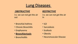
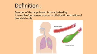
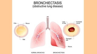
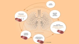
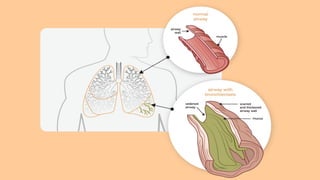
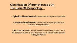
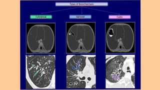
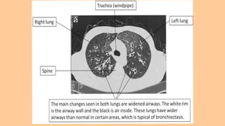
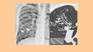
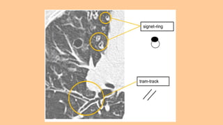
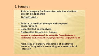
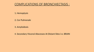
BRONCHIECTASIS An
Obstructive Lung Disease
Presented By :
Dr. Maryum Malik - Resident Medicine ( R1 )
Sahiwal Teaching Hospital , Sahiwal
Lung Diseases
OBSTRUCTIVE
i-e. we can not get the air
out
• Bronchial Asthma
• Chronic Bronchitis
• Emphysema
• Bronchiectasis
• Bronchiolitis
RESTRICTIVE
i-e. we can not get the air
in
• ILD
• Sarcoidosis
• Scoliosis
• Obesity
• Neuromuscular Disease
Definition :
Disorder of the large bronchi characterized by
irreversible/permanent abnormal dilation & destruction of
bronchial walls.
Pathophysiology Of
Bronchiectasis :
• Microorganisms trigger inflammatory
response i-e. release of Neutrophils,
Lymphocytes/Macrophages
• Neutrophils alters function of ciliary
epithelium that changes the cilial beat
frequency & mucous gland hypersecretion
• Impaired muco ciliary clearance & Airway
mucous hypersecretion leads to microbial
colonization/infection
• Neutrophils releases inf. cytokines +
elastases & matrix metalloprotienases that
destroy bronchial elastin & other supporting
lung structures
• In response to this a cycle of intense Chronic
Inflammation occurs resulting in BRONCHIAL
DILATION & DESTRUCTION.
its pathophysiology is
commonly described as
distinct phases of
infection and chronic
inflammation. The
interaction between
these phases establishes
a vicious circle.
Classification Of Bronchiectasis On
The Basis Of Morphology :
1. Cylindrical bronchiectasis: bronchi are enlarged and cylindrical.
2. Varicose bronchiectasis: bronchi are irregular with areas of
dilatation and constriction.
3. Saccular or cystic: dilated bronchi form clusters of cysts. This is
the most severe form of bronchiectasis and is often found in patients
with cystic fibrosis
Aetioloy ( Causes ) Of
Bronchiectasis :
1. CONGENITAL :
• Cystic Fibrosis ( Most Common Cause In US & All Western
Countries ! ) : CFTR gene defect > Defective Ion Transport > Airway
Surface Liquid Depletion > Impaired Muco ciliary Clearance
• Ciliary Dysfunction Syndromes : Absent dynein arm in cilia
* Primary Ciliary Dyskinesia - Immotile Cilia Syndrome
* Kartagener's Syndrome - Triad Of Situs Inversus , Chronic
Sinusitis & Bronchiectasis
• Alpha-1 antitrypsin (AAT) deficiency : Disruption of Protease
- Anti protease balance > Neutrophils Elastases & Proteases
destroy lung structure
• Primary Hypogammaglobulinemia
2. ACQUIRED :
A. CHILDREN -
• Primary Tuberculosis ( Most Common Cause World Wide + 3rd World
Countries i-e, Egypt / Developing Countries )
• Severe Infections In Infancy : Whooping Cough/Pertussis , Measles
• Inhaled Foreign Body
B. ADULTS -
• Pulmonary Tuberculosis ( Most Common Cause World Wide + 3rd
World Countries i-e, Egypt / Developing Countries )
• Suppurative Pneumonia
• Mycobacterium Avium Complex MAC : Non Tuberculous
Mycobacterium
• Allergic Bronchopulmonary Aspergillosis ABPA
Kartagener's Syndrome Visual Map
CLINICAL FINDINGS :
A. SYMPTOMS -
• Chronic Cough + Production Of Copious Amounts Of Purulent
Sputum i-e. Foul smelling sputum ( Classic Symptom! )
• Hemoptysis - resulting from airway damage
• Pleuritic Chest Pain - d/t inflammatory changes in the airways
• Dyspnea , Weight Loss , Anemia & Other Systemic Manifestations
are common.
B. SIGNS -
• Coarse Crackles at the Lung Bases
• Wheezing
• Clubbing - More frequent in Moderate to Severe Cases
DIAGNOSIS :
1. History + Physical Examination - Compatible History Of Chronic
Respiratory Symptoms i-e. Daily Cough & Purulent Sputum Production
2. Sputum Analysis - Bacteriological & Mycological Ex. Of Sputum
3. Radiological Examinations
a. Plain Chest X-ray -
Tram-track opacities i-e. dilated bronchi with thickened walls (tram-
track sign) or as ring-like markings. Scattered Irregular Opacities ,
Atelectasis & Focal consolidation may be present. Overall there appears
to be an increase in bronchovascular markings.
b. HRCT Chest ( Most Sensitive & Specific Test For Diagnosing
Bronchiectasis ) -
• Tram-Track Appearance : Lack of narrowing in the caliber of bronchus
towards periphery i-e. dilated bronchi with thickened walls
• Signet Ring Sign : Bronchus & Pul. Artery should be of same size but in
BE Bronchus is markedly dilated than Pul. Artery
4. Pulmonary Function Tests PFTs - Obstructive Pattern i-e. Low
FEV1/FVC ( Normal : 0.75 - 0.85 )
5. Tests To Identify Underlying Illness -
6. Assessment Of Ciliary Function -
Electron microscopy
Healthcare provider can use a special microscope, called an electron
microscope, to look at samples of airway cilia. This test can show
whether there are any problems with the structure of your cilia or how
they are working.
MANAGEMENT :
2. Antibiotics :
3. Surgery :
COMPLICATIONS OF BRONCHIECTASIS :
1. Hemoptysis
2. Cor Pulmonale
3. Amyloidosis
4. Secondary Visceral Abscesses At Distant Sites i-e. BRAIN
BRONCHIECTASIS and its causes and management.pptx

BRONCHIECTASIS and its causes and management.pptx

  • 1.
    BRONCHIECTASIS An Obstructive LungDisease Presented By : Dr. Maryum Malik - Resident Medicine ( R1 ) Sahiwal Teaching Hospital , Sahiwal
  • 2.
    Lung Diseases OBSTRUCTIVE i-e. wecan not get the air out • Bronchial Asthma • Chronic Bronchitis • Emphysema • Bronchiectasis • Bronchiolitis RESTRICTIVE i-e. we can not get the air in • ILD • Sarcoidosis • Scoliosis • Obesity • Neuromuscular Disease
  • 3.
    Definition : Disorder ofthe large bronchi characterized by irreversible/permanent abnormal dilation & destruction of bronchial walls.
  • 5.
    Pathophysiology Of Bronchiectasis : •Microorganisms trigger inflammatory response i-e. release of Neutrophils, Lymphocytes/Macrophages • Neutrophils alters function of ciliary epithelium that changes the cilial beat frequency & mucous gland hypersecretion • Impaired muco ciliary clearance & Airway mucous hypersecretion leads to microbial colonization/infection • Neutrophils releases inf. cytokines + elastases & matrix metalloprotienases that destroy bronchial elastin & other supporting lung structures • In response to this a cycle of intense Chronic Inflammation occurs resulting in BRONCHIAL DILATION & DESTRUCTION. its pathophysiology is commonly described as distinct phases of infection and chronic inflammation. The interaction between these phases establishes a vicious circle.
  • 8.
    Classification Of BronchiectasisOn The Basis Of Morphology : 1. Cylindrical bronchiectasis: bronchi are enlarged and cylindrical. 2. Varicose bronchiectasis: bronchi are irregular with areas of dilatation and constriction. 3. Saccular or cystic: dilated bronchi form clusters of cysts. This is the most severe form of bronchiectasis and is often found in patients with cystic fibrosis
  • 10.
    Aetioloy ( Causes) Of Bronchiectasis : 1. CONGENITAL : • Cystic Fibrosis ( Most Common Cause In US & All Western Countries ! ) : CFTR gene defect > Defective Ion Transport > Airway Surface Liquid Depletion > Impaired Muco ciliary Clearance • Ciliary Dysfunction Syndromes : Absent dynein arm in cilia * Primary Ciliary Dyskinesia - Immotile Cilia Syndrome * Kartagener's Syndrome - Triad Of Situs Inversus , Chronic Sinusitis & Bronchiectasis • Alpha-1 antitrypsin (AAT) deficiency : Disruption of Protease - Anti protease balance > Neutrophils Elastases & Proteases destroy lung structure • Primary Hypogammaglobulinemia
  • 11.
    2. ACQUIRED : A.CHILDREN - • Primary Tuberculosis ( Most Common Cause World Wide + 3rd World Countries i-e, Egypt / Developing Countries ) • Severe Infections In Infancy : Whooping Cough/Pertussis , Measles • Inhaled Foreign Body B. ADULTS - • Pulmonary Tuberculosis ( Most Common Cause World Wide + 3rd World Countries i-e, Egypt / Developing Countries ) • Suppurative Pneumonia • Mycobacterium Avium Complex MAC : Non Tuberculous Mycobacterium • Allergic Bronchopulmonary Aspergillosis ABPA
  • 12.
  • 13.
    CLINICAL FINDINGS : A.SYMPTOMS - • Chronic Cough + Production Of Copious Amounts Of Purulent Sputum i-e. Foul smelling sputum ( Classic Symptom! ) • Hemoptysis - resulting from airway damage • Pleuritic Chest Pain - d/t inflammatory changes in the airways • Dyspnea , Weight Loss , Anemia & Other Systemic Manifestations are common. B. SIGNS - • Coarse Crackles at the Lung Bases • Wheezing • Clubbing - More frequent in Moderate to Severe Cases
  • 14.
    DIAGNOSIS : 1. History+ Physical Examination - Compatible History Of Chronic Respiratory Symptoms i-e. Daily Cough & Purulent Sputum Production 2. Sputum Analysis - Bacteriological & Mycological Ex. Of Sputum 3. Radiological Examinations a. Plain Chest X-ray - Tram-track opacities i-e. dilated bronchi with thickened walls (tram- track sign) or as ring-like markings. Scattered Irregular Opacities , Atelectasis & Focal consolidation may be present. Overall there appears to be an increase in bronchovascular markings. b. HRCT Chest ( Most Sensitive & Specific Test For Diagnosing Bronchiectasis ) - • Tram-Track Appearance : Lack of narrowing in the caliber of bronchus towards periphery i-e. dilated bronchi with thickened walls • Signet Ring Sign : Bronchus & Pul. Artery should be of same size but in BE Bronchus is markedly dilated than Pul. Artery
  • 15.
    4. Pulmonary FunctionTests PFTs - Obstructive Pattern i-e. Low FEV1/FVC ( Normal : 0.75 - 0.85 ) 5. Tests To Identify Underlying Illness -
  • 16.
    6. Assessment OfCiliary Function - Electron microscopy Healthcare provider can use a special microscope, called an electron microscope, to look at samples of airway cilia. This test can show whether there are any problems with the structure of your cilia or how they are working.
  • 20.
  • 23.
  • 27.
  • 29.
    COMPLICATIONS OF BRONCHIECTASIS: 1. Hemoptysis 2. Cor Pulmonale 3. Amyloidosis 4. Secondary Visceral Abscesses At Distant Sites i-e. BRAIN