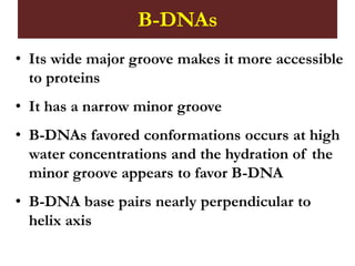
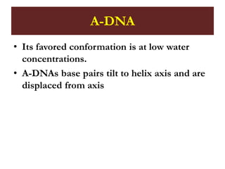
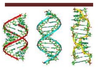
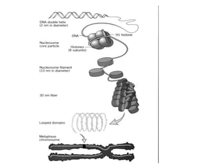
DNA Higher organization
(Tertiary) Structure
By
R Wanyama
Polynucleotides (1)
• Polynucleotides are formed by the
condensation of two or more nucleotides.
The condensation most commonly occurs
between the alcohol of a 5'-phosphate of
one nucleotide and the 3'-hydroxyl of a
second, with the elimination of H2O,
forming a phosphodiester bond. The
formation of phosphodiester bonds in DNA
and RNA exhibits directionality.
Polynucleotides (1)
• The primary structure of DNA and RNA
(the linear arrangement of the nucleotides)
proceeds in the 5'—>3' direction. The
common representation of the primary
structure of DNA or RNA molecules is to
write the nucleotide sequences from left to
right synonymous with the 5'—>3' direction
as shown:
• 5'–pGpApTpC–3'
DNA: structure
• Like RNAs, deoxyribonucleic acids (DNAs)
are polymeric molecules consisting of
nucleotide building blocks.
• Instead of ribose, however, DNA contains 2-
deoxyribose, and the uracil base in RNA is
replaced by thymine.
Tertiary Structures of DNA
• Tertiary structure is the locations of the atoms
in three-dimensional space, taking into
consideration geometrical and steric constraints
• A higher order than the secondary structure in
which large-scale folding in a linear polymer
occurs and the entire chain is folded into a
specific 3-dimensional shape
Tertiary Structures of DNA
• There are 4 areas in which the structural
forms of DNA can differ
• Handedness - right or left
• Length of the helix turn
• Number of base pairs per turn
• Difference in size between the major and
minor grooves
Tertiary Structures of DNA
• The tertiary arrangement of DNA's double
helix in space includes
– B-DNA,
– A-DNA
– Z-DNA
• B-DNA is the most common form of DNA in
vivo and is more narrow, elongated helix than
A-DNA
B-DNAs
• Its wide major groove makes it more accessible
to proteins
• It has a narrow minor groove
• B-DNAs favored conformations occurs at high
water concentrations and the hydration of the
minor groove appears to favor B-DNA
• B-DNA base pairs nearly perpendicular to
helix axis
• The sugar pucker which determines the
shape of the α-helix, whether the helix will
exist in the A-form or in the B-form occurs
at the C2'-end
A-DNA
• Is shorter and wider than helix B
• Most RNA and RNA-DNA duplex in this
form
• A-DNA has a deep, narrow major groove
which does not make it easily accessible to
proteins
• On the other hand, its wide, shallow minor
groove makes it accessible to proteins but
with lower information content than the
major groove
A-DNA
• Its favored conformation is at low water
concentrations.
• A-DNAs base pairs tilt to helix axis and are
displaced from axis
Z-DNA
• Is a relatively rare left-handed double-helix
• Given the proper sequence and superhelical
tension, it can be formed in vivo but its
function is unclear
• It has a more narrow, more elongated helix
than A or B.
• Z-DNA's major groove is not really groove and
it has a narrow minor groove
• The most favored conformation occurs when
there are high salt concentrations
Forms of DNA
Each of us has enough DNA to
reach from here to the sun and
back, more than 300 times. How
is all of that DNA packaged so
tightly into chromosomes and
squeezed into a tiny nucleus?
The Problem
• Spacing between base pairs ≈3.4Å
• For human genome, approximately 3.2 billion
base pairs
• Total length≈ 3.4×10-10×3.2×109×2 ≈2.2m
• Diameter of a nucleus: 5~10×10-6m
• Access to genetic information must be
regulated
• DNA are packaged with associated proteins
into chromosomes
DNA, Histones, and Chromatin (1)
• The answer to this question lies in the fact
that certain proteins compact chromosomal
DNA into the eukaryotic nucleus
• These proteins are called histones, and the
resulting DNA-protein complex is
called chromatin
• Histones are a family of small, positively
charged proteins termed H1, H2A, H2B, H3,
and H4
DNA, Histones, and Chromatin (2)
• DNA is negatively charged, due to the
phosphate groups in its phosphate-
sugar backbone, so histones bind with DNA
very tightly
• Packaged DNA must provide controlled
access to regions required for gene
expression
HISTONES
• Main packaging proteins
• 5 classes: H1, H2A, H2B, H3, H4.
• Rich in Lysine and Arginine
The Nucleosome: The Unit of
Chromatin
• The basic repeating structural (and
functional) unit of chromatin is the
nucleosome, which contains nine histone
proteins and about 166 base pairs of DNA
• Two, each of the histones H2A, H2B, H3,
and H4 come together to form a histone
octamer, which binds and wraps about 1.7
turns of DNA, or about 146 base pairs
• The addition of one H1 protein wraps
another 20 base pairs, resulting in two full
turns around the octamer
• Obviously, 166 base pairs are not very much,
as each chromosome contains over 100
million base pairs of DNA on average
• Therefore, every chromosome contains
hundreds of thousands of nucleosomes, and
these nucleosomes are joined by the DNA
that runs between them
• This joining DNA is referred to as linker
DNA
• Each chromosome is thus a long chain of
nucleosomes, which gives the appearance of
a string of beads when viewed using an
electron microscope
Chromatin is Coiled into Higher-
Order Structures (1)
• The packaging of DNA into nucleosomes
shortens the fiber length about sevenfold. In
other words, a piece of DNA that is 1 meter
long will become a "string-of-beads"
chromatin fiber just 14 centimeters (about 6
inches) long
• Despite this shortening, a half-foot of
chromatin is still much too long to fit into
the nucleus, which is typically only 10-20
microns in diameter
Chromatin is Coiled into Higher-
Order Structures (2)
• Therefore, chromatin is further coiled into an
even shorter, thicker fiber, termed the "30-nm
fiber," because it is ~30 nanometers in
diameter
• The precise structure of the 30-nm fiber is
not yet known
• ~40 folds in packaging
• Chromatin structure beyond nucleosomes is
poorly understood
DNA tertiary structure 2022.pdf

DNA tertiary structure 2022.pdf

  • 1.
    DNA Higher organization (Tertiary)Structure By R Wanyama
  • 2.
    Polynucleotides (1) • Polynucleotidesare formed by the condensation of two or more nucleotides. The condensation most commonly occurs between the alcohol of a 5'-phosphate of one nucleotide and the 3'-hydroxyl of a second, with the elimination of H2O, forming a phosphodiester bond. The formation of phosphodiester bonds in DNA and RNA exhibits directionality.
  • 3.
    Polynucleotides (1) • Theprimary structure of DNA and RNA (the linear arrangement of the nucleotides) proceeds in the 5'—>3' direction. The common representation of the primary structure of DNA or RNA molecules is to write the nucleotide sequences from left to right synonymous with the 5'—>3' direction as shown: • 5'–pGpApTpC–3'
  • 4.
    DNA: structure • LikeRNAs, deoxyribonucleic acids (DNAs) are polymeric molecules consisting of nucleotide building blocks. • Instead of ribose, however, DNA contains 2- deoxyribose, and the uracil base in RNA is replaced by thymine.
  • 6.
    Tertiary Structures ofDNA • Tertiary structure is the locations of the atoms in three-dimensional space, taking into consideration geometrical and steric constraints • A higher order than the secondary structure in which large-scale folding in a linear polymer occurs and the entire chain is folded into a specific 3-dimensional shape
  • 7.
    Tertiary Structures ofDNA • There are 4 areas in which the structural forms of DNA can differ • Handedness - right or left • Length of the helix turn • Number of base pairs per turn • Difference in size between the major and minor grooves
  • 8.
    Tertiary Structures ofDNA • The tertiary arrangement of DNA's double helix in space includes – B-DNA, – A-DNA – Z-DNA • B-DNA is the most common form of DNA in vivo and is more narrow, elongated helix than A-DNA
  • 9.
    B-DNAs • Its widemajor groove makes it more accessible to proteins • It has a narrow minor groove • B-DNAs favored conformations occurs at high water concentrations and the hydration of the minor groove appears to favor B-DNA • B-DNA base pairs nearly perpendicular to helix axis
  • 10.
    • The sugarpucker which determines the shape of the α-helix, whether the helix will exist in the A-form or in the B-form occurs at the C2'-end
  • 11.
    A-DNA • Is shorterand wider than helix B • Most RNA and RNA-DNA duplex in this form • A-DNA has a deep, narrow major groove which does not make it easily accessible to proteins • On the other hand, its wide, shallow minor groove makes it accessible to proteins but with lower information content than the major groove
  • 12.
    A-DNA • Its favoredconformation is at low water concentrations. • A-DNAs base pairs tilt to helix axis and are displaced from axis
  • 13.
    Z-DNA • Is arelatively rare left-handed double-helix • Given the proper sequence and superhelical tension, it can be formed in vivo but its function is unclear • It has a more narrow, more elongated helix than A or B. • Z-DNA's major groove is not really groove and it has a narrow minor groove • The most favored conformation occurs when there are high salt concentrations
  • 14.
  • 15.
    Each of ushas enough DNA to reach from here to the sun and back, more than 300 times. How is all of that DNA packaged so tightly into chromosomes and squeezed into a tiny nucleus?
  • 16.
    The Problem • Spacingbetween base pairs ≈3.4Å • For human genome, approximately 3.2 billion base pairs • Total length≈ 3.4×10-10×3.2×109×2 ≈2.2m • Diameter of a nucleus: 5~10×10-6m • Access to genetic information must be regulated • DNA are packaged with associated proteins into chromosomes
  • 17.
    DNA, Histones, andChromatin (1) • The answer to this question lies in the fact that certain proteins compact chromosomal DNA into the eukaryotic nucleus • These proteins are called histones, and the resulting DNA-protein complex is called chromatin • Histones are a family of small, positively charged proteins termed H1, H2A, H2B, H3, and H4
  • 18.
    DNA, Histones, andChromatin (2) • DNA is negatively charged, due to the phosphate groups in its phosphate- sugar backbone, so histones bind with DNA very tightly • Packaged DNA must provide controlled access to regions required for gene expression
  • 19.
    HISTONES • Main packagingproteins • 5 classes: H1, H2A, H2B, H3, H4. • Rich in Lysine and Arginine
  • 20.
    The Nucleosome: TheUnit of Chromatin • The basic repeating structural (and functional) unit of chromatin is the nucleosome, which contains nine histone proteins and about 166 base pairs of DNA • Two, each of the histones H2A, H2B, H3, and H4 come together to form a histone octamer, which binds and wraps about 1.7 turns of DNA, or about 146 base pairs
  • 21.
    • The additionof one H1 protein wraps another 20 base pairs, resulting in two full turns around the octamer • Obviously, 166 base pairs are not very much, as each chromosome contains over 100 million base pairs of DNA on average
  • 22.
    • Therefore, everychromosome contains hundreds of thousands of nucleosomes, and these nucleosomes are joined by the DNA that runs between them • This joining DNA is referred to as linker DNA • Each chromosome is thus a long chain of nucleosomes, which gives the appearance of a string of beads when viewed using an electron microscope
  • 23.
    Chromatin is Coiledinto Higher- Order Structures (1) • The packaging of DNA into nucleosomes shortens the fiber length about sevenfold. In other words, a piece of DNA that is 1 meter long will become a "string-of-beads" chromatin fiber just 14 centimeters (about 6 inches) long • Despite this shortening, a half-foot of chromatin is still much too long to fit into the nucleus, which is typically only 10-20 microns in diameter
  • 24.
    Chromatin is Coiledinto Higher- Order Structures (2) • Therefore, chromatin is further coiled into an even shorter, thicker fiber, termed the "30-nm fiber," because it is ~30 nanometers in diameter • The precise structure of the 30-nm fiber is not yet known • ~40 folds in packaging • Chromatin structure beyond nucleosomes is poorly understood