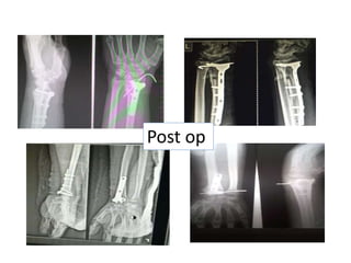
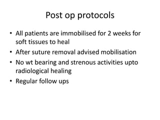
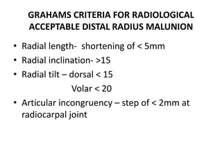
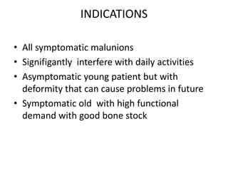
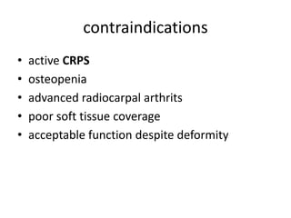
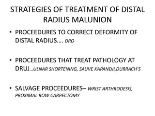
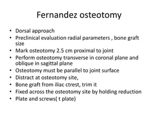
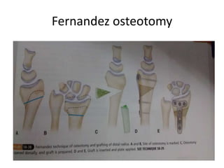
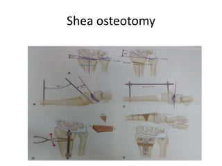
MANAGEMENT OF DISTAL RADIUS MALUNION BY
CORRECTIVE OSTEOTOMY AND BONE GRAFTING
WITH DEFINITIVE FIXATION
By
Dr kishore vemula
Final year postgraduate
Department of orthopaedics
SVRRGGH.
Case 1
Case 2 case 3
Intra op
Post op
Post op protocols
• All patients are immobilised for 2 weeks for
soft tissues to heal
• After suture removal advised mobilisation
• No wt bearing and strenous activities upto
radiological healing
• Regular follow ups
MANAGEMENT OF DISTAL RADIUS MALUNION
BY CORRECTIVE OSTEOTOMY AND BONE
GRAFTING WITH DEFINITIVE FIXATION
Introduction
• Despite improvements in treatment since
early 1980’s malunion remains a common
cause of residual disability in distal radius
fractures
• “Wrist will enjoy perfect freedom in all its
motions and can be completely exempt from
pain’” colles observation in 1814
• Malunion can be caused by failure to achieve
or maintain accurate reduction or by
inadequate duration or type of
immobilisation.
• Rduction is difficult in communition,
osteoporosis
• Old age is commonly assosciated with
malunions
Malunion - deformities
• Extraarticular -Decrease in
• Radial length
• Decrease in radial
inclination
• Loss of normal volar
tilt
• Intraarticlar - articular step
≥2mm
• DRUJ incongruency or instability. -
Step of 1-2mm at distal radioulnar joint
or combination of these can occur
Clinical features
• Decrease in Radial length- druj pathology, pain at druj
• Decrease in radial inclination– impaired ulnar deviation
• Loss of normal volar tilt
– Dorsal tilt- deformity, decreased wrist flexion,
carpal instabiliy pattern
– Excessive Volar tilt – deformity, decreased
extension, mid carpal instability
. Articular step--- radiocarpal arthritis pain at wrist
.druj instability--- pain at dista radio ulnar joint
Clinical features
• Excessive dorsal
angulation ≥15-20
for long time can
alter the wrist
biomecanics and can
cause DISI pattern
instabiliy pattern
• Excessive volar
angulation ≥20 can
leas to VISI pattern.
Clinical features
• Excessive dorsal tilt can cause median nerve
compression
• Long term- attritional rupture of EPL tendon
Predictors of poor outcomes
fernandez et al
• Distal radius articular step of
• DRUJ step of 1-2mm
• Doral tilt morethan 15-20
• Volar tilt of morethan 20
• Radial length of lessthan 6mm
• Radial inclination of lessthan 10
≥2mm
Radiographic evaluation
• Ap/ lateral views in neutral rotation
• Cotralateral wrist also for measurement and
comparision
• Ct san- for articular step
• Mri- itegrity of TFCC & Intercarpal ligaments
Operative treatment
• Not all patients of distal radius malunion
requires surgery
• Not indicated in patients with
– Minimally symptomatic
– Not interfering daily activities
– Malunion in acceptable range
– Very old age
Asymptomatic patient with even gross deformity in
old age--- not indicated
GRAHAMS CRITERIA FOR RADIOLOGICAL
ACCEPTABLE DISTAL RADIUS MALUNION
• Radial length- shortening of < 5mm
• Radial inclination- >15
• Radial tilt – dorsal < 15
Volar < 20
• Articular incongruency – step of < 2mm at
radiocarpal joint
INDICATIONS
• All symptomatic malunions
• Signifigantly interfere with daily activities
• Asymptomatic young patient but with
deformity that can cause problems in future
• Symptomatic old with high functional
demand with good bone stock
contraindications
• active CRPS
• osteopenia
• advanced radiocarpal arthrits
• poor soft tissue coverage
• acceptable function despite deformity
STRATEGIES OF TREATMENT OF DISTAL
RADIUS MALUNION
• PROCEEDURES TO CORRECT DEFORMITY OF
DISTAL RADIUS…. DRO
• PROCEEDURES THAT TREAT PATHOLOGY AT
DRUJ…ULNAR SHORTENING, SAUVE KAPANDJI,DURRACH’S
• SALVAGE PROCEEDURES– WRIST ARTHRODESIS,
PROXIMAL ROW CARPECTOMY
TREATMENT OF EXTRAARTICULAR
MALUNION
• 1. FERNANADEZ OSTEOTOMY…
for dorsal angulation
open wedge metaphyseal osteotomy with
bone grafting and internal fixation with plate.
• 2. SHEA OSTEOTOMY
for volar angulation
open wedge metaphyseal osteotomy with
bone grafting and internal fixation with plate
• 3.INTRAMEDULLARY FIXATION WITH micronail
• 4. EXTERNAL FIXATION
Fernandez osteotomy
• Dorsal approach
• Preclinical evaluation radial parameters , bone graft
size
• Mark osteotomy 2.5 cm proximal to joint
• Perform osteotomy transverse in coronal plane and
oblique in sagittal plane
• Osteotomy must be parallel to joint surface
• Distract at osteotomy site,
• Bone graft from iliac ctrest, trim it
• Fixed across the osteotomy site by holding reduction
• Plate and screws( t plate)
Fernandez osteotomy
Shea osteotomy
• Volar henry approach
• Preclinical evaluation radial parameters , bone graft
size
• Mark osteotomy 2.5 cm proximal to joint
• Perform osteotomy transverse in coronal plane and
oblique in sagittal plane
• Osteotomy must be parallel to joint surface
• Distract at osteotomy site,
• Bone graft from iliac ctrest, trim it
• Fixed across the osteotomy site by holding reduction
• Plate and screws( t plate)
Shea osteotomy
Proceedures to correct DRUJ
incongruency
• These proceedures may require either single
or in combination with distal radius osteotomy
based on maintainance of DRUJ congruency
after DRO
• DRUJ that maintained with DRO alone can be
left with DRO alone.
• DRUJ not maintained with DRO alone may
require these proceedures.
Proceedures to correct DRUJ
incongruency
• DRUJ preservation surgeries;
Ulnar shortening osteotomy
• DRUJ ablation sugeries;
1.Darrach’s pcedure
2.Bowers arthroplasty- partial
resection of distal ulna
3.Sauve- kapandji Proceedure - druj
fusion + prox ulnarpsseudoarhosis
•Thank u…..
Every one…

Distal radius malunion , correction

  • 1.
    MANAGEMENT OF DISTALRADIUS MALUNION BY CORRECTIVE OSTEOTOMY AND BONE GRAFTING WITH DEFINITIVE FIXATION By Dr kishore vemula Final year postgraduate Department of orthopaedics SVRRGGH.
  • 2.
  • 3.
  • 4.
  • 5.
  • 6.
    Post op protocols •All patients are immobilised for 2 weeks for soft tissues to heal • After suture removal advised mobilisation • No wt bearing and strenous activities upto radiological healing • Regular follow ups
  • 7.
    MANAGEMENT OF DISTALRADIUS MALUNION BY CORRECTIVE OSTEOTOMY AND BONE GRAFTING WITH DEFINITIVE FIXATION
  • 8.
    Introduction • Despite improvementsin treatment since early 1980’s malunion remains a common cause of residual disability in distal radius fractures • “Wrist will enjoy perfect freedom in all its motions and can be completely exempt from pain’” colles observation in 1814
  • 9.
    • Malunion canbe caused by failure to achieve or maintain accurate reduction or by inadequate duration or type of immobilisation. • Rduction is difficult in communition, osteoporosis • Old age is commonly assosciated with malunions
  • 10.
    Malunion - deformities •Extraarticular -Decrease in • Radial length • Decrease in radial inclination • Loss of normal volar tilt • Intraarticlar - articular step ≥2mm • DRUJ incongruency or instability. - Step of 1-2mm at distal radioulnar joint or combination of these can occur
  • 11.
    Clinical features • Decreasein Radial length- druj pathology, pain at druj • Decrease in radial inclination– impaired ulnar deviation • Loss of normal volar tilt – Dorsal tilt- deformity, decreased wrist flexion, carpal instabiliy pattern – Excessive Volar tilt – deformity, decreased extension, mid carpal instability . Articular step--- radiocarpal arthritis pain at wrist .druj instability--- pain at dista radio ulnar joint
  • 12.
    Clinical features • Excessivedorsal angulation ≥15-20 for long time can alter the wrist biomecanics and can cause DISI pattern instabiliy pattern • Excessive volar angulation ≥20 can leas to VISI pattern.
  • 13.
    Clinical features • Excessivedorsal tilt can cause median nerve compression • Long term- attritional rupture of EPL tendon
  • 14.
    Predictors of pooroutcomes fernandez et al • Distal radius articular step of • DRUJ step of 1-2mm • Doral tilt morethan 15-20 • Volar tilt of morethan 20 • Radial length of lessthan 6mm • Radial inclination of lessthan 10 ≥2mm
  • 15.
    Radiographic evaluation • Ap/lateral views in neutral rotation • Cotralateral wrist also for measurement and comparision • Ct san- for articular step • Mri- itegrity of TFCC & Intercarpal ligaments
  • 16.
    Operative treatment • Notall patients of distal radius malunion requires surgery • Not indicated in patients with – Minimally symptomatic – Not interfering daily activities – Malunion in acceptable range – Very old age Asymptomatic patient with even gross deformity in old age--- not indicated
  • 17.
    GRAHAMS CRITERIA FORRADIOLOGICAL ACCEPTABLE DISTAL RADIUS MALUNION • Radial length- shortening of < 5mm • Radial inclination- >15 • Radial tilt – dorsal < 15 Volar < 20 • Articular incongruency – step of < 2mm at radiocarpal joint
  • 18.
    INDICATIONS • All symptomaticmalunions • Signifigantly interfere with daily activities • Asymptomatic young patient but with deformity that can cause problems in future • Symptomatic old with high functional demand with good bone stock
  • 19.
    contraindications • active CRPS •osteopenia • advanced radiocarpal arthrits • poor soft tissue coverage • acceptable function despite deformity
  • 20.
    STRATEGIES OF TREATMENTOF DISTAL RADIUS MALUNION • PROCEEDURES TO CORRECT DEFORMITY OF DISTAL RADIUS…. DRO • PROCEEDURES THAT TREAT PATHOLOGY AT DRUJ…ULNAR SHORTENING, SAUVE KAPANDJI,DURRACH’S • SALVAGE PROCEEDURES– WRIST ARTHRODESIS, PROXIMAL ROW CARPECTOMY
  • 21.
    TREATMENT OF EXTRAARTICULAR MALUNION •1. FERNANADEZ OSTEOTOMY… for dorsal angulation open wedge metaphyseal osteotomy with bone grafting and internal fixation with plate. • 2. SHEA OSTEOTOMY for volar angulation open wedge metaphyseal osteotomy with bone grafting and internal fixation with plate • 3.INTRAMEDULLARY FIXATION WITH micronail • 4. EXTERNAL FIXATION
  • 22.
    Fernandez osteotomy • Dorsalapproach • Preclinical evaluation radial parameters , bone graft size • Mark osteotomy 2.5 cm proximal to joint • Perform osteotomy transverse in coronal plane and oblique in sagittal plane • Osteotomy must be parallel to joint surface • Distract at osteotomy site, • Bone graft from iliac ctrest, trim it • Fixed across the osteotomy site by holding reduction • Plate and screws( t plate)
  • 23.
  • 24.
    Shea osteotomy • Volarhenry approach • Preclinical evaluation radial parameters , bone graft size • Mark osteotomy 2.5 cm proximal to joint • Perform osteotomy transverse in coronal plane and oblique in sagittal plane • Osteotomy must be parallel to joint surface • Distract at osteotomy site, • Bone graft from iliac ctrest, trim it • Fixed across the osteotomy site by holding reduction • Plate and screws( t plate)
  • 25.
  • 26.
    Proceedures to correctDRUJ incongruency • These proceedures may require either single or in combination with distal radius osteotomy based on maintainance of DRUJ congruency after DRO • DRUJ that maintained with DRO alone can be left with DRO alone. • DRUJ not maintained with DRO alone may require these proceedures.
  • 27.
    Proceedures to correctDRUJ incongruency • DRUJ preservation surgeries; Ulnar shortening osteotomy • DRUJ ablation sugeries; 1.Darrach’s pcedure 2.Bowers arthroplasty- partial resection of distal ulna 3.Sauve- kapandji Proceedure - druj fusion + prox ulnarpsseudoarhosis
  • 28.