
More Related Content
What's hot
What's hot (20)
Similar to Nonunion ppt
Similar to Nonunion ppt (20)
More from Saurabh Chahar
More from Saurabh Chahar (15)
Recently uploaded
Recently uploaded (20)
Nonunion ppt
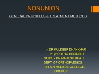
- 1. NONUNION GENERAL PRINCIPLES & TREATMENT METHODS :- DR KULDEEP DHANKHAR 3rd yr ORTHO RESIDENT GUIDE:- DR MAHESH BHATI DEPT. OF ORTHOPAEDICS DR S.N.MEDICAL COLLEGE, JODHPUR
- 3. DEFINITIONS:- Nonunion: (somewhat arbitrary) A fracture that has not and is not going to heal Delayed union: A fracture that requires more time than usual to heal Shows progression over time
- 4. DEFINITIONS:- Nonunion: A fracture that is a minimum of 9 months post occurrence and is not healed and has not shown radiographic progression for 3 months (FDA 1986) Not pragmatic Prolonged morbidity Narcotic abuse Work-related and/or emotional impairment
- 5. Definitions (pragmatic):- Nonunion: A fracture that has no potential to heal without further intervention -:9 months elapsed time with no healing progress for 3 months.
- 6. Classification:- Paley’s Classification Muller and Weber’s Classification
- 7. Muller & Weber’s classification:- Amount of callus at the fracture site 1. Hypervascular Nonunion 2. Avascular Nonunion
- 8. 1.Hypervascular 1. Hypertrophic:- -Elephant foot - Horse hoof 2. Oligotrophic 2.Avascular 1. Torsion Wedge 2. Comminuted 3. Defect 4. Atrophic
- 9. 1.Hypertrophic: Vascularized Callus formation present on x-ray Elephant’s foot - abundant callus Horse’s hoof - less abundant callus Typically only needs stability to consolidate! Elephant foot Horse hoof
- 10. 2.Oligotrophic:- Some/minimal callus on x-ray Not an aggressive healing response, but not completely void of biologic activity Vascularity is present on bone scan Oligotrophic
- 11. 3.Atrophic:- No evidence of callous formation on x-ray Ischemic or cold on bone scan Atrophic
- 13. Pseudarthrosis:- Typically has adequate vascularity Excessive motion/instability False joint forms over significant time
- 14. Etiology of Nonunion:- Host factors Fracture/Injury factors Initial treatment of injury factors Complicating factor = Infection
- 15. Etiology of Nonunion – (Host Factors):- Smoking Diabetes/Endocrinopathy Thyroid/ parathyroid disorders, hypogonadism [testosterone deficiency], Vit D deficiency, others Malnutrition Medications Steroids, Chemotherapy, Bispohosphonates Bone quality, vascular status Balance, compliance with weight bearing restrictions Psychiatric conditions, dementia
- 16. • Smoking:- Decreases peripheral oxygen tension Dampens peripheral blood flow Well documented difficulties in wound healing in patients who smoke • Retrospective studies show time to union • Higher infection and nonunion rates
- 17. • Diabetes (Neuropathic Fractures):- Best studied in ankle and pilon fractures: Complicated diabetics – those with end organ disease – neuropathy, PVD, renal dysfunction Increased rates of infection and soft tissue complications Increased rates of nonunion, time to union significantly longer Prolonged NWB required Inability to control response to trauma can result in hyperemia, osteopenia, and osteoclastic bone resorption Charcot arthropathy
- 18. • Malnutrition:- Adequate protein and energy is required for wound healing Screening test: serum albumin total lymphocyte count Albumin less than 3.5 and lymphocytes less than 1,500 cells/ml is significant
- 19. • Etiology of Nonunion – (Fracture/Injury Factors):- High energy injury Fracture mechanism – MVC vs fall from standing Open or closed fracture Bone loss Soft tissue injury Bone involved and anatomic location
- 20. • Fracture Pattern:- Fracture patterns in higher energy injuries (i.e.: comminution, bone loss, or segmental patterns) have a higher degree of soft tissue and bone ischemia • Traumatic Soft Tissue Disruption:- • Incidence of nonunion is increased with open fractures • More severe open fracture (i.e. Gustillo III B vs Grade I) have higher incidence of nonunion
- 21. • Etiology of Nonunion –(Initial Treatment Factors):- Nonunion may occur after completely appropriate treatment of a fracture, or after less than appropriate treatment Was appropriate management performed initially? Operative vs non-operative? Was the stability achieved initially appropriate? Consider: Bone and anatomic location (shaft vs metaphysis) Patient – host status, compliance with care
- 22. • Etiology of Nonunion –(Initial Treatment Factors):- After operative treatment….. Was the appropriate implant and technique employed? (Fixation strategy) Relative vs absolute stability? Direct vs indirect reduction? Implant size/length, number of screws, locking vs conventional Location of incisions. Signs of poor dissection? Iatrogenic soft tissue disruption, devascularization of bone
- 24. Etiology: Surgeon Excessive soft tissue stripping •Improper or unstable fixation -Absolute stability- • Gap due to distraction or poor reduction -Relative stability = Excessive motion
- 25. • Etiology of Nonunion –(Initial Treatment Factors):- Is the current construct too flexible or too stiff? Implant too short? Bridge plating of a simple pattern with lack of compression? Why did the current treatment fail? Understanding the mode of failure for the initial procedure helps with planning the nonunion surgery
- 26. • Anatomic Location of Fractures:- Some areas of skeleton are at risk for nonunion due to anatomic vascular considerations i.e.: Proximal 5th metatarsal, femoral neck, carpal scaphoid Open diaphyseal tibia fractures are the classic example with high rates of nonunion throughout the literature
- 27. • Infection:- May be obvious Open draining wounds, erythema, inadequate soft tissue coverage Subclinical is more difficult High index of suspicion ESR, CRP may indicate infection and provide baseline values to follow after debridement and antibiotic therapy • Must be dealt with….. • Debridement, debridement, debridement • Multiple cultures. Identify the bacteria • Infected bone requires stability to resolve infection • May achieve union in the presence of infection with appropriate treatment
- 28. • Evaluation OF PATIENTS:- History of injury and prior treatment Medical history and co-morbidities Physical examination Including deformity! Imaging modalities Patient needs, goals, expectations
- 29. • Patient Evaluation – History of Injury:- Date and nature of original injury (high or low energy) Open or closed injury? Number of prior surgical procedures History of drainage or wound healing difficulties? Prior infection? Identify antibiotics used and bacteria cultured (if possible) Written timeline in complex cases Current symptoms – pain, deformity, motion problems, chronic drainage Ability to work and perform ADL’s
- 30. • Patient Evaluation – Medical History:- Diabetes, endocrinopathies, vit D, etc Physiologic age – co-morbidities Heart disease, COPD, kidney/liver disease Nutrition Smoking Medications Ambulatory/functional status now and prior to original injury
- 31. • Patient Evaluation –(Physical Exam):- Appearance of limb Color, skin quality, prior incisions, skin grafts Erythema or drainage Range of motion of all joints Pain – location and contributing factors Strength, ability to bear weight Vascular status and sensation (complete neurovascular exam) Deformity Clinically = Length, alignment, AND rotation
- 32. • Patient Evaluation –(Imaging):- Any injury-related imaging available – plain film and CT Serial plain radiographs from injury to present are extremely helpful (hard to get) Most current imaging – orthogonal x-rays, typically diagnostic for nonunion Healing of 3 out of 4 cortices without pain is typically considered union. Oblique view may be helpful for radiographic diagnosis of nonunion CT can be helpful but metal artifact can make it difficult
- 33. • Patient Evaluation –(Imaging Tomography):- Linear tomograms Helpful if metallic hardware present Helps to identify persistent fracture line in: Hyptrophic nonunions in which x-rays are not diagnostic and pain persists at fracture site CT and MRI are replacing linear tomography Still a good option if available at your institution
- 34. • Patient Evaluation- (Radionuclide Scanning):- Technetium - 99 diphosphonate Detects repairable process in bone ( not specific) Gallium - 67 citrate Accumulates at site of inflammation (not specific) Sequential technetium or gallium scintigraphy Only 50-60% accuracy in subclinical ostoemyelitis
- 35. • Indium III - Labeled Leukocyte Scan:- Infected Nonunion? Good with acute osteomyelitis, but less effective in diagnosing chronic or subacute bone infections Sensitivity 83-86%, specificity 84-86% Technique is superior to technetium and gallium to identify infection
- 36. MRI:- Infected Nonunion? Abnormal marrow with increased signal on T2 and low signal on T1 Can identify and follow sinus tacts and sequestrum diagnostic sensitivity of 100%, specificity 63%, accuracy 93%
- 37. Nonunion:-Diagnosis • Persistent Pain • Non physiologic motion • Progressive deformity • No radiographic evidence of healing • Failing implants
- 38. • Patient Evaluation –(Goals & Expectations):- What are the patient’s goals and needs? Household ambulation vs marathon runner Pain relief expectations Range of motion expectations Long standing nonunions may have stiff adjacent joints Risks to neurovascular structures (radial nerve in humerus nonunion)
- 41. Electrical Stimulation:- Applied mechanical stress on bone generates electrical potentials Compression = electronegative potentials = bone formation Tension = electropositive potentials = bone resorption Basic science suggests e-stim upregulates TGF-β and BMP’s suggesting osteoinduction
- 42. Three Modalities of Electric bone Growth Stimulators:- 1. Direct current - implantation of cathode in bone and anode on skin 2. Inductive coupling – pulsed electromagnetic field with device on skin 3. Capacitive coupling - electrodes placed on skin, alternating current Conflicting and inconclusive evidence
- 43. Contraindication to Electric Stimulation:- Synovial pseudoarthrosis Electric stimulation does not address associated problems of angulation, malrotation and shortening – deformity!!
- 44. Unanswered Questions:-?? When is electric stimulation indicated? Which fracture types are indicated? What are the efficacy rates? What time after injury is best for application?
- 45. Ultrasound:- Piezoelectric transducer generates an acoustic pressure wave Some evidence to show faster healing in fresh fractures Evidence is moderate to poor in quality with conflicting results
- 46. Extracorporeal Shock Wave Therapy:- Single impulse acoustic wave with a high amplitude and short wavelength. Microtrauma induced in bone thought to stimulate neovascularization and cell differentiation Clinical studies are of a poor level and no strong evidence for use in nonunions is available
- 47. Operative Treatment:- Debridement and hardware removal Plate osteosynthesis Intramedullary nailing External fixation Autogenous bone graft Bone marrow aspirate Allograft bone Demineralized bone matrix BMP’s Platelet concentrates
- 48. Autogenous Bone Marrow Aspirate:- Typically from the iliac crest Transplant osteoprogenitor and mesenchymal stem cells to nonunion site Osteoinductive, not osteoconductive Level III and IV studies available Positive correlation between number of progenitor cells in aspirate and amount of callous
- 49. BMP’s:- rhBMP-2 and rhBMP-7 have been shown to be equivalent to autologous iliac crest graft for delayed reconstruction of tibial bone defects May be a good alternative to ICBG for the management of nonunion Very expensive!! rhBMP-2 inserted at the time of definitive wound closure for high grade (3A or 3B) open tibia fractures- unclear effect on re-operation and infection rates because literature conflicting
- 50. Autogenous Bone Grafting:- Considered the “gold standard” Osteoinductive - contain proteins and other factors promoting vascular ingrowth and healing Osteogenic – contains viable osteoblasts, progenitor cells, mesenchymal stem cells Osteoconductive - contains a scaffolding for which new bone growth can occur
- 51. Surgical/Fixation Strategy:- Define nonunion type Hyper-, oligo-, atrophic, or pseudarthrosis Fracture location – diaphysis vs metaphysis Infected vs Aseptic Deformity? Patient/host factors Goals and expectations
- 52. Plate Osteosynthesis:- Correction of malalignment Osteotomy may be required, planning always required Compression in hypertrophic cases Immediate mobilization, likely NWB Requires adequate soft tissue coverage More dissection required for plating and osteotomy in deformity correction Bone graft as needed
- 53. Plate Osteosynthesis:- Soft tissue and bony dissection are extremely important! Preserve periosteum and muscular attachment to bone Concept of “working window” Only expose the necessary amount of bone to do the case, maintain vascularity
- 54. Plate Osteosynthesis: Osteoperiosteal Decortication Management of the bone… Do not simply elevate the periosteum off the bone!! Use a sharp chisel or osteotome to elevate an osteoperiosteal flap Sharp chisel and a mallet to take some good, vascularized bone with the periosteum Provides excellent environment for bone graft to produce callous as the elevated bone remains vascularized by the periosteum
- 55. Intramedullary Nailing:- Mechanically stabilizes long bone nonunions as a load sharing implant May allow for early weight bearing Must manage malalignment Starting and ending points, entrance and exit angle of each fragment Initially destroys endosteal blood supply (will recover) but increase periosteal blood supply
- 56. Intramedullary Nailing… Can be performed without direct exposure or dissection of the fracture soft tissue envelope Or can be performed in conjunction with an open exposure of the nonunion site and bone grafting Not applicable in articular nonunions and malunions
- 57. IM Nail Dynamization:- Removal of interlocking bolt(s) to allow for axial compression at nonunion with weight bearing •Commonly performed technique for nonunion management when IM nail is in place •Extremely limited data to support this technique •83% success rate in tibial nonunion management
- 58. IM Exchange Nailing:- Replacing IM nail with larger IM nail increased stability (r4) •Medullary reaming reactive vascularity •limited data to support this technique (stronger than dynamization data) •90% success rate in tibial nonunion management
- 59. External Fixation:- Excellent for gradual malalignment correction Useful in the management of infected nonunions Allows for repeat debridements while providing stability Soft tissue coverage without contaminated hardware in wound Allows for bone transport for large intercalary defects Can generate large compressive forces at nonunion Allows mobilization of joints May be bulky and difficult for patients to manage Pin infections common In complex cases, may be good for limb salvage but may require a long period of time 1.LRS technique(monorail) 2.Ilizarov technique
- 60. Nonunions: (Summary) • Definition of delayed unions/nonunions and natural course of fracture healing • Causes of disturbed bone healing such as vascularity, instability, and infection • Principles of treatment applied based on types of nonunion: - stabilization - enhancement of biology - eradication of infection if any • How to prevent delayed unions/nonunions: - biological fixation in original operation - early recognition of delayed union