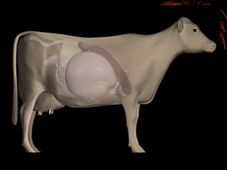
Abomasal Displacement in Dairy
Cows: Old to Recent Advances
Dr. Mohamed Tharwat
Prof. of Vet. Internal Medicine,
Zagazig University
Post
Calving
Metabolic
Problems
0 1 2 3 4 5 6 7 8 9 10 11 12
1 2 3 4 5
Periods
Month
Freshening
Body Stores
Used for
Milk Production
Body Stores
Regained for
Next Lactation
Dry Period
Rumen
Rehab
Nutrient and Milk Yield Relationships in the
Lactation and Gestation Cycle
Dry Matter Intake
Metabolic disorders often occur in a predictable
sequence or cascade
Significance of these Problems
Reduced Milk
Production
Reduced
Fertility
Loss of
Animal
Post Calving
Metabolic Problems
What is happening to the cow
post calving?
• Milk production increases quicker
than appetite
• Cows lose weight
• Cows adjusting to new diet
• Stress levels increase
• Combating Infection
Post Metabolic Problems
Primary Metabolic
• Hypocalcaemia
• Hypomagnesaemia
• Post-Parturient
Haemoglobinuria
• Acetonaemia
• Fatty Liver
Syndrome
Secondary
Metabolic
• Displaced
Abomasum
• Retained
Membranes
• Lactic Acidosis
Displaced Abomasum
Displaced Abomasum (DA)
• Refers to repositioning of abomasum from the right
ventral abdominal wall
• Twisting and displacement slows or stops flow of
digesta.
• Leads to bloat appearance
• “Pinging” detected with stethoscope by thumping
the cow near the last rib and listening on the left
flank.
• Displacements may be left or right sided (RDA,
LDA)
• Most are left-sided.
Displaced Abomasums
• Left displaced abomasum (LDA)
is most common in early lactation
• Right displaced abomasum (RDA)
occurs throughout lactation
• Right torsion of the abomasum
(RTA)
Abomasum
• True Stomach
• Site of gastric juice production
• Site of protein digestion and
amino acid absorption
• Breakdown to peptides
and amino acids by the
enzyme.
Types of nutrients?
Digestive Process
Nonprotein N
(NPN)
Feed
proteins FatsCarbohydrates
Cellulose Starches
Hemicellulose Sugars
Microbial protein
(essential AA)
Volatile fatty
acids (VFA’s)
VFA’s
RUMEN/
RETICULUM
RUP
Microbial protein
Peptides
OMASUM
ABOMASUM
SMALL
INTESTINE
Peptides
Amino acids
Fats
Fatty acids &
glycerol
Glucose
Glucose
RUP
LIVER
Glucose
= microbial action; = main site of absorption = some absorptionRDP = rumen degraded protein; RUP = rumen undegraded protein;
Outline: Displaced Abomasum
• History and Signalment
• Pathophysiology
• Diagnosis
• Clinical signs, clin path, R/O’s
• Treatment
• Non-surgical
• Surgical
• Ancillary care (Fluids, …)
• Risk Factors for LDA
History of DA
• Age: older lactating dairy cattle
• Timing: 80% occur during first month after
parturition
• Nutrition:
• Dry cow rations: inadequate fiber
• Fresh cow: excess carbohydrates/ inadequate fiber
Concurrent disease:
40% of DA’s have retained placenta,
mastitis, or metritis
Risk Factors for LDA
• High-production Dairy Cows
• High concentrate, low roughage
diet
• Large body size
• Limited exercise
• Post-partum
• Abomasal atony
Normal location of abomasum
Digestive Tract
Esophagus
Rumen
Omasum
Reticulum
Abomasum
Pancreas
Liver
Gall
bladder
Cecum
Small intestine
Large
intestine
Rectum
Left view bovine
stomach
Why does the abomasum displace?
(1) Abomasal atony
(2) Increased abomasal gas production
(1) + (2) => abomasum moves (LDA,RDA)
Normal position of abomasum Left displacement
Displacing
Abomasum
In Action
LDA
Cross Section Abdominal Cavity
A
B
C
D
B
C
D
A
Abomasum = A
Rumen = B
C = Omasum
D = Liver
A typical area of ping outlined in a cow with an LDA
Why does abomasal atony occur?
• Hypocalcaemia
• Inadequate effective fiber
• VFA’s reach abomasum => abomasal
hypomotility => HCl refluxes back into
rumen => systemic metabolic alkalosis
• Endotoxemia
• Released during mastitis/metritis
RUMEN
HAY
RUMEN
VFA
pH
Abomasal atony
Growth Rate
(all Bacteria)
Fermentable CHO
Taken from “Digestive Physiology of Herbivores”
http://arbl.cvmbs.colostate.edu/hbooks/pathphys/digestion/herbivores/
Why increased gas productn?
Diet Type Gas volume
(methane,O2,N2)
Hay 800 ml/hr
Concentrate 3 lb 1100 ml/hr
Concentrate 15 lb 2200 ml/hr
Clinical Pathology
• Normal CBC
• Metabolic alkalosis(slight)
• Hypo
• Ca
• K
• Cl
• Ketosis (mild)
• Dehydration
• Hypoglycemia (maybe)
• Hyperbilirubinemia
Clinical Signs of DA’s
• Normal TPR (most cases)
• Partial anorexia (“off feed”)
• Hypogalactia (“down in milk” ~ 5-10
lb/day)
• Depression
• Secondary ketosis
• mild to moderate
• Scant stool
• firm/loose
• undigested particles
Clinical Signs (continued)
• Paralumbar fossa:
• “Slab-sided” abdomen
• Visualize / Palpate
• Rectal palpation (can’t)
• Mild colic
• Mild hypocalcemia
• Hypotonic rumen
• Cold ears, widely dilated pupils
Clinical Signs (continued)
• LDA: Ping & Splash
• Ascult and percuss
• Ping high pitched
• Ballottement for splash
of fluid
• All pings are not
created equal – rumen
ping
Note: ~15% of LDA’s DO NOT PING or ping
sporadically
Differential Diagnosis
• LDA
• Ketosis (non-pinging LDA)
• Rumen ping
• RDA
• Ketosis (non-pinging RDA)
• Other Right-sided pings:
• Uterus, cecum, peritoneum, colon, rectum
• “Off feed” ping
Right-sided pings
Treatment of Displaced Abomasum
Therapeutic Goals
• Return abomasum to proper position
• Create a permanent attachment
• Correct electrolyte, acid-base, & hydration
deficits
• Treat other concurrent diseases
Rolling
• The cow is cast and laid on her back, then
rolled vigorously to the right and the roll
stopped abruptly in the hope that the
abomasum will free itself.
• Bring the cow to sternal position & allow to
stand
• Auscultate the left thorax to ensure LDA is
relieved
• Starvation and water restriction for 2 days
before rolling may be advisable.
Rolling Technique
• Advantages
• Quick & easy technique
• No invasive surgery
• Disadvantages
• >50% redisplace
• If RDA or RTA are present, can
exacerbate problems
Surgical correction
Left abomasal displacement
Left abomasal
displacement
1) Left flank approach.
Left flank approach.
Left abomasal displacement
Left flank approach.
Left abomasal displacement
Left flank approach.
Left abomasal displacement
• Left flank approach.
• Left Para costal incision as for exploratory
laparotomy or rumenotomy but to a slightly
lower site.
• With along thread 1.5 m length of non-
absorbable suture material (polyamide no. 3-
4) with straight long triangular needle 5-6
continuous ford interlocking sutures are made
in the greater omentum and wall of
abomasum.
• Evacuate the gas from the abomasum with
needle connected to a plastic tube.
Left abomasal displacement
•The abomasum is repositioned in its
normal position.
•The two ends of the thread are passes
through the ventral abdominal wall
slightly to the right of midline by two
needles.
•The two ends are then tied together.
•The flank incision is closed.
•The abomaso-omental skin suture is
removed after two weeks
Left abomasal displacement
Right flank approach
Percutaneous fixation (toggle or bar suture)
Left abomasal displacement
Percutaneous fixation
(toggle or bar suture)
Percutaneous fixation
(toggle or bar suture)
Percutaneous fixation
(toggle or bar suture)
• Percutaneous fixation (toggle or bar suture)
• The lower abdominal wall is prepared aseptically
• The animal is turned into dorsal recumbancy.
• The surgeon confirms the presence of tympanic
abomasum ventrally.
• Trocar and cannula are inserted firmly through the
abdominal wall and abomasum caudal to the xiphoid and
right to the midline.
• The trocar is pulled out and toggle is inserted.
• The cannula is removed and another toggle is applied by
the same way caudal to the first.
• The two are tied together.
Left abomasal displacement
• Its incidence is 1:8 to left displacement.
• The same steps for diagnosis for left displacement
except that it is done of course on the right.
• Corrective surgery of RDA is generally more
difficult and the patient progress is guarded to
unfavorable.
• Longstanding cases is not indicated for surgery
specially with abomasal volvulus (RTA).
• Corrected through:
• Right paralumbar approach.
• Ventral abdominal approach (open surgery).
Right abomasal displacement
Paramedian approach
Replacement Fluids
• Isotonic Saline, Lactated Ringer’s IV
to replace deficit
• K, Ca salts as needed to correct
electrolyte imbalances
• Free-choice oral fluids with NaCl, KCl
Bad prognostic indicators
• Chloride level equal to or below 79mEq/l.
• Pulse rate equal to or greater than 100.
• Base excess.
• Tachycardia and decreased temperature.
• Anion gap of equal to or over 30 mEq/l.
• Large abomasal fluid volume.
• Blue abomasal color at surgery.
• Decreased GI motility post surgery.
Control
• Feeding large quantity of forages at late
pregnancy.
• Ensure daily exercise.
• Minimize dietary alterations near
parturition.
• Decease the amount of grain and corn
silage fed prepartum, while other forages
are fed ad libitum.
Questions
are
welcome?

Displaced abomasum

  • 2.
    Abomasal Displacement inDairy Cows: Old to Recent Advances Dr. Mohamed Tharwat Prof. of Vet. Internal Medicine, Zagazig University
  • 3.
  • 4.
    0 1 23 4 5 6 7 8 9 10 11 12 1 2 3 4 5 Periods Month Freshening Body Stores Used for Milk Production Body Stores Regained for Next Lactation Dry Period Rumen Rehab Nutrient and Milk Yield Relationships in the Lactation and Gestation Cycle Dry Matter Intake
  • 5.
    Metabolic disorders oftenoccur in a predictable sequence or cascade
  • 6.
    Significance of theseProblems Reduced Milk Production Reduced Fertility Loss of Animal Post Calving Metabolic Problems
  • 7.
    What is happeningto the cow post calving? • Milk production increases quicker than appetite • Cows lose weight • Cows adjusting to new diet • Stress levels increase • Combating Infection
  • 8.
    Post Metabolic Problems PrimaryMetabolic • Hypocalcaemia • Hypomagnesaemia • Post-Parturient Haemoglobinuria • Acetonaemia • Fatty Liver Syndrome Secondary Metabolic • Displaced Abomasum • Retained Membranes • Lactic Acidosis
  • 9.
  • 10.
    Displaced Abomasum (DA) •Refers to repositioning of abomasum from the right ventral abdominal wall • Twisting and displacement slows or stops flow of digesta. • Leads to bloat appearance • “Pinging” detected with stethoscope by thumping the cow near the last rib and listening on the left flank. • Displacements may be left or right sided (RDA, LDA) • Most are left-sided.
  • 11.
    Displaced Abomasums • Leftdisplaced abomasum (LDA) is most common in early lactation • Right displaced abomasum (RDA) occurs throughout lactation • Right torsion of the abomasum (RTA)
  • 12.
    Abomasum • True Stomach •Site of gastric juice production • Site of protein digestion and amino acid absorption • Breakdown to peptides and amino acids by the enzyme.
  • 13.
  • 14.
    Digestive Process Nonprotein N (NPN) Feed proteinsFatsCarbohydrates Cellulose Starches Hemicellulose Sugars Microbial protein (essential AA) Volatile fatty acids (VFA’s) VFA’s RUMEN/ RETICULUM RUP Microbial protein Peptides OMASUM ABOMASUM SMALL INTESTINE Peptides Amino acids Fats Fatty acids & glycerol Glucose Glucose RUP LIVER Glucose = microbial action; = main site of absorption = some absorptionRDP = rumen degraded protein; RUP = rumen undegraded protein;
  • 15.
    Outline: Displaced Abomasum •History and Signalment • Pathophysiology • Diagnosis • Clinical signs, clin path, R/O’s • Treatment • Non-surgical • Surgical • Ancillary care (Fluids, …) • Risk Factors for LDA
  • 16.
    History of DA •Age: older lactating dairy cattle • Timing: 80% occur during first month after parturition • Nutrition: • Dry cow rations: inadequate fiber • Fresh cow: excess carbohydrates/ inadequate fiber Concurrent disease: 40% of DA’s have retained placenta, mastitis, or metritis
  • 17.
    Risk Factors forLDA • High-production Dairy Cows • High concentrate, low roughage diet • Large body size • Limited exercise • Post-partum • Abomasal atony
  • 18.
  • 19.
  • 28.
  • 29.
    Why does theabomasum displace? (1) Abomasal atony (2) Increased abomasal gas production (1) + (2) => abomasum moves (LDA,RDA) Normal position of abomasum Left displacement
  • 30.
  • 31.
  • 32.
    Cross Section AbdominalCavity A B C D B C D A Abomasum = A Rumen = B C = Omasum D = Liver
  • 33.
    A typical areaof ping outlined in a cow with an LDA
  • 49.
    Why does abomasalatony occur? • Hypocalcaemia • Inadequate effective fiber • VFA’s reach abomasum => abomasal hypomotility => HCl refluxes back into rumen => systemic metabolic alkalosis • Endotoxemia • Released during mastitis/metritis
  • 50.
  • 51.
  • 52.
    VFA pH Abomasal atony Growth Rate (allBacteria) Fermentable CHO
  • 53.
    Taken from “DigestivePhysiology of Herbivores” http://arbl.cvmbs.colostate.edu/hbooks/pathphys/digestion/herbivores/
  • 54.
    Why increased gasproductn? Diet Type Gas volume (methane,O2,N2) Hay 800 ml/hr Concentrate 3 lb 1100 ml/hr Concentrate 15 lb 2200 ml/hr
  • 55.
    Clinical Pathology • NormalCBC • Metabolic alkalosis(slight) • Hypo • Ca • K • Cl • Ketosis (mild) • Dehydration • Hypoglycemia (maybe) • Hyperbilirubinemia
  • 56.
    Clinical Signs ofDA’s • Normal TPR (most cases) • Partial anorexia (“off feed”) • Hypogalactia (“down in milk” ~ 5-10 lb/day) • Depression • Secondary ketosis • mild to moderate • Scant stool • firm/loose • undigested particles
  • 57.
    Clinical Signs (continued) •Paralumbar fossa: • “Slab-sided” abdomen • Visualize / Palpate • Rectal palpation (can’t) • Mild colic • Mild hypocalcemia • Hypotonic rumen • Cold ears, widely dilated pupils
  • 58.
    Clinical Signs (continued) •LDA: Ping & Splash • Ascult and percuss • Ping high pitched • Ballottement for splash of fluid • All pings are not created equal – rumen ping Note: ~15% of LDA’s DO NOT PING or ping sporadically
  • 59.
    Differential Diagnosis • LDA •Ketosis (non-pinging LDA) • Rumen ping • RDA • Ketosis (non-pinging RDA) • Other Right-sided pings: • Uterus, cecum, peritoneum, colon, rectum • “Off feed” ping
  • 60.
  • 71.
  • 72.
    Therapeutic Goals • Returnabomasum to proper position • Create a permanent attachment • Correct electrolyte, acid-base, & hydration deficits • Treat other concurrent diseases
  • 73.
    Rolling • The cowis cast and laid on her back, then rolled vigorously to the right and the roll stopped abruptly in the hope that the abomasum will free itself. • Bring the cow to sternal position & allow to stand • Auscultate the left thorax to ensure LDA is relieved • Starvation and water restriction for 2 days before rolling may be advisable.
  • 74.
    Rolling Technique • Advantages •Quick & easy technique • No invasive surgery • Disadvantages • >50% redisplace • If RDA or RTA are present, can exacerbate problems
  • 75.
  • 77.
  • 78.
    Left flank approach. Leftabomasal displacement
  • 79.
    Left flank approach. Leftabomasal displacement
  • 84.
    Left flank approach. Leftabomasal displacement
  • 85.
    • Left flankapproach. • Left Para costal incision as for exploratory laparotomy or rumenotomy but to a slightly lower site. • With along thread 1.5 m length of non- absorbable suture material (polyamide no. 3- 4) with straight long triangular needle 5-6 continuous ford interlocking sutures are made in the greater omentum and wall of abomasum. • Evacuate the gas from the abomasum with needle connected to a plastic tube. Left abomasal displacement
  • 86.
    •The abomasum isrepositioned in its normal position. •The two ends of the thread are passes through the ventral abdominal wall slightly to the right of midline by two needles. •The two ends are then tied together. •The flank incision is closed. •The abomaso-omental skin suture is removed after two weeks Left abomasal displacement
  • 87.
  • 90.
    Percutaneous fixation (toggleor bar suture) Left abomasal displacement
  • 91.
  • 92.
  • 93.
  • 99.
    • Percutaneous fixation(toggle or bar suture) • The lower abdominal wall is prepared aseptically • The animal is turned into dorsal recumbancy. • The surgeon confirms the presence of tympanic abomasum ventrally. • Trocar and cannula are inserted firmly through the abdominal wall and abomasum caudal to the xiphoid and right to the midline. • The trocar is pulled out and toggle is inserted. • The cannula is removed and another toggle is applied by the same way caudal to the first. • The two are tied together. Left abomasal displacement
  • 100.
    • Its incidenceis 1:8 to left displacement. • The same steps for diagnosis for left displacement except that it is done of course on the right. • Corrective surgery of RDA is generally more difficult and the patient progress is guarded to unfavorable. • Longstanding cases is not indicated for surgery specially with abomasal volvulus (RTA). • Corrected through: • Right paralumbar approach. • Ventral abdominal approach (open surgery). Right abomasal displacement
  • 106.
  • 117.
    Replacement Fluids • IsotonicSaline, Lactated Ringer’s IV to replace deficit • K, Ca salts as needed to correct electrolyte imbalances • Free-choice oral fluids with NaCl, KCl
  • 119.
    Bad prognostic indicators •Chloride level equal to or below 79mEq/l. • Pulse rate equal to or greater than 100. • Base excess. • Tachycardia and decreased temperature. • Anion gap of equal to or over 30 mEq/l. • Large abomasal fluid volume. • Blue abomasal color at surgery. • Decreased GI motility post surgery.
  • 120.
    Control • Feeding largequantity of forages at late pregnancy. • Ensure daily exercise. • Minimize dietary alterations near parturition. • Decease the amount of grain and corn silage fed prepartum, while other forages are fed ad libitum.
  • 121.