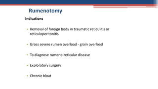
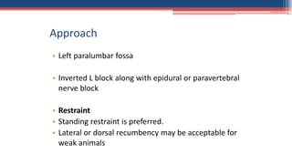
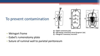
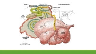
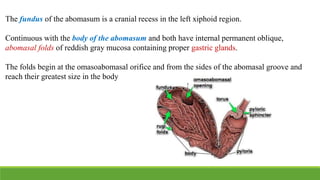
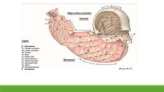
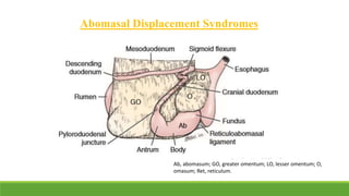
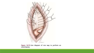
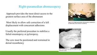
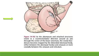
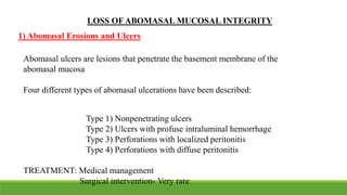
Surgical management of
Abdominal Affections in
bovines
Anatomy
Rumen
The three non-glandular forestomach compartments in the cow are the rumen, reticulum, and
omasum
Rumen occupies most of the left abdomen
Extends from ribs seven to eight to the pelvis
Common affections
• Bloat
• Vagal Indigestion
• Impaction
• Hernia
• TRP
• TRPC
Rumenotomy
Indications
• Removal of foreign body in traumatic reticulitis or
reticuloperitonitis
• Gross severe rumen overload - grain overload
• To diagnose rumeno-reticular disease
• Exploratory surgery
• Chronic bloat
Approach
• Left paralumbar fossa
• Inverted L block along with epidural or paravertebral
nerve block
• Restraint
• Standing restraint is preferred.
• Lateral or dorsal recumbency may be acceptable for
weak animals
Laparotomy – left
• Make paracostal incision 15 cm long about 5 cm
behind last rib, starting 10 cm below lumbar
transverse processes
• Incise skin in single movement and continue scalpel
incision through subcutaneous fat and fascia to
expose abdominal wall musculature
To prevent contamination
• Weingart frame
• Gabel’s rumenotomy plate
• Suture of ruminal wall to parietal peritoneum
• Rumen forceps are hooked
on the dorsal and ventral
aspect of the rumen board
• This allows exteriorization
of a portion of the rumen
wall
Method
Weingart frame method
• Place the frame
• Exteriose the rumen
• Place a sterile cloth or rubber drape or shroud completely around the
exteriorised rumen between the frame and the abdominal wall
• Incise as required
Gabel’s rumenotomy plate
• Explore the rumen for any foreign body
• Remove solid matter if any
• Explore cranially and ventrally for reticulum at
rumeno-reticular pillar
• Place a magnet in the rumen in the case of hardware
disease
Closure of rumen
Flush the area with large volumes of warm sterile saline to clean it and
avoid peritoneal contamination.
Close the rumen incision with No. 2 or 3 absorbable suture
material.
Use a continuous inverting pattern (Cushing, Lembert, or Guard
pattern).
Remove the rumen-to-skin sutures.
Oversew the ruminal incision with a second layer in a fashion similar to
the first.
• Laparotomy site is closed in 4 layers
• It is wise to close the ventral aspect of the skin incision with two to three simple
interrupted sutures.
• The possibility of incisional infection is obvious, and drainage can be easily obtained
by removing these ventral two-to-three sutures if necessary
Post-operative care
• Analgesics
• Antibiotics
• Fluid therapy
• Mild osmotic laxative, may assist in prompting gut motility
• Light diet to animal for few days after the surgery
Normal position : Over the ventral midline caudal to the xiphoid process and
from both left and right paramedian regions lateral to the midline site
DEVELOPMENTAL ANATOMY -
ABOMASUM
Most distal of the four stomach compartments
Neonatal life- Abomasum is the primary functioning stomach compartment
At birth – largest of the four compartments – volume twice that of combined
ruminoreticulum
Fills right cranioventral abdomen – extending caudally on and to the right of
midline to a point beyond the 13 th rib
Young calf- bypassing the under developed rumen
 Thin-walled and capable of great distension and displacement
 Capacity- upto 28L
 Parietal surface and part of the greater curvature – ventral abdominal wall
 Greater omentum – Separates the greater curvature from intestines
 Visceral surface- Lies on the rumen
 Lesser curvature – Around the omasum
The fundus of the abomasum is a cranial recess in the left xiphoid region.
Continuous with the body of the abomasum and both have internal permanent oblique,
abomasal folds of reddish gray mucosa containing proper gastric glands.
The folds begin at the omasoabomasal orifice and from the sides of the abomasal groove and
reach their greatest size in the body
The folds diminish toward the pyloric part which begins at the angle of the abomasum
and consists of the pyloric antrum, pyloric canal, and pylorus.
Lined by wrinkled yellowish mucosa containing pyloric glands.
The pyloric sphincter and the torus pyloricus that bulges from the lesser curvature into
the pylorus can close off the flow from the abomasum to the duodenum
ABOMASAL FUNCTION
 Only stomach compartment – glandular mucosa
 In the adult ruminant, the abomasum functions in a manner similar to that of the
simple stomach of monogastric animals.
 The luminal pH is maintained at a level 3.0 in healthy cattle
 Secrets digestive juices including HCl, pepsin, and rennin
 Alkaline chyme stimulates the abomasal emptying and acidic chyme inhibit
emptying via release of local peptides and hormones
Motility
Abomasal outflow reflects a balance between propulsive abomasal contraction
and a braking action at the gastroduodenal juncture- Duodenal brake
Composition
of chyme
Gastrin and
somatostatin
Volume of
material
entering the
duodenum Inhibit abomasal outflow via
duodenal brake
 Narcotic and alpha-2 adrenergic agents such as xylazine hydrochloride may also
inhibit abomasal outflow by affecting the duodenal brake.
 Abomasal motility is regulated by sympathetic and parasympathetic pathways as
well as the enteric nervous system.
 The enteric pathways may be the most important method of control
 Vagal nerve function plays a role in normal abomasal motility
SURGICALAFFECTIONS OF THE ABOMASUM
Two major categories of abomasal disorders;
Disorders recognized because of altered
abomasal outflow
Conditions associated with loss of abomasal wall
integrity
Altered Abomasal Outflow
The disorders can be grouped into two categories:
Associated with repositioning of the
abdomen in the abdominal cavity
(i.e., displacements)
Occur without a significant
change in abomasal position
Eg: Abomasal impaction
Abomasal Displacement Syndromes
Ab, abomasum; GO, greater omentum; LO, lesser omentum; O,
omasum; Ret, reticulum.
Three syndromes of abomasal displacement are commonly recognized:
Left displacement of the abomasum (LDA)
Right dilation/displacement of the abomasum (RDA)
Rightside volvulus of the abomasum (RVA)
Most common in highproduction dairy cows but also appear sporadically in
calves, dairy bulls, and beef cattle.
LDA - Most common of the recognized displacement syndromes- 85% to
96% of all displacing syndromes
Left Abomasal Displacement (LDA)
Condition in which the body of the abomasum relocates to the left side of the
midline between the rumen and left body wall
Still maintaining flow without complete luminal obstruction
Predisposing factors:
1) Reduction in effective abomasal motility
2) Increase in accumulation of gas in the abomasum
Others:
Stage of Lactation - The majority of LDAs in adult dairy cows occurs early in the
lactation period - transitional period
Anatomy - In late pregnancy the large uterus displaces the abomasum into a more
cranial and transverse position with a greater amount of the abomasum lying to the
left of the midline on the abdominal floor.
Decreased feed intake reduces the volume of the rumen, and the expanding uterus
can displace the rumen dorsally.
With calving, the sudden reduction in the volume of the uterus leaves a void in the
abdomen that may allow the abomasum to slide more easily to the left under the smaller
rumen.
Twin pregnancies may increase the risk of LDA by virtue of a more dramatic size change
of the uterus in the periparturient period.
Genetics - Breed difference in the motilin gene and its expression of the peptide hormone
motilin.
Nutrition - Large amounts of concentrates and low fiber intake in the dry period
Metabolism - Increased insulin resistance in late pregnancy high plasma glucose
and insulin levels Abomasal motility
Management and Environment-
Housing at high density in complex systems
Overconditioning in the dry period
Increased incidence reported in cold seasons
CLINICAL SIGNS AND DIAGNOSIS
 Simultaneous auscultation and percussion - tympanic ping on the left side
 Slab-side abdomen
 Liptac test - pH less than 3.5 indicates a displaced abomasum
 Ultrasonographic examinations - 3.5- to 5-MHz linear or convex
transducer in the standing cow
 Assessment of electrolyte and acid-base disturbances - Decreased Na, Cl,
K, and Cl in 92% LDA cases
Normal topography of left abdominal viscera, cow, and left displacement of abomasum
A) Normal topography of left abdominal viscera
B) Left displacement of abomasum
Illustration by Dr. Gheorghe Constantinescu. Adapted, with permission, from DeLahunta and
Habel, Applied Veterinary Anatomy, W.B. Saunders, 1986.
Left lateral view
Cross-section, Caudocranial view
Left Abomasal Displacement (LDA)
Closed Procedures Open Surgical Procedures
 Right-Paralumbar Fossa (Flank) Omentopexy
 Right-paramedian abomasopexy
 Left-Paralumbar Fossa (Flank) Abomasopexy
 Laparoscopic Abomasopexy
 Rolling
 Blind Tack
 Toggle Pin
 Standing right-paralumbar fossa (flank) approach is probably the most versatile
approach
 Only the most distal pyloric region can be visualized at the level of the incision by
placing caudal traction on the greater omentum
 Approach is not indicated when direct access to the abomasal fundus or body is needed
Right-Paralumbar Fossa (Flank) Omentopexy
Preparation
 The right flank should be clipped and prepared for aseptic surgery
 A right-paravertebral or inverted-L block is recommended for analgesia
Surgical approach
 15- to 20-cm vertical (or up to 20-degree craniodorsal to caudoventral) incision should
be made through the body wall in the cranial right-paralumbar fossa
 The incision starts at 8- to 10-cm ventral to the transverse processes of the second or
third lumbar vertebra and extends ventrally
When the peritoneal cavity is entered , in case of LDA , the duodenum will be
ventrad instead of its horizontal position
Abomasal repositioning
12- 13 gauge needle with a length of sterile tube attached
The needle is carried caudal to the rumen to the most distal part of the displaced
abomasum- inserted obliquely to the abomasal wall
Pressure is applied firmly with the forearm and the hand to release the gas
Needle is withdrawn carefully
Abomasum is returned its normal position following the peritoneal surfaces ventrally
with the hand b/w the rumen and the bodywall
Omentopexy
A 6- to 8-cm vertical fold of thick greater omentum located no more than 3 to 4 cm caudal to
the pyloroduodenal juncture is identified
The omentum is stabilized by incorporating a 1.5-cm fold of omentum with three horizontal
mattress sutures that pass through all three muscle layers and the peritoneum 2 cm cranial to
the incision
The first layer of incisional closure - begin ventrally , include the transversus abdominis muscle
and peritoneum as well as a small bite of omentum upto the ventral third of the suture line
The internal and external oblique muscle layer and skin are apposed as in a routine flank
laparotomy
Approach provides the most direct access to the
greatest surface area of the abomasum
Most likely to allow safe correction of a left
displacement with concurrent adhesions
Usually the preferred procedure to stabilize a
failed omentopexy or pyloropexy.
The cow must be positioned and restrained in
dorsal recumbency
Right-paramedian abomasopexy
A 15- to 20-cm incision should be made parallel and 3
to 4 cm to the right of the midline, extending caudally
from a point 4 to 5 cm caudal to the xiphoid
Six distinct layers: the skin, subcutaneous fascia , thick
external fascia of the rectus sheath, rectus abdominis,
thinner internal fascia of the rectus sheath, and
peritoneum.
Procedure
Abomasal repositioning
The fibrous core adheshions is transected at the body wall
Bring to normal position
Abomasopexy
The optimum site for pexy is a 10- to 12-cm section on the serosal surface, 2 to 4 cm to
the right of the insertion of the greater omentum and extending caudally from a site 5 to
8 cm caudal to the reticuloabomasal ligament
Partial or total decompression of gas from the abomasum
The standard suture pattern for an abomasopexy - simple continuous
pattern initiated at the caudal aspect of the incision through the internal
layer of the rectus sheath and peritoneum
Inclusion of the peritoneum is specifically indicated in this approach -
enhance the stability of the adhesion at the incision
Seromuscular layer of the abomasum should be incorporated in at least
six subsequent bites with the peritoneum and internal fascia
A No. 1– to No. 3–gauge nonabsorbable nonreactive suture material is recommended
for the first layer of closure to establish the most permanent adhesion
Monofilament such as nylon or polypropylene is preferable
Braided and coated nonabsorbable materials will cause very stable adhesions in the
healthy cow but carry an increased risk of infection or fistulation
Complications
Incisional haemorrhage
Dehiscence
Herniation or fistulation
Redisplacement
Intestinal or uterine volvulus associated with shifting of
viscera during recovery from dorsal recumbency
Left-Paralumbar Fossa (Flank) Abomasopexy
Provides the some access to the greater curvature and parietal surface of the abomasum
when it is in a left-displaced position
Safest method of stabilization for left displacements in cows in the last trimester of
pregnancy
Accurate repositioning can be challenging and requires experience
Not indicated for right displacements or volvulus
Approach
A 15- to 20-cm vertical incision in the left flank 2 to 4 cm caudal to the last rib
The ventral aspect of the standard incision should be at the level of the
caudoventral curve of the costochondral arch
Stabilization
A straight needle should be threaded on each end of a 1- to 2-m length of No. 2
monofilament nonabsorbable suture
One needle should be used to take 5 to 8 bites in a continuous pattern through
the seromuscular layers of the parietal surface of the abomasum 2 to 3 cm away
from but parallel to the attachment of the greater omentum and as far cranially
on the abomasum as possible.
The needle attached to the most cranial aspect of the suture line in the abomasum should
be guarded in the right hand and carried ventrally along the left body wall to a site 3 to 4
cm to the right of the midline and 4 to 5 cm caudal to the sternum.
After successful placement of the cranial suture, the procedure should be repeated with
the needle on the caudal end of the suture, taking care to avoid crossing sutures or
perforating omentum or other viscera that can be present on the ventral body wall
The bites should be placed at the center of the suture segment, and two long ends of
suture are left
Once both suture ends have been passed through the ventral body wall and stabilized
with hemostats
Decompress the gas from the abomasum with a 10- to 14-gauge needle with attached
tubing
The surgeon should then manually push the abomasum to the ventral body wall as
the assistant maintains tension on the sutures
Tying of the knot - assistant
Closure of the incision is routine
If prophylactic antibiotics were administered before surgery, no
postoperative antibiotics are indicated.
To avoid fistulation, exposed suture must be cut and allowed to retract into
the abdomen once a stable adhesion has been allowed to form but before the
process of fistulation has begun—optimally between 14 and 21 days after
surgery
Postoperative care
Abomasal Displacement (RDA) And Volvulus (RVA) To The
Right
Right sided abomasal dilation in a cow
Float dorsally with a relatively flat or folded lesser omentum (RDA)
or
To twist on the lesser omentum that supports it
Abomasal volvulus
Illustration by Dr. Gheorghe Constantinescu. Adapted,
with permission, from DeLahunta and Habel, Applied
Veterinary Anatomy, W.B. Saunders, 1986.
Right displaced abomasum with volvulus, cow
A) Normal topography of right
abdominal viscera, cow. B) Right
displacement of abomasum with
volvulus.
Right-Flank Omentopexy/Pyloropexy
Right-Paramedian Abomasopexy
SURGICAL MANAGEMENT
Placing the left forearm medial to the distended abomasal
body (A) and rocking the distended body laterally,
ventrally (B), and finally caudally (C) to free the
duodenum from its site of entrapment ventral to the
abomaso omasal juncture
Correction of a counter clockwise abomasal volvulus by
abomasal manipulation from a right-paralumbar fossa
approach
ABOMASAL OUTFLOW OBSTRUCTIONS
WITHOUT DISPLACEMENT
Mixed Mechanical and Functional Obstructions: Abomasal
Impactions
Distention of a viscus beyond its normal volume with contents that have less
fluid content than normal
Potential mechanical causes
Abnormal or dry luminal contents (hair, placenta, sand, gravel, and poor-
quality roughage , restricted water intake) that lodge in the pyloric region
Mural lesions (lymphosarcoma, fibrosis) - prevent normal contraction and
dilation of the pyloric antrum or cranial duodenum
Extraluminal lesions (adhesions, masses) - distort or compress the abomasal
outflow tract
Surgical approaches
1) Rumenotomy to access content in the lumen of the omasum such as foreign
bodies (plastic materials, etc.) or deliver lubricants via the omasum into the
abomasum
2) Right-paracostal or right-paramedian approach - allow access to the
extraluminal and intraluminal cause of impaction and examination of most of the
abomasum and to allow emptying of the abomasum through abomasotomy
3) A right-flank celiotomy - allows access to the pylorus and cranial duodenum
LOSS OF ABOMASAL MUCOSAL INTEGRITY
Abomasal ulcers are lesions that penetrate the basement membrane of the
abomasal mucosa
Four different types of abomasal ulcerations have been described:
Type 1) Nonpenetrating ulcers
Type 2) Ulcers with profuse intraluminal hemorrhage
Type 3) Perforations with localized peritonitis
Type 4) Perforations with diffuse peritonitis
TREATMENT: Medical management
Surgical intervention- Very rare
1) Abomasal Erosions and Ulcers
2) Abomasal Fistulas
Tracts that communicate from the lumen of the abomasum to the skin surface or
occasionally to the lumen of another viscera (reticulum, rumen, and omasum), organ
(liver), or body cavity (thorax).
Fistulas between the abomasum and skin are most commonly recognized as a
technique-related complication of right-paramedian abomasopexy or of blind tack
technique or toggle pin fixation
The primary predisposing factor that leads to fistulation after abomasopexy is
penetration of abomasal mucosa, particularly with multifilament nonabsorbable
materials that trap organisms between filaments.
Treatment
Treatment goals are to adequately stabilize the cow before surgery, resect the fistulous
tract, and close the affected viscera.
Preparation
• Correction of fluid imbalances
• Preoperative antibiotics are indicated because of the potential for contamination of
adjacent tissues during surgery
• The cow should be restrained in dorsal recumbency under general anesthesia or cast
and restrained with ropes
Procedure
An elliptical or fusiform incision should be made around the tract through the skin
and body wall
The dissection continues into the peritoneal cavity
Separate adhered omentum and, occasionally, other structures from the tract
Once the tract and attached abomasum have been freed , the tract and abomasum
should be elevated to the incision.
Stay sutures should be placed in the abomasal wall 2 to 4 cm from either end of
the tract sharply incised and the tract removed
The resulting defect in the abomasum should be closed in a simple continuous
pattern followed by an inverting layer - polyglactin 910 or polyglycolic acid
The areas should be copiously lavaged
Closure of the body wall - Use of 18-gauge, stainless steel wire in a through-and-
through vertical or horizontal mattress pattern provides the most secure closure
under these circumstances
Postoperative Management
 Antibiotics
 Analgesics
 Supportive therapy, including fluids and calcium
 Nonsteroidal antiinflammatory agents are recommended for 2 to 3 days after
surgery
 The incision should be cleaned daily
SURGICAL AFFECTIONS OF
INTESTINE - RUMINANTS
 Small intestine of the cow - 27 to 49 meters
 Descending duodenum – at entry into the abdomen - right paralumbar fossa -
interposed between the mesoduodenum dorsally and the greater omentum ventrally
 The jejunum is 26 to 48 meters long and is tightly coiled at the edge of the sheet like
mesentery that suspends it
 The distal jejunum and proximal ileum are suspended by a narrow, mobile portion
of the mesentery- distal flange
 The ileum consists of proximal coiled and distal straight segments that form the
terminal portion of the small intestine
SURGICAL ANATOMY
SURGICALAPPROACH
Exploration of the bovine gastrointestinal tract is most commonly performed via a
right paralumbar fossa laparotomy
General anesthesia is preferred in valuable cattle to minimize the risk of aspiration
pneumonia and ingesta contamination of the abdomen during surgery
Recumbent surgery under sedation and local anesthesia is also possible
SMALL-INTESTINAL ACCIDENTS
Obstructive diseases of the bovine small-intestinal tract
1) Nonstrangulating (simple and functional)
2) Strangulating
Nonstrangulating simple obstructions - less common - usually caused by a
trichobezoar, phytobezoar, or enterolith
Strangulating lesions of the small intestine - intussusception and volvulus, and
although relatively uncommon, they still occur regularly
Clinical Signs
 Abdominal pain manifested by treading and stretching out
Kicking at the abdomen
 Affected animals are depressed and anorexic
Decreased rumen contractions
Precipitous drop in milk production
The heart rate is usually elevated associated with pain and hypovolemia.
 Fluid accumulates in the bowel proximal to the obstruction -> abdominal
distention, usually low and bilateral.
Succussion of the abdomen yields a fluid wave
Manure becomes scant or absent
In some cases melena is passed, presumably from sloughing of devitalized
intestine mixed with mucus and fecal material
Abdominal-fluid analysis
Obtained from an avascular portion of the abdomen either
close to the midline or paramedian in front of the udder
Normal peritoneal fluid in cattle - clear and light yellow with
<5000 nucleated cells/µL and total protein <2.6 g/dL
Elevations in total protein (>2.5 g/dL) and cell count
(>10,000 cell/µL) - indicative of inflammation
Large volumes of abdominal fluid - indicate a grave prognosis
associated with diffuse peritonitis, (even though nucleated cell
counts may remain within the normal range)
Cloudy, serosanguinous, or foul-smelling fluid - possible
ischemia or other intestinal accident - poor prognosis
Ultrasound and rectal examination
Very helpful in identifying fluid pockets in the
abdomen, distended viscera, and in some cases,
the actual cause of the obstruction
Ultrasound is especially useful in small calves
when a rectal examination is not possible
Functional and Non-strangulating Obstruction
 Usually secondary to motility dysfunction often associated with enteritis
 The duodenum is most commonly affected
 Obstruction may result from duodenitis, ulcers, electrolyte disturbances,
stricture, foreign bodies, obstruction by displacement of viscera etc
 Obstruction associated with omento- or pyloropexy, and extraluminal
compression by abscessation or neoplasia
DUODENAL OUTFLOW OBSTRUCTION
 Causes - inflammation of the duodenum that results from ulcers,
penetrating foreign bodies, intraluminal or extraluminal masses, or
adhesions in the vicinity of the sigmoid flexure
 Have bilateral ventral abdominal distention
 Cattle with identified duodenal lesions all had marked fluid and electrolyte
disturbances
Treatment
 Removal of any identified obstructing lesions (adhesions, masses)
 If the lesion cannot be removed or identified, a duodenal bypass around the site
of obstruction needs to be done
 The cranial part of the duodenum is anastamosed to the descending duodenum
usually in a side to-side manner
 Supportive fluid and/or antibiotic therapy
STRANGULATING OBSTRUCTION
The invagination of a portion of intestine (intussusceptum) into the lumen of
the adjacent bowel (intussuscipiens)
This action drags the mesentery and associated blood vessels of the
intussusceptum into the neighboring bowel, creating an intestinal obstruction
The affected bowel becomes nonviable because of its compromised blood
supply and peritonitis results
Intussusception
Untreated cattle usually die 5 to 8 days after the onset of clinical signs.
Causes
 Altered normal intestinal peristalsis including enteritis, intestinal
parasitism, mural granuloma, abscess or hematoma, neoplasia
 Sudden diet changes, and medications affecting gastrointestinal motility
 Overeating on lush pasture
CLINICAL SIGNS
 An increased prevalence in Brown Swiss cattle relative to Holsteins was
found and a decreased risk existed for Hereford cattle
 Calves 1 to 2 months of age were at greater risk for developing
intussusception
 The most common locations of intussusception - small intestine (84%),
colocolic (11%), and ileocolic (2%)
 Animals with intussusceptions distal to the ileum were more likely to be
calves
 The length and mobility of the jejunal mesenteric attachments, especially the
distal third, may be why the majority of cattle have jejunojejunal or
jejunoileal intussusceptions
 Fluid therapy
 Nonsteroidal antiinflammatory drugs
 Calcium solutions
 Broad-spectrum antimicrobials are indicated
 An epidural should be given before a standing procedure if the cow is
straining
 Local infiltration analgesia using 2% lidocaine hydrochloride
Preoperative Management
 A right-paralumbar fossa celiotomy provides the best exposure to the intestinal
tract distal to the pylorus – Standing position
Surgical Management
 Distended bowel should be handled gently, cupping the hands during
suspension, using careful manipulations to prevent injury or rupture.
 The color and quantity of peritoneal fluid should be noted
 An intussusception usually presents as tightly coiled loops of firm,
distended intestine
 The lesion is typically nonreducible, and attempts to manipulate the
bowel are contraindicated because it may be friable and rupture is a
risk
 The vasculature should be ligated close to the affected
bowel to avoid impinging on the blood supply of
adjacent bowel.
 An end-to-end anastomosis was performed following
the jejunoileal resection
 Before the resection, a Penrose drain is placed
proximal and distal to the diseased bowel to minimize
spillage of intestinal contents
 The area should be isolated from the rest of the
abdomen with moist, sterile towels or laparotomy
pads.
 After the anastomosis, the mesenteric defect is closed
using 2-0 absorbable suture.
 Site rinsed copiously with sterile fluids, and the
bowel replaced into the abdomen.
Segmental Small-Intestinal Volvulus
 Twisting of a segment of intestine upon itself- creating an
obstruction and strangulation of the blood supply- volvulus of the
intestine and torsion of the mesentery
 The cause of the twist is unknown but may be secondary to ileus.
 Because of the long mesentery of the distal jejunum and ileum—the
so-called distal flange—these sections of intestine are more mobile
and prone to volvulus
 All ages can be affected
 Feces are passed initially, then become scant, and finally absent or
mucoid
 Rectal and ultrasound examinations usually show distended small
intestine often wedged in the pelvic inlet
 Cows become tachycardic and dehydrated
 Initially a hypochloremic metabolic alkalosis is typical -later bowel may
become nonviable and a metabolic acidosis results
 Abdominal pain is apparent with signs similar to—but generally more
severe than—those of cattle with intussusception
 Abdominal distention develops as the proximal intestine fills with gas
and fluid
Treatment
 Cattle should be hydrated and prepared for surgery.
 Perioperative antibiotics and nonsteroidal antiinflammatory drugs are
indicated.
 A right-paralumbar fossa celiotomy is performed
 The proximal intestine usually is greatly distended with fluid and gas.
 The twisted bowel feels turgid and, if its location is distal, the intestine may
be wedged up in the pelvic inlet.
 With the animal in lateral recumbency, exteriorizing the majority of the small
intestine, correcting any displacement, and checking its orientation are possible
 In the standing animal, the bowel is gently untwisted as it is delivered to the incision
SURGERY OF THE CECUM
 The cecum is a large, mobile tube with a
blind apex directed caudally
 Cranially, the cecum is continuous with
the proximal loop of the ascending colon
(PLAC), through the cecocolic orifice
 The cecum is attached dorsally to the
PLAC by the short cecocolic fold and
ventrally to the ileum by the ileocecal fold
ANATOMY
CECAL DILATION/DISLOCATION
Hypocalcemia and/or an inhibitory effect of elevated VFA concentrations in
the cecum on cecal motility
Diets excessively rich in rumen-resistant starch increased carbohydrate
fermentation in the large intestine development of spontaneous cecal
dilation and dislocation (CDD)
Cecal dilation is distention of the cecum without a twist.
Rotation along its long axis is called cecal torsion
Rotation in the area of the ICC junction or the PLAC—when viewed from
the right side of the cow—is termed clockwise or counterclockwise twist or
volvulus
 At ultrasonography of the right-paralumbar fossa, the dilated cecum
closest to the abdominal wall appears as tense tubular hollow organ
with a diameter of about 15 to 20 cm
SURGICAL MANAGEMENT
TYPHLOTOMY
 Right-flank approach
 Preferably in the standing animal under local anesthesia.
 The abdomen is opened through a 25-cm incision that starts dorsally about 8 cm
below the lateral processes of the lumbar vertebrae and 8 cm cranial to the tuber
coxae, extending slightly oblique in a cranioventral direction parallel to the
internal oblique abdominal muscle
 The apex of the cecum is isolated from the rest of the abdomen
 Typhlotomy is performed at the most ventral location
 Digesta are first passively drained from the extraabdominal part of the
cecum and then gently milked from the intraabdominal part of the cecum
and the PLAC to the incision site
 The exteriorized cecum is rinsed with copious amounts of prewarmed 0.9%
saline solution
 The incision site closed with a simple inverting continuous or an inverting
seromuscular suture pattern (3-0 or 2-0 monofilament absorbable suture
material)
 The exteriorized sections are again copiously rinsed and placed back into
their physiologic position within the supraomental recess.
 The cecum is evaluated again 10 minutes later, and if it has refilled, a second
typhlotomy is done to relieve the cecum and PLAC of digesta that may have
accumulated - by propulsion from the ileum or reflux from the spiral colon.
 The typhlotomy site is finally oversewn twice (one layer should be in an inverting
pattern).
 At this point, the orientation of spiral loop of the ascending colon should be axial to
the cecum and PLAC
 Closure of the abdominal wall is performed in a routine manner.
CECAL INTUSSUSCEPTIONS
 Why adult cattle have a low occurrence of intussusception in the cecal region
is that they have a fat filled mesentery that maintains the relationship of the
various segments of the intestine
 Calves mesenteric fat is usually minimal, which allows increased mobility of
the slings of the intestine
 Four different types of intussusceptions involving the cecum have been
described
1) Cecocecal
2) Cecocolic
3) Ileocecocolic
4) Ileocecal
 78% of the cases occurred within the first 4 weeks of life
 80% had a history of severe diarrhea with a mean duration of 1 week
SYMPTOMS AND DIAGNOSIS
 Moderate to severe depression
 Partial to complete anorexia
 Abdominal distention accentuated in the right flank
 Mild signs of abdominal pain
 Scant amounts of dark-red feces and mucous strands may be present.
 Radiography and ultrasonography - used as diagnostic aids to identify distended
bowel in young calves where a rectal examination cannot be performed.
 The definitive diagnosis is usually made during exploratory celiotomy.
 Tachycardia and dehydration may be evident
 Auscultation performed simultaneously with percussion identifies variable small pings
and superficial splashing sounds of fluid-filled bowel in the right flank when performed
simultaneously with succussion.
 Dehydration and acid/base imbalances should be corrected before surgery
 Perioperative antimicrobials should also be administered
 The calf is restrained in left-lateral recumbency - exploratory celiotomy - right
flank - local or general anesthesia
 The affected bowel is exteriorized, and the intussusception manually reduced if
possible
 Depending on the type of intussusception, cecal amputation and resection of the
ileum and proximal loop of the ascending colon may be indicated
SURGICAL MANAGEMENT
REFERENCES
Farm Animal Surgery by Susan L. Fubini, Norm G. Ducharme Second edition
Ruminant Surgery – R.P.S Tyagi and Jit Singh
Bovine Anatomy- Klaus-Dieter Budras/Robert E. Habel
Atlas of large animal surgery - A. W.Kersjes, F.Nemeth and L. J.E.Rutgers
Abomasal surgery

Abomasal surgery

  • 1.
    Surgical management of AbdominalAffections in bovines
  • 2.
    Anatomy Rumen The three non-glandularforestomach compartments in the cow are the rumen, reticulum, and omasum Rumen occupies most of the left abdomen Extends from ribs seven to eight to the pelvis
  • 4.
    Common affections • Bloat •Vagal Indigestion • Impaction • Hernia • TRP • TRPC
  • 5.
    Rumenotomy Indications • Removal offoreign body in traumatic reticulitis or reticuloperitonitis • Gross severe rumen overload - grain overload • To diagnose rumeno-reticular disease • Exploratory surgery • Chronic bloat
  • 6.
    Approach • Left paralumbarfossa • Inverted L block along with epidural or paravertebral nerve block • Restraint • Standing restraint is preferred. • Lateral or dorsal recumbency may be acceptable for weak animals
  • 7.
  • 8.
    • Make paracostalincision 15 cm long about 5 cm behind last rib, starting 10 cm below lumbar transverse processes • Incise skin in single movement and continue scalpel incision through subcutaneous fat and fascia to expose abdominal wall musculature
  • 9.
    To prevent contamination •Weingart frame • Gabel’s rumenotomy plate • Suture of ruminal wall to parietal peritoneum
  • 10.
    • Rumen forcepsare hooked on the dorsal and ventral aspect of the rumen board • This allows exteriorization of a portion of the rumen wall
  • 11.
    Method Weingart frame method •Place the frame • Exteriose the rumen • Place a sterile cloth or rubber drape or shroud completely around the exteriorised rumen between the frame and the abdominal wall • Incise as required
  • 12.
  • 13.
    • Explore therumen for any foreign body • Remove solid matter if any • Explore cranially and ventrally for reticulum at rumeno-reticular pillar • Place a magnet in the rumen in the case of hardware disease
  • 14.
    Closure of rumen Flushthe area with large volumes of warm sterile saline to clean it and avoid peritoneal contamination. Close the rumen incision with No. 2 or 3 absorbable suture material. Use a continuous inverting pattern (Cushing, Lembert, or Guard pattern). Remove the rumen-to-skin sutures. Oversew the ruminal incision with a second layer in a fashion similar to the first.
  • 15.
    • Laparotomy siteis closed in 4 layers • It is wise to close the ventral aspect of the skin incision with two to three simple interrupted sutures. • The possibility of incisional infection is obvious, and drainage can be easily obtained by removing these ventral two-to-three sutures if necessary
  • 16.
    Post-operative care • Analgesics •Antibiotics • Fluid therapy • Mild osmotic laxative, may assist in prompting gut motility • Light diet to animal for few days after the surgery
  • 17.
    Normal position :Over the ventral midline caudal to the xiphoid process and from both left and right paramedian regions lateral to the midline site
  • 19.
    DEVELOPMENTAL ANATOMY - ABOMASUM Mostdistal of the four stomach compartments Neonatal life- Abomasum is the primary functioning stomach compartment At birth – largest of the four compartments – volume twice that of combined ruminoreticulum Fills right cranioventral abdomen – extending caudally on and to the right of midline to a point beyond the 13 th rib Young calf- bypassing the under developed rumen
  • 21.
     Thin-walled andcapable of great distension and displacement  Capacity- upto 28L  Parietal surface and part of the greater curvature – ventral abdominal wall  Greater omentum – Separates the greater curvature from intestines  Visceral surface- Lies on the rumen  Lesser curvature – Around the omasum
  • 22.
    The fundus ofthe abomasum is a cranial recess in the left xiphoid region. Continuous with the body of the abomasum and both have internal permanent oblique, abomasal folds of reddish gray mucosa containing proper gastric glands. The folds begin at the omasoabomasal orifice and from the sides of the abomasal groove and reach their greatest size in the body
  • 23.
    The folds diminishtoward the pyloric part which begins at the angle of the abomasum and consists of the pyloric antrum, pyloric canal, and pylorus. Lined by wrinkled yellowish mucosa containing pyloric glands. The pyloric sphincter and the torus pyloricus that bulges from the lesser curvature into the pylorus can close off the flow from the abomasum to the duodenum
  • 25.
    ABOMASAL FUNCTION  Onlystomach compartment – glandular mucosa  In the adult ruminant, the abomasum functions in a manner similar to that of the simple stomach of monogastric animals.  The luminal pH is maintained at a level 3.0 in healthy cattle  Secrets digestive juices including HCl, pepsin, and rennin  Alkaline chyme stimulates the abomasal emptying and acidic chyme inhibit emptying via release of local peptides and hormones
  • 26.
    Motility Abomasal outflow reflectsa balance between propulsive abomasal contraction and a braking action at the gastroduodenal juncture- Duodenal brake Composition of chyme Gastrin and somatostatin Volume of material entering the duodenum Inhibit abomasal outflow via duodenal brake
  • 27.
     Narcotic andalpha-2 adrenergic agents such as xylazine hydrochloride may also inhibit abomasal outflow by affecting the duodenal brake.  Abomasal motility is regulated by sympathetic and parasympathetic pathways as well as the enteric nervous system.  The enteric pathways may be the most important method of control  Vagal nerve function plays a role in normal abomasal motility
  • 28.
    SURGICALAFFECTIONS OF THEABOMASUM Two major categories of abomasal disorders; Disorders recognized because of altered abomasal outflow Conditions associated with loss of abomasal wall integrity
  • 29.
    Altered Abomasal Outflow Thedisorders can be grouped into two categories: Associated with repositioning of the abdomen in the abdominal cavity (i.e., displacements) Occur without a significant change in abomasal position Eg: Abomasal impaction
  • 30.
    Abomasal Displacement Syndromes Ab,abomasum; GO, greater omentum; LO, lesser omentum; O, omasum; Ret, reticulum.
  • 31.
    Three syndromes ofabomasal displacement are commonly recognized: Left displacement of the abomasum (LDA) Right dilation/displacement of the abomasum (RDA) Rightside volvulus of the abomasum (RVA)
  • 32.
    Most common inhighproduction dairy cows but also appear sporadically in calves, dairy bulls, and beef cattle. LDA - Most common of the recognized displacement syndromes- 85% to 96% of all displacing syndromes Left Abomasal Displacement (LDA) Condition in which the body of the abomasum relocates to the left side of the midline between the rumen and left body wall Still maintaining flow without complete luminal obstruction
  • 33.
    Predisposing factors: 1) Reductionin effective abomasal motility 2) Increase in accumulation of gas in the abomasum Others: Stage of Lactation - The majority of LDAs in adult dairy cows occurs early in the lactation period - transitional period Anatomy - In late pregnancy the large uterus displaces the abomasum into a more cranial and transverse position with a greater amount of the abomasum lying to the left of the midline on the abdominal floor. Decreased feed intake reduces the volume of the rumen, and the expanding uterus can displace the rumen dorsally.
  • 34.
    With calving, thesudden reduction in the volume of the uterus leaves a void in the abdomen that may allow the abomasum to slide more easily to the left under the smaller rumen. Twin pregnancies may increase the risk of LDA by virtue of a more dramatic size change of the uterus in the periparturient period. Genetics - Breed difference in the motilin gene and its expression of the peptide hormone motilin. Nutrition - Large amounts of concentrates and low fiber intake in the dry period Metabolism - Increased insulin resistance in late pregnancy high plasma glucose and insulin levels Abomasal motility
  • 35.
    Management and Environment- Housingat high density in complex systems Overconditioning in the dry period Increased incidence reported in cold seasons
  • 36.
  • 37.
     Simultaneous auscultationand percussion - tympanic ping on the left side  Slab-side abdomen  Liptac test - pH less than 3.5 indicates a displaced abomasum  Ultrasonographic examinations - 3.5- to 5-MHz linear or convex transducer in the standing cow  Assessment of electrolyte and acid-base disturbances - Decreased Na, Cl, K, and Cl in 92% LDA cases
  • 38.
    Normal topography ofleft abdominal viscera, cow, and left displacement of abomasum A) Normal topography of left abdominal viscera B) Left displacement of abomasum Illustration by Dr. Gheorghe Constantinescu. Adapted, with permission, from DeLahunta and Habel, Applied Veterinary Anatomy, W.B. Saunders, 1986.
  • 39.
  • 40.
    Left Abomasal Displacement(LDA) Closed Procedures Open Surgical Procedures  Right-Paralumbar Fossa (Flank) Omentopexy  Right-paramedian abomasopexy  Left-Paralumbar Fossa (Flank) Abomasopexy  Laparoscopic Abomasopexy  Rolling  Blind Tack  Toggle Pin
  • 41.
     Standing right-paralumbarfossa (flank) approach is probably the most versatile approach  Only the most distal pyloric region can be visualized at the level of the incision by placing caudal traction on the greater omentum  Approach is not indicated when direct access to the abomasal fundus or body is needed Right-Paralumbar Fossa (Flank) Omentopexy
  • 42.
    Preparation  The rightflank should be clipped and prepared for aseptic surgery  A right-paravertebral or inverted-L block is recommended for analgesia Surgical approach  15- to 20-cm vertical (or up to 20-degree craniodorsal to caudoventral) incision should be made through the body wall in the cranial right-paralumbar fossa  The incision starts at 8- to 10-cm ventral to the transverse processes of the second or third lumbar vertebra and extends ventrally
  • 43.
    When the peritonealcavity is entered , in case of LDA , the duodenum will be ventrad instead of its horizontal position
  • 44.
    Abomasal repositioning 12- 13gauge needle with a length of sterile tube attached The needle is carried caudal to the rumen to the most distal part of the displaced abomasum- inserted obliquely to the abomasal wall Pressure is applied firmly with the forearm and the hand to release the gas Needle is withdrawn carefully Abomasum is returned its normal position following the peritoneal surfaces ventrally with the hand b/w the rumen and the bodywall
  • 45.
    Omentopexy A 6- to8-cm vertical fold of thick greater omentum located no more than 3 to 4 cm caudal to the pyloroduodenal juncture is identified The omentum is stabilized by incorporating a 1.5-cm fold of omentum with three horizontal mattress sutures that pass through all three muscle layers and the peritoneum 2 cm cranial to the incision
  • 47.
    The first layerof incisional closure - begin ventrally , include the transversus abdominis muscle and peritoneum as well as a small bite of omentum upto the ventral third of the suture line The internal and external oblique muscle layer and skin are apposed as in a routine flank laparotomy
  • 48.
    Approach provides themost direct access to the greatest surface area of the abomasum Most likely to allow safe correction of a left displacement with concurrent adhesions Usually the preferred procedure to stabilize a failed omentopexy or pyloropexy. The cow must be positioned and restrained in dorsal recumbency Right-paramedian abomasopexy
  • 49.
    A 15- to20-cm incision should be made parallel and 3 to 4 cm to the right of the midline, extending caudally from a point 4 to 5 cm caudal to the xiphoid Six distinct layers: the skin, subcutaneous fascia , thick external fascia of the rectus sheath, rectus abdominis, thinner internal fascia of the rectus sheath, and peritoneum. Procedure
  • 50.
    Abomasal repositioning The fibrouscore adheshions is transected at the body wall Bring to normal position Abomasopexy The optimum site for pexy is a 10- to 12-cm section on the serosal surface, 2 to 4 cm to the right of the insertion of the greater omentum and extending caudally from a site 5 to 8 cm caudal to the reticuloabomasal ligament Partial or total decompression of gas from the abomasum
  • 51.
    The standard suturepattern for an abomasopexy - simple continuous pattern initiated at the caudal aspect of the incision through the internal layer of the rectus sheath and peritoneum Inclusion of the peritoneum is specifically indicated in this approach - enhance the stability of the adhesion at the incision Seromuscular layer of the abomasum should be incorporated in at least six subsequent bites with the peritoneum and internal fascia
  • 52.
    A No. 1–to No. 3–gauge nonabsorbable nonreactive suture material is recommended for the first layer of closure to establish the most permanent adhesion Monofilament such as nylon or polypropylene is preferable Braided and coated nonabsorbable materials will cause very stable adhesions in the healthy cow but carry an increased risk of infection or fistulation
  • 53.
    Complications Incisional haemorrhage Dehiscence Herniation orfistulation Redisplacement Intestinal or uterine volvulus associated with shifting of viscera during recovery from dorsal recumbency
  • 54.
    Left-Paralumbar Fossa (Flank)Abomasopexy Provides the some access to the greater curvature and parietal surface of the abomasum when it is in a left-displaced position Safest method of stabilization for left displacements in cows in the last trimester of pregnancy Accurate repositioning can be challenging and requires experience Not indicated for right displacements or volvulus
  • 55.
    Approach A 15- to20-cm vertical incision in the left flank 2 to 4 cm caudal to the last rib The ventral aspect of the standard incision should be at the level of the caudoventral curve of the costochondral arch Stabilization A straight needle should be threaded on each end of a 1- to 2-m length of No. 2 monofilament nonabsorbable suture One needle should be used to take 5 to 8 bites in a continuous pattern through the seromuscular layers of the parietal surface of the abomasum 2 to 3 cm away from but parallel to the attachment of the greater omentum and as far cranially on the abomasum as possible.
  • 56.
    The needle attachedto the most cranial aspect of the suture line in the abomasum should be guarded in the right hand and carried ventrally along the left body wall to a site 3 to 4 cm to the right of the midline and 4 to 5 cm caudal to the sternum. After successful placement of the cranial suture, the procedure should be repeated with the needle on the caudal end of the suture, taking care to avoid crossing sutures or perforating omentum or other viscera that can be present on the ventral body wall The bites should be placed at the center of the suture segment, and two long ends of suture are left
  • 57.
    Once both sutureends have been passed through the ventral body wall and stabilized with hemostats Decompress the gas from the abomasum with a 10- to 14-gauge needle with attached tubing The surgeon should then manually push the abomasum to the ventral body wall as the assistant maintains tension on the sutures Tying of the knot - assistant Closure of the incision is routine
  • 58.
    If prophylactic antibioticswere administered before surgery, no postoperative antibiotics are indicated. To avoid fistulation, exposed suture must be cut and allowed to retract into the abdomen once a stable adhesion has been allowed to form but before the process of fistulation has begun—optimally between 14 and 21 days after surgery Postoperative care
  • 59.
    Abomasal Displacement (RDA)And Volvulus (RVA) To The Right Right sided abomasal dilation in a cow Float dorsally with a relatively flat or folded lesser omentum (RDA) or To twist on the lesser omentum that supports it Abomasal volvulus
  • 60.
    Illustration by Dr.Gheorghe Constantinescu. Adapted, with permission, from DeLahunta and Habel, Applied Veterinary Anatomy, W.B. Saunders, 1986. Right displaced abomasum with volvulus, cow A) Normal topography of right abdominal viscera, cow. B) Right displacement of abomasum with volvulus.
  • 61.
  • 64.
    Placing the leftforearm medial to the distended abomasal body (A) and rocking the distended body laterally, ventrally (B), and finally caudally (C) to free the duodenum from its site of entrapment ventral to the abomaso omasal juncture Correction of a counter clockwise abomasal volvulus by abomasal manipulation from a right-paralumbar fossa approach
  • 65.
    ABOMASAL OUTFLOW OBSTRUCTIONS WITHOUTDISPLACEMENT Mixed Mechanical and Functional Obstructions: Abomasal Impactions Distention of a viscus beyond its normal volume with contents that have less fluid content than normal Potential mechanical causes Abnormal or dry luminal contents (hair, placenta, sand, gravel, and poor- quality roughage , restricted water intake) that lodge in the pyloric region Mural lesions (lymphosarcoma, fibrosis) - prevent normal contraction and dilation of the pyloric antrum or cranial duodenum Extraluminal lesions (adhesions, masses) - distort or compress the abomasal outflow tract
  • 66.
    Surgical approaches 1) Rumenotomyto access content in the lumen of the omasum such as foreign bodies (plastic materials, etc.) or deliver lubricants via the omasum into the abomasum 2) Right-paracostal or right-paramedian approach - allow access to the extraluminal and intraluminal cause of impaction and examination of most of the abomasum and to allow emptying of the abomasum through abomasotomy 3) A right-flank celiotomy - allows access to the pylorus and cranial duodenum
  • 67.
    LOSS OF ABOMASALMUCOSAL INTEGRITY Abomasal ulcers are lesions that penetrate the basement membrane of the abomasal mucosa Four different types of abomasal ulcerations have been described: Type 1) Nonpenetrating ulcers Type 2) Ulcers with profuse intraluminal hemorrhage Type 3) Perforations with localized peritonitis Type 4) Perforations with diffuse peritonitis TREATMENT: Medical management Surgical intervention- Very rare 1) Abomasal Erosions and Ulcers
  • 68.
    2) Abomasal Fistulas Tractsthat communicate from the lumen of the abomasum to the skin surface or occasionally to the lumen of another viscera (reticulum, rumen, and omasum), organ (liver), or body cavity (thorax). Fistulas between the abomasum and skin are most commonly recognized as a technique-related complication of right-paramedian abomasopexy or of blind tack technique or toggle pin fixation The primary predisposing factor that leads to fistulation after abomasopexy is penetration of abomasal mucosa, particularly with multifilament nonabsorbable materials that trap organisms between filaments.
  • 69.
    Treatment Treatment goals areto adequately stabilize the cow before surgery, resect the fistulous tract, and close the affected viscera. Preparation • Correction of fluid imbalances • Preoperative antibiotics are indicated because of the potential for contamination of adjacent tissues during surgery • The cow should be restrained in dorsal recumbency under general anesthesia or cast and restrained with ropes
  • 70.
    Procedure An elliptical orfusiform incision should be made around the tract through the skin and body wall The dissection continues into the peritoneal cavity Separate adhered omentum and, occasionally, other structures from the tract Once the tract and attached abomasum have been freed , the tract and abomasum should be elevated to the incision. Stay sutures should be placed in the abomasal wall 2 to 4 cm from either end of the tract sharply incised and the tract removed
  • 71.
    The resulting defectin the abomasum should be closed in a simple continuous pattern followed by an inverting layer - polyglactin 910 or polyglycolic acid The areas should be copiously lavaged Closure of the body wall - Use of 18-gauge, stainless steel wire in a through-and- through vertical or horizontal mattress pattern provides the most secure closure under these circumstances
  • 73.
    Postoperative Management  Antibiotics Analgesics  Supportive therapy, including fluids and calcium  Nonsteroidal antiinflammatory agents are recommended for 2 to 3 days after surgery  The incision should be cleaned daily
  • 74.
  • 75.
     Small intestineof the cow - 27 to 49 meters  Descending duodenum – at entry into the abdomen - right paralumbar fossa - interposed between the mesoduodenum dorsally and the greater omentum ventrally  The jejunum is 26 to 48 meters long and is tightly coiled at the edge of the sheet like mesentery that suspends it  The distal jejunum and proximal ileum are suspended by a narrow, mobile portion of the mesentery- distal flange  The ileum consists of proximal coiled and distal straight segments that form the terminal portion of the small intestine SURGICAL ANATOMY
  • 76.
    SURGICALAPPROACH Exploration of thebovine gastrointestinal tract is most commonly performed via a right paralumbar fossa laparotomy General anesthesia is preferred in valuable cattle to minimize the risk of aspiration pneumonia and ingesta contamination of the abdomen during surgery Recumbent surgery under sedation and local anesthesia is also possible
  • 77.
    SMALL-INTESTINAL ACCIDENTS Obstructive diseasesof the bovine small-intestinal tract 1) Nonstrangulating (simple and functional) 2) Strangulating Nonstrangulating simple obstructions - less common - usually caused by a trichobezoar, phytobezoar, or enterolith Strangulating lesions of the small intestine - intussusception and volvulus, and although relatively uncommon, they still occur regularly
  • 78.
    Clinical Signs  Abdominalpain manifested by treading and stretching out Kicking at the abdomen  Affected animals are depressed and anorexic Decreased rumen contractions Precipitous drop in milk production The heart rate is usually elevated associated with pain and hypovolemia.  Fluid accumulates in the bowel proximal to the obstruction -> abdominal distention, usually low and bilateral.
  • 79.
    Succussion of theabdomen yields a fluid wave Manure becomes scant or absent In some cases melena is passed, presumably from sloughing of devitalized intestine mixed with mucus and fecal material
  • 80.
    Abdominal-fluid analysis Obtained froman avascular portion of the abdomen either close to the midline or paramedian in front of the udder Normal peritoneal fluid in cattle - clear and light yellow with <5000 nucleated cells/µL and total protein <2.6 g/dL Elevations in total protein (>2.5 g/dL) and cell count (>10,000 cell/µL) - indicative of inflammation Large volumes of abdominal fluid - indicate a grave prognosis associated with diffuse peritonitis, (even though nucleated cell counts may remain within the normal range) Cloudy, serosanguinous, or foul-smelling fluid - possible ischemia or other intestinal accident - poor prognosis
  • 81.
    Ultrasound and rectalexamination Very helpful in identifying fluid pockets in the abdomen, distended viscera, and in some cases, the actual cause of the obstruction Ultrasound is especially useful in small calves when a rectal examination is not possible
  • 82.
    Functional and Non-strangulatingObstruction  Usually secondary to motility dysfunction often associated with enteritis  The duodenum is most commonly affected  Obstruction may result from duodenitis, ulcers, electrolyte disturbances, stricture, foreign bodies, obstruction by displacement of viscera etc  Obstruction associated with omento- or pyloropexy, and extraluminal compression by abscessation or neoplasia
  • 83.
    DUODENAL OUTFLOW OBSTRUCTION Causes - inflammation of the duodenum that results from ulcers, penetrating foreign bodies, intraluminal or extraluminal masses, or adhesions in the vicinity of the sigmoid flexure  Have bilateral ventral abdominal distention  Cattle with identified duodenal lesions all had marked fluid and electrolyte disturbances
  • 84.
    Treatment  Removal ofany identified obstructing lesions (adhesions, masses)  If the lesion cannot be removed or identified, a duodenal bypass around the site of obstruction needs to be done  The cranial part of the duodenum is anastamosed to the descending duodenum usually in a side to-side manner  Supportive fluid and/or antibiotic therapy
  • 85.
    STRANGULATING OBSTRUCTION The invaginationof a portion of intestine (intussusceptum) into the lumen of the adjacent bowel (intussuscipiens) This action drags the mesentery and associated blood vessels of the intussusceptum into the neighboring bowel, creating an intestinal obstruction The affected bowel becomes nonviable because of its compromised blood supply and peritonitis results Intussusception
  • 86.
    Untreated cattle usuallydie 5 to 8 days after the onset of clinical signs. Causes  Altered normal intestinal peristalsis including enteritis, intestinal parasitism, mural granuloma, abscess or hematoma, neoplasia  Sudden diet changes, and medications affecting gastrointestinal motility  Overeating on lush pasture
  • 87.
  • 88.
     An increasedprevalence in Brown Swiss cattle relative to Holsteins was found and a decreased risk existed for Hereford cattle  Calves 1 to 2 months of age were at greater risk for developing intussusception  The most common locations of intussusception - small intestine (84%), colocolic (11%), and ileocolic (2%)  Animals with intussusceptions distal to the ileum were more likely to be calves  The length and mobility of the jejunal mesenteric attachments, especially the distal third, may be why the majority of cattle have jejunojejunal or jejunoileal intussusceptions
  • 89.
     Fluid therapy Nonsteroidal antiinflammatory drugs  Calcium solutions  Broad-spectrum antimicrobials are indicated  An epidural should be given before a standing procedure if the cow is straining  Local infiltration analgesia using 2% lidocaine hydrochloride Preoperative Management
  • 90.
     A right-paralumbarfossa celiotomy provides the best exposure to the intestinal tract distal to the pylorus – Standing position Surgical Management  Distended bowel should be handled gently, cupping the hands during suspension, using careful manipulations to prevent injury or rupture.  The color and quantity of peritoneal fluid should be noted  An intussusception usually presents as tightly coiled loops of firm, distended intestine  The lesion is typically nonreducible, and attempts to manipulate the bowel are contraindicated because it may be friable and rupture is a risk
  • 91.
     The vasculatureshould be ligated close to the affected bowel to avoid impinging on the blood supply of adjacent bowel.  An end-to-end anastomosis was performed following the jejunoileal resection  Before the resection, a Penrose drain is placed proximal and distal to the diseased bowel to minimize spillage of intestinal contents  The area should be isolated from the rest of the abdomen with moist, sterile towels or laparotomy pads.  After the anastomosis, the mesenteric defect is closed using 2-0 absorbable suture.  Site rinsed copiously with sterile fluids, and the bowel replaced into the abdomen.
  • 92.
    Segmental Small-Intestinal Volvulus Twisting of a segment of intestine upon itself- creating an obstruction and strangulation of the blood supply- volvulus of the intestine and torsion of the mesentery  The cause of the twist is unknown but may be secondary to ileus.  Because of the long mesentery of the distal jejunum and ileum—the so-called distal flange—these sections of intestine are more mobile and prone to volvulus  All ages can be affected
  • 93.
     Feces arepassed initially, then become scant, and finally absent or mucoid  Rectal and ultrasound examinations usually show distended small intestine often wedged in the pelvic inlet  Cows become tachycardic and dehydrated  Initially a hypochloremic metabolic alkalosis is typical -later bowel may become nonviable and a metabolic acidosis results  Abdominal pain is apparent with signs similar to—but generally more severe than—those of cattle with intussusception  Abdominal distention develops as the proximal intestine fills with gas and fluid
  • 94.
    Treatment  Cattle shouldbe hydrated and prepared for surgery.  Perioperative antibiotics and nonsteroidal antiinflammatory drugs are indicated.  A right-paralumbar fossa celiotomy is performed  The proximal intestine usually is greatly distended with fluid and gas.  The twisted bowel feels turgid and, if its location is distal, the intestine may be wedged up in the pelvic inlet.
  • 95.
     With theanimal in lateral recumbency, exteriorizing the majority of the small intestine, correcting any displacement, and checking its orientation are possible  In the standing animal, the bowel is gently untwisted as it is delivered to the incision
  • 96.
    SURGERY OF THECECUM  The cecum is a large, mobile tube with a blind apex directed caudally  Cranially, the cecum is continuous with the proximal loop of the ascending colon (PLAC), through the cecocolic orifice  The cecum is attached dorsally to the PLAC by the short cecocolic fold and ventrally to the ileum by the ileocecal fold ANATOMY
  • 97.
    CECAL DILATION/DISLOCATION Hypocalcemia and/oran inhibitory effect of elevated VFA concentrations in the cecum on cecal motility Diets excessively rich in rumen-resistant starch increased carbohydrate fermentation in the large intestine development of spontaneous cecal dilation and dislocation (CDD) Cecal dilation is distention of the cecum without a twist. Rotation along its long axis is called cecal torsion
  • 98.
    Rotation in thearea of the ICC junction or the PLAC—when viewed from the right side of the cow—is termed clockwise or counterclockwise twist or volvulus
  • 99.
     At ultrasonographyof the right-paralumbar fossa, the dilated cecum closest to the abdominal wall appears as tense tubular hollow organ with a diameter of about 15 to 20 cm
  • 100.
    SURGICAL MANAGEMENT TYPHLOTOMY  Right-flankapproach  Preferably in the standing animal under local anesthesia.  The abdomen is opened through a 25-cm incision that starts dorsally about 8 cm below the lateral processes of the lumbar vertebrae and 8 cm cranial to the tuber coxae, extending slightly oblique in a cranioventral direction parallel to the internal oblique abdominal muscle
  • 101.
     The apexof the cecum is isolated from the rest of the abdomen  Typhlotomy is performed at the most ventral location  Digesta are first passively drained from the extraabdominal part of the cecum and then gently milked from the intraabdominal part of the cecum and the PLAC to the incision site  The exteriorized cecum is rinsed with copious amounts of prewarmed 0.9% saline solution  The incision site closed with a simple inverting continuous or an inverting seromuscular suture pattern (3-0 or 2-0 monofilament absorbable suture material)  The exteriorized sections are again copiously rinsed and placed back into their physiologic position within the supraomental recess.
  • 103.
     The cecumis evaluated again 10 minutes later, and if it has refilled, a second typhlotomy is done to relieve the cecum and PLAC of digesta that may have accumulated - by propulsion from the ileum or reflux from the spiral colon.  The typhlotomy site is finally oversewn twice (one layer should be in an inverting pattern).  At this point, the orientation of spiral loop of the ascending colon should be axial to the cecum and PLAC  Closure of the abdominal wall is performed in a routine manner.
  • 104.
    CECAL INTUSSUSCEPTIONS  Whyadult cattle have a low occurrence of intussusception in the cecal region is that they have a fat filled mesentery that maintains the relationship of the various segments of the intestine  Calves mesenteric fat is usually minimal, which allows increased mobility of the slings of the intestine  Four different types of intussusceptions involving the cecum have been described 1) Cecocecal 2) Cecocolic 3) Ileocecocolic 4) Ileocecal
  • 106.
     78% ofthe cases occurred within the first 4 weeks of life  80% had a history of severe diarrhea with a mean duration of 1 week SYMPTOMS AND DIAGNOSIS  Moderate to severe depression  Partial to complete anorexia  Abdominal distention accentuated in the right flank  Mild signs of abdominal pain  Scant amounts of dark-red feces and mucous strands may be present.
  • 107.
     Radiography andultrasonography - used as diagnostic aids to identify distended bowel in young calves where a rectal examination cannot be performed.  The definitive diagnosis is usually made during exploratory celiotomy.  Tachycardia and dehydration may be evident  Auscultation performed simultaneously with percussion identifies variable small pings and superficial splashing sounds of fluid-filled bowel in the right flank when performed simultaneously with succussion.
  • 108.
     Dehydration andacid/base imbalances should be corrected before surgery  Perioperative antimicrobials should also be administered  The calf is restrained in left-lateral recumbency - exploratory celiotomy - right flank - local or general anesthesia  The affected bowel is exteriorized, and the intussusception manually reduced if possible  Depending on the type of intussusception, cecal amputation and resection of the ileum and proximal loop of the ascending colon may be indicated SURGICAL MANAGEMENT
  • 109.
    REFERENCES Farm Animal Surgeryby Susan L. Fubini, Norm G. Ducharme Second edition Ruminant Surgery – R.P.S Tyagi and Jit Singh Bovine Anatomy- Klaus-Dieter Budras/Robert E. Habel Atlas of large animal surgery - A. W.Kersjes, F.Nemeth and L. J.E.Rutgers

Editor's Notes

  • #37  Although often called diarrhea, cows with an LDA alone typically have an increase in fecal fluid content but a reduction in overall fecal volume.
  • #38 The ping is usually centered over the last few ribs on the left side on a line from the elbow to the tuber coxae.
  • #43 Location of the ventral end of the incision is critical for proper placement of the omentopexy and should be placed 3 to 4 cm caudal to the caudal curve of the last rib
  • #45 Before attempting to reposition the abomasum, decompression of the gas in the abomasum is recommended to facilitate manipulation and minimize tension and omental tearing
  • #46 , from cranial to cadual and the from caudal to cranial through the omental fold, and back through the peritoneum and all three muscle layers 2 cm cranial to the incision
  • #51 The fibrous core adheshions is transected at the body wall; this procedure is done blindly with a large scissors with a long handle that are kept as close to the body wall as possible.