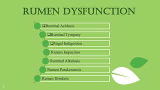
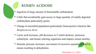
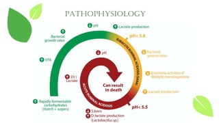
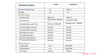
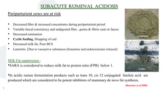
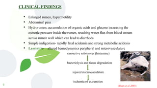
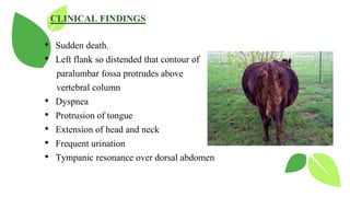
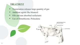
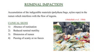
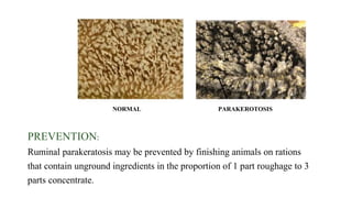
RUMEN
DYSFUNCTION
SUBMITTED BY,
PRASANTH M.NAIR
21-P-AN-11
ANIMAL NUTRITION
ICAR-NDRI
AP-712 DIGESTIVE PHYSIOLOGY
RUMEN
⊷ Rumen is a fermentation vat par excellence providing an anaerobic
environment, constant temperature, pH and good mixing
⊷ The rumen through its strong musculature allows mixing and churning
of digesta. The movement of the rumen mixes the contents, promoting
turnover and accessibility of the coarser forage particles for
regurgitation, cud chewing, size reduction, and microbial digestion
2
3
Ruminal Acidosis
Ruminal Tympany
Vagal Indigestion
Rumen Impaction
Ruminal Alkalosis
Rumen Parakeratosis
Rumen Drinkers
RUMEN DYSFUNCTION
RUMEN ACIDOSIS
• Ingestion of large amount of fermentable carbohydrate
• Cattle that accidentally gain access to large quantity of readily digested
carbohydrate particularly grains
• Change in microbial population particularly Gram-positive bacteria like
Streptococcus bovis
• Lactic acid increases, pH decreases to 5 which destroys protozoa,
cellulolytic and lactate utilizing organisms and impairs rumen motility
• Osmotic pressure increases, movement of excessive quantity of fluid in
rumen resulting in dehydration.
(Hernandez et al., 2014)
PATHOPHYSIOLOGY
Ruminal acidosis Acute Subacute
Presence of clinical signs Yes May be
Mortality Yes No
Ruminal changes
(1) Rumen pH Below 5.4 Below 6
(2) Lactic acid Increase (50–120 mM) Normal (0–5 Mm)
(3) Volatile fatty acids (VFA) Decrease (<100 mM) Increase (150 – 225mM)
(4) Gram negative bacteria Decrease Normal
(5) Gram positive bacteria Increase Normal
(6) Streptococcus bovis Increase Normal
(7) Lactobacillus spp. Increase Normal
(8) Lactic acid producers Increase Increase
(9) Lactic acid consumers
Blood parameters
Decrease Increase
(1) Blood pH Low Borderline
(2) Bicarbonate Low Borderline
(3) Lactate Increase Normal
(Nagaraja et al.,2007)
7
SUBACUTE RUMINAL ACIDOSIS
Periparturient cows are at risk
• Decreased fibre & increased concentrates during periparturient period
• Variable faecal consistency and undigested fiber , grains & fibrin casts in faeces
• Decreased rumination
• Cyclic feeding, Dropping of cud
• Decreased milk fat, Poor BCS
• Laminitis {Due to vasoactive substances (histamine and endotoxins)are released}
Milk Fat suppression ;
•SARA is considered to reduce milk fat to protein ratio (FPR) below 1.
•In acidic rumen fermentation products such as trans 10, cis 12 conjugated linoleic acid are
produced which are considered to be potent inhibitors of mammary de novo fat synthesis.
(Bauman et al.2008)
NORMAL
SARA
8
CLINICAL FINDINGS
• Enlarged rumen, hypermotility
• Abdominal pain
• Hydrorumen; accumulation of organic acids and glucose increasing the
osmotic pressure inside the rumen, resulting water flux from blood stream
across rumen wall which can lead to diarrhoea
• Simple indigestion- rapidly fatal acidemia and strong metabolic acidosis
• Laminitis- altered hemodynamics peripheral and microvasculature
vasoactive substances (histamine)
bacteriolysis and tissue degradation
injured microvasculature
ischemia of extremities
(Kleen et al.,2003)
9
MANAGEMENT
• Slowly degradable starch sources such as maize pose a lower risk than
sugars, wheat or barley
• More rapidly digestible fiber encourages high dry matter intake and allows
higher milk yield to be maintained, lowering the risk of acidosis
• Sufficient dietary sodium is required to absorb VFAs and the requirement is
likely to increase when lactate is present in the rumen
(Russell et al., 2017)
• Probiotics (Saccharomyces cerevisiae, L. plantarum) reduces organic acid
accumulation and might decrease the risk of subacute ruminal acidosis
(SARA) (Hiroko et al., 2016)
TREATMENT
⊷ Restricted water intake for 24 hrs
⊷ 5% Sodium carbonate solution- intravenous
⊷ Balanced electrolyte solution.
⊷ Antibiotics, Vit B complex.
10
(Morgan et al., 2009)
RUMINAL TYMPANY/ BLOAT
Distension of ruminoreticulum by accumulated gases, because of failure of
eructation mechanism
⊷ Breed :-feedlot bloat is higher in Holstein cattle than in beef cattle
⊷ Feed factors: Feed particle size : - Affect foam stability
11
1. Primary Bloat
Inability to eructate
Froth and foam
2. Secondary bloat
Choke (esophageal
obstruction)
Neoplastic growth
Disease affecting vagal
neve functions
(Vogel and Parrott, 1994)
12
PRIMARY BLOAT/ FROTHY BLOAT PATHOPHYSIOLOGY
Intake of legumes alfa alfa (saponins)
Entrapment of gases in a stable foam
Small gas bubbles
Intra ruminal pressure
Receptors in cardia signal medulla
Esophageal sphincter will not relax
Eructation reflex can’t complete
Gas cannot escape
• Fine particles arising from fragmentation and rupture of chloroplasts that prevent the coalescence of gas bubbles.
• Ruminal bacteria produce an excessive amount of exopolysaccharides or bacterial slime forms stable foam.
(Majak et al., 2003)
CLINICAL FINDINGS
• Sudden death.
• Left flank so distended that contour of
paralumbar fossa protrudes above
vertebral column
• Dyspnea
• Protrusion of tongue
• Extension of head and neck
• Frequent urination
• Tympanic resonance over dorsal abdomen
14
• Pasture management; Grasses or bloat resistant legumes such as sainfoin,
birdsfoot trefoil.
• Use of grass or silage–legume mixtures.
• Crop maturity; risk is highest at the vegetative and pre-bud stage of growth.
(Thompson et al., 2000)
• Feed additives; ionophores like monensin and lasalocid, various mineral
mixtures, poloxalene prevent bloat. (Majak et al., 2015)
MANAGEMENT
15
TREATMENT
• Trocarisation releases large quantity of gas
• Antibloat agents like bloatosil
• Oils and non absorbed surfactants
• Use of Dimethicone, Polaxalene
Ruminal impaction
Accumulation of the indigestible materials (polythene bags, nylon rope) in the
rumen which interferes with the flow of ingesta.
(Abdullahi et al., 1984)
CLINICAL SIGNS
1) Absence of rumination
2) Reduced ruminal motility
3) Distension of rumen
4) Passing of scanty or no faeces
17
Pathophysiology of ruminal impaction due to plastic materials in ruminants
(Conditions mentioned in yellow box indicate the probable outcome after chronic exposure to the
products of chemical leaching in rumen)
18
TREATMENT
• As clinical signs in animals suffering from ruminal impaction due to plastic materials
are nonspecific, diagnosis of ruminal impaction due to plastic materials is a real
challenge to clinicians.
• Exploratory rumenotomy is the only choice for both diagnosis and treatment
(Tyagi and Singh, 2004)
(Rumen impacted by plastic waste materials)
VAGAL INDIGESTION (HOFLUND’S SYNDROME)
Gradual development of abdominal distension as a result of
lesions affecting vagus nerve
Injury,
inflammation, or
pressure on vagus
nerve
Trumatic
reticuloperitonitis,
reticular abscesses,
diaphramatic hernia
and hepatic abscesses
TYPE
1
• Localized
peritonitis,
chronic
pneumonia
• Inflammatory
lesions in the
vicinity of vagus
nerve
• Failure of
eructation
TYPE
2
• Failure of
omasal
transport
• Conditions
preventing
ingesta from
passing through
omasal canal
into abomasum
TYPE
3
• Indigestion of
late gestation
• Enlarged uterus
shift the
abomasum
cranially
• Inhibits normal
functioning
TYPE
4
• TRP, abomasal
volvulus
• Secondary
abomasal
impaction
TYPES OF VAGAL INDIGESTION
Clinical findings
1) Gradual development of abomasal distension secondary to
ruminoreticulum distension.
2) Distension of dorsal and ventral sac
L shaped rumen on rectal examination
3) Left dorsal and left and right
ventral distension of abdomen
Papple shaped abdomen
(Pear + Apple) from behind
4) Sticky faeces and faecal output decreases
5) Milk production decreases
6) Strength of ruminal contraction decreases
Normal ‘L’ shape of rumen
22
NORMAL PAPPLE SHAPE
23
• Left flank rumenotomy for type 1 and type 2 vagal indigestion
• Right flank rumenotomy for type 3 and type 4 vagal indigestion.
• Transfaunation
• Surfectancts such as poloxalene can be used
Treatment
24
Increased ammonia in the rumen causes alkalinization. This excess ammonia leads
to alkalosis (pH 7.5 plus) and rumen stasis (paralysis of the rumen wall), thus
preventing eructation and the animal suffers from bloat.
Causes
 Increased protein feeding, Urea toxicity, nitrate toxicity
 Ruminal putrefaction
 Feeding putrefied roughage (e.g. Rain feed putrefied paddy straw) or
putrefied vegetable waste
 Drinking sewage water. Grass and water contaminated with manure.
RUMINAL ALKALOSIS
CLINICAL SIGNS
MILD CASES
⊷ •Main complaint :Bad smell in
milk.
⊷ •Relapsing foamy tympany
⊷ •Reduced fat in milk
⊷ •Reduced semen quality, mastitis,
staggering gait & recumbency
SEVERE CASES
⊷ •Tympany
⊷ •Recumbency
⊷ •Regurgitation of rumen
contents and bad smell
⊷ •Nervous signs.
25
RUMEN LIQUOR
pH 7.5-8.5, dark, ammoniacal/ putrid smell, few motile protozoa
26
TREATMENT
1) Changing ruminal pH and stopping further ammonia production. This can be done by
administration of acetic acid- 4%, 100-500 ml (5-10 ml/kg body wt). Commonly vinegar bottles are
available as 1 litre, commercial lactic acid preparations are also used
(Walter Grünberg, 2009)
2) Preferably intra ruminal antibiotic is advised to check the growth of unwanted microbes,
Sterptopenicillin- 1 large dose.
3) Liver tonics- Play a vital role. B-complex preparation with liver extract is more effective to
improve to impaired liver function.
4) Cud transplantation for rejuvenation of micro flora.
RUMEN DRINKERS
Ruminal drinkers refers to calves that develop chronic indigestion because milk is deposited
into the rumen as a result of failure of the reticular groove reflex during drinking.
The esophageal groove allows milk and milk replacers to bypass the rumen, reticulum and
omasum and to flow directly into the abomasum.
(Gygi et al., 2006)
• Common in bucket fed calves 2-8wk old.
• Calves that “gulp” milk rather than “sip” milk are at greatest risk.
Fermentation of milk retained in rumen
Production of acetic acid, lactate and butyric acid
pH
Parakeratosis of ruminal mucosa develops
(Choudhary et al., 2010)
Aetiology and pathogenesis
Clinical signs
• Calves show in appetence, ventral abdomen distension.
• Clay- like feces that may adhere to the tail, perineum and hindlegs
(Gentle et al .,2004)
• Rancid- smelling grayish white fermented material can be obtained by stomach
tube from the rumen.
• Ruminal pH less than 6 leads to systemic acid-base balance disturbance.
Diagnostic feature
• Fluid-splashing sounds, audible on auscultation over the left flank while the calf
is drinking.
Management
30
• A rubber nipple floating on the surface of bucket-fed milk may prevent
syndrome (Choudhary et al., 2010)
• Sodium bicarbonate effective alkalinizing agent.
RUMINAL PARAKERATOSIS
• Characterized by hardening and enlargement of papillae of the rumen.
• Common in animals fed on; high concentrate ration
heat treated alfalfa pellets
calves having prolonged ruminal acidosis
• Papillae are enlarged and hardened
• Papillae of the anterior ventral sac are commonly affected
• Affected papillae contain excessive layers of keratinized epithelial cells
• Interfering with feed absorption, efficiency of feed utilization
(Steele et al., 2009)
32
PREVENTION:
Ruminal parakeratosis may be prevented by finishing animals on rations
that contain unground ingredients in the proportion of 1 part roughage to 3
parts concentrate.
NORMAL PARAKEROTOSIS
33
THANKS

RUMEN DYSFUNCTIONS.pptx

  • 1.
    RUMEN DYSFUNCTION SUBMITTED BY, PRASANTH M.NAIR 21-P-AN-11 ANIMALNUTRITION ICAR-NDRI AP-712 DIGESTIVE PHYSIOLOGY
  • 2.
    RUMEN ⊷ Rumen isa fermentation vat par excellence providing an anaerobic environment, constant temperature, pH and good mixing ⊷ The rumen through its strong musculature allows mixing and churning of digesta. The movement of the rumen mixes the contents, promoting turnover and accessibility of the coarser forage particles for regurgitation, cud chewing, size reduction, and microbial digestion 2
  • 3.
    3 Ruminal Acidosis Ruminal Tympany VagalIndigestion Rumen Impaction Ruminal Alkalosis Rumen Parakeratosis Rumen Drinkers RUMEN DYSFUNCTION
  • 4.
    RUMEN ACIDOSIS • Ingestionof large amount of fermentable carbohydrate • Cattle that accidentally gain access to large quantity of readily digested carbohydrate particularly grains • Change in microbial population particularly Gram-positive bacteria like Streptococcus bovis • Lactic acid increases, pH decreases to 5 which destroys protozoa, cellulolytic and lactate utilizing organisms and impairs rumen motility • Osmotic pressure increases, movement of excessive quantity of fluid in rumen resulting in dehydration. (Hernandez et al., 2014)
  • 5.
  • 6.
    Ruminal acidosis AcuteSubacute Presence of clinical signs Yes May be Mortality Yes No Ruminal changes (1) Rumen pH Below 5.4 Below 6 (2) Lactic acid Increase (50–120 mM) Normal (0–5 Mm) (3) Volatile fatty acids (VFA) Decrease (<100 mM) Increase (150 – 225mM) (4) Gram negative bacteria Decrease Normal (5) Gram positive bacteria Increase Normal (6) Streptococcus bovis Increase Normal (7) Lactobacillus spp. Increase Normal (8) Lactic acid producers Increase Increase (9) Lactic acid consumers Blood parameters Decrease Increase (1) Blood pH Low Borderline (2) Bicarbonate Low Borderline (3) Lactate Increase Normal (Nagaraja et al.,2007)
  • 7.
    7 SUBACUTE RUMINAL ACIDOSIS Periparturientcows are at risk • Decreased fibre & increased concentrates during periparturient period • Variable faecal consistency and undigested fiber , grains & fibrin casts in faeces • Decreased rumination • Cyclic feeding, Dropping of cud • Decreased milk fat, Poor BCS • Laminitis {Due to vasoactive substances (histamine and endotoxins)are released} Milk Fat suppression ; •SARA is considered to reduce milk fat to protein ratio (FPR) below 1. •In acidic rumen fermentation products such as trans 10, cis 12 conjugated linoleic acid are produced which are considered to be potent inhibitors of mammary de novo fat synthesis. (Bauman et al.2008) NORMAL SARA
  • 8.
    8 CLINICAL FINDINGS • Enlargedrumen, hypermotility • Abdominal pain • Hydrorumen; accumulation of organic acids and glucose increasing the osmotic pressure inside the rumen, resulting water flux from blood stream across rumen wall which can lead to diarrhoea • Simple indigestion- rapidly fatal acidemia and strong metabolic acidosis • Laminitis- altered hemodynamics peripheral and microvasculature vasoactive substances (histamine) bacteriolysis and tissue degradation injured microvasculature ischemia of extremities (Kleen et al.,2003)
  • 9.
    9 MANAGEMENT • Slowly degradablestarch sources such as maize pose a lower risk than sugars, wheat or barley • More rapidly digestible fiber encourages high dry matter intake and allows higher milk yield to be maintained, lowering the risk of acidosis • Sufficient dietary sodium is required to absorb VFAs and the requirement is likely to increase when lactate is present in the rumen (Russell et al., 2017) • Probiotics (Saccharomyces cerevisiae, L. plantarum) reduces organic acid accumulation and might decrease the risk of subacute ruminal acidosis (SARA) (Hiroko et al., 2016)
  • 10.
    TREATMENT ⊷ Restricted waterintake for 24 hrs ⊷ 5% Sodium carbonate solution- intravenous ⊷ Balanced electrolyte solution. ⊷ Antibiotics, Vit B complex. 10 (Morgan et al., 2009)
  • 11.
    RUMINAL TYMPANY/ BLOAT Distensionof ruminoreticulum by accumulated gases, because of failure of eructation mechanism ⊷ Breed :-feedlot bloat is higher in Holstein cattle than in beef cattle ⊷ Feed factors: Feed particle size : - Affect foam stability 11 1. Primary Bloat Inability to eructate Froth and foam 2. Secondary bloat Choke (esophageal obstruction) Neoplastic growth Disease affecting vagal neve functions (Vogel and Parrott, 1994)
  • 12.
    12 PRIMARY BLOAT/ FROTHYBLOAT PATHOPHYSIOLOGY Intake of legumes alfa alfa (saponins) Entrapment of gases in a stable foam Small gas bubbles Intra ruminal pressure Receptors in cardia signal medulla Esophageal sphincter will not relax Eructation reflex can’t complete Gas cannot escape • Fine particles arising from fragmentation and rupture of chloroplasts that prevent the coalescence of gas bubbles. • Ruminal bacteria produce an excessive amount of exopolysaccharides or bacterial slime forms stable foam. (Majak et al., 2003)
  • 13.
    CLINICAL FINDINGS • Suddendeath. • Left flank so distended that contour of paralumbar fossa protrudes above vertebral column • Dyspnea • Protrusion of tongue • Extension of head and neck • Frequent urination • Tympanic resonance over dorsal abdomen
  • 14.
    14 • Pasture management;Grasses or bloat resistant legumes such as sainfoin, birdsfoot trefoil. • Use of grass or silage–legume mixtures. • Crop maturity; risk is highest at the vegetative and pre-bud stage of growth. (Thompson et al., 2000) • Feed additives; ionophores like monensin and lasalocid, various mineral mixtures, poloxalene prevent bloat. (Majak et al., 2015) MANAGEMENT
  • 15.
    15 TREATMENT • Trocarisation releaseslarge quantity of gas • Antibloat agents like bloatosil • Oils and non absorbed surfactants • Use of Dimethicone, Polaxalene
  • 16.
    Ruminal impaction Accumulation ofthe indigestible materials (polythene bags, nylon rope) in the rumen which interferes with the flow of ingesta. (Abdullahi et al., 1984) CLINICAL SIGNS 1) Absence of rumination 2) Reduced ruminal motility 3) Distension of rumen 4) Passing of scanty or no faeces
  • 17.
    17 Pathophysiology of ruminalimpaction due to plastic materials in ruminants (Conditions mentioned in yellow box indicate the probable outcome after chronic exposure to the products of chemical leaching in rumen)
  • 18.
    18 TREATMENT • As clinicalsigns in animals suffering from ruminal impaction due to plastic materials are nonspecific, diagnosis of ruminal impaction due to plastic materials is a real challenge to clinicians. • Exploratory rumenotomy is the only choice for both diagnosis and treatment (Tyagi and Singh, 2004) (Rumen impacted by plastic waste materials)
  • 19.
    VAGAL INDIGESTION (HOFLUND’SSYNDROME) Gradual development of abdominal distension as a result of lesions affecting vagus nerve Injury, inflammation, or pressure on vagus nerve Trumatic reticuloperitonitis, reticular abscesses, diaphramatic hernia and hepatic abscesses
  • 20.
    TYPE 1 • Localized peritonitis, chronic pneumonia • Inflammatory lesionsin the vicinity of vagus nerve • Failure of eructation TYPE 2 • Failure of omasal transport • Conditions preventing ingesta from passing through omasal canal into abomasum TYPE 3 • Indigestion of late gestation • Enlarged uterus shift the abomasum cranially • Inhibits normal functioning TYPE 4 • TRP, abomasal volvulus • Secondary abomasal impaction TYPES OF VAGAL INDIGESTION
  • 21.
    Clinical findings 1) Gradualdevelopment of abomasal distension secondary to ruminoreticulum distension. 2) Distension of dorsal and ventral sac L shaped rumen on rectal examination 3) Left dorsal and left and right ventral distension of abdomen Papple shaped abdomen (Pear + Apple) from behind 4) Sticky faeces and faecal output decreases 5) Milk production decreases 6) Strength of ruminal contraction decreases Normal ‘L’ shape of rumen
  • 22.
  • 23.
    23 • Left flankrumenotomy for type 1 and type 2 vagal indigestion • Right flank rumenotomy for type 3 and type 4 vagal indigestion. • Transfaunation • Surfectancts such as poloxalene can be used Treatment
  • 24.
    24 Increased ammonia inthe rumen causes alkalinization. This excess ammonia leads to alkalosis (pH 7.5 plus) and rumen stasis (paralysis of the rumen wall), thus preventing eructation and the animal suffers from bloat. Causes  Increased protein feeding, Urea toxicity, nitrate toxicity  Ruminal putrefaction  Feeding putrefied roughage (e.g. Rain feed putrefied paddy straw) or putrefied vegetable waste  Drinking sewage water. Grass and water contaminated with manure. RUMINAL ALKALOSIS
  • 25.
    CLINICAL SIGNS MILD CASES ⊷•Main complaint :Bad smell in milk. ⊷ •Relapsing foamy tympany ⊷ •Reduced fat in milk ⊷ •Reduced semen quality, mastitis, staggering gait & recumbency SEVERE CASES ⊷ •Tympany ⊷ •Recumbency ⊷ •Regurgitation of rumen contents and bad smell ⊷ •Nervous signs. 25 RUMEN LIQUOR pH 7.5-8.5, dark, ammoniacal/ putrid smell, few motile protozoa
  • 26.
    26 TREATMENT 1) Changing ruminalpH and stopping further ammonia production. This can be done by administration of acetic acid- 4%, 100-500 ml (5-10 ml/kg body wt). Commonly vinegar bottles are available as 1 litre, commercial lactic acid preparations are also used (Walter Grünberg, 2009) 2) Preferably intra ruminal antibiotic is advised to check the growth of unwanted microbes, Sterptopenicillin- 1 large dose. 3) Liver tonics- Play a vital role. B-complex preparation with liver extract is more effective to improve to impaired liver function. 4) Cud transplantation for rejuvenation of micro flora.
  • 27.
    RUMEN DRINKERS Ruminal drinkersrefers to calves that develop chronic indigestion because milk is deposited into the rumen as a result of failure of the reticular groove reflex during drinking. The esophageal groove allows milk and milk replacers to bypass the rumen, reticulum and omasum and to flow directly into the abomasum. (Gygi et al., 2006)
  • 28.
    • Common inbucket fed calves 2-8wk old. • Calves that “gulp” milk rather than “sip” milk are at greatest risk. Fermentation of milk retained in rumen Production of acetic acid, lactate and butyric acid pH Parakeratosis of ruminal mucosa develops (Choudhary et al., 2010) Aetiology and pathogenesis
  • 29.
    Clinical signs • Calvesshow in appetence, ventral abdomen distension. • Clay- like feces that may adhere to the tail, perineum and hindlegs (Gentle et al .,2004) • Rancid- smelling grayish white fermented material can be obtained by stomach tube from the rumen. • Ruminal pH less than 6 leads to systemic acid-base balance disturbance. Diagnostic feature • Fluid-splashing sounds, audible on auscultation over the left flank while the calf is drinking.
  • 30.
    Management 30 • A rubbernipple floating on the surface of bucket-fed milk may prevent syndrome (Choudhary et al., 2010) • Sodium bicarbonate effective alkalinizing agent.
  • 31.
    RUMINAL PARAKERATOSIS • Characterizedby hardening and enlargement of papillae of the rumen. • Common in animals fed on; high concentrate ration heat treated alfalfa pellets calves having prolonged ruminal acidosis • Papillae are enlarged and hardened • Papillae of the anterior ventral sac are commonly affected • Affected papillae contain excessive layers of keratinized epithelial cells • Interfering with feed absorption, efficiency of feed utilization (Steele et al., 2009)
  • 32.
    32 PREVENTION: Ruminal parakeratosis maybe prevented by finishing animals on rations that contain unground ingredients in the proportion of 1 part roughage to 3 parts concentrate. NORMAL PARAKEROTOSIS
  • 33.