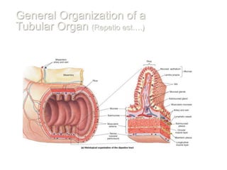
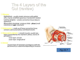
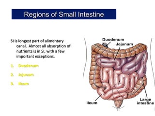
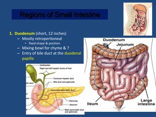
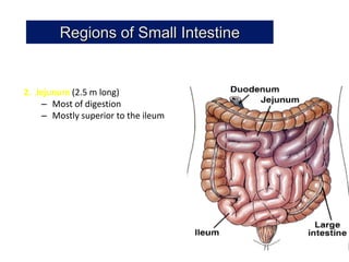
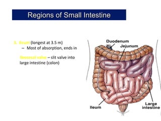
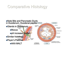
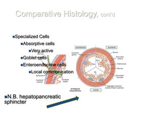
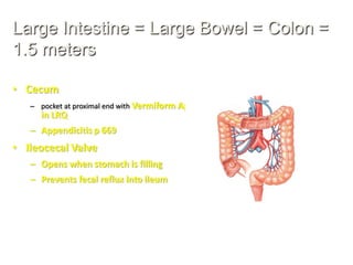
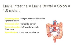
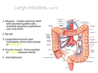
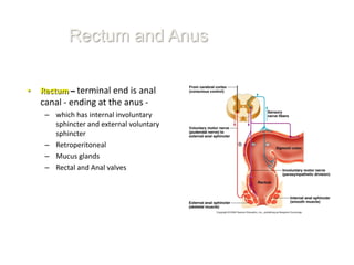
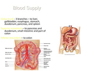
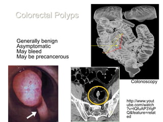
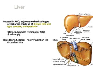
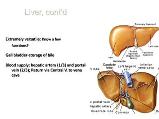
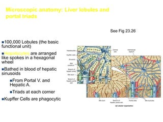
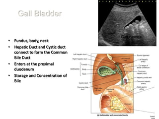
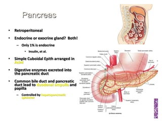
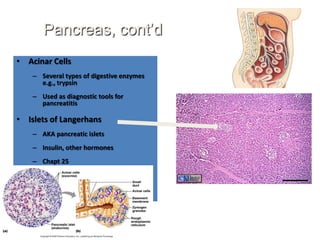
Ch 23: Digestive
      System, Part 2
Objectives:
Discuss the general functions and
anatomy of the digestive tract,
including accessory structures.

First, an overview of the tubular
nature of the digestive system.

Describe the individual organs of
the system, including a
discussion of the gross and
microscopic anatomy.


                                               Developed by
                                    John Gallagher, MS, DVM
General Organization of a
Tubular Organ (Repetio est….)
The 4 Layers of the
               Gut (review)
1) Mucosa
    Epithelium - usually simple columnar with goblet
    cells; may be stratified squamous if protection needed
    Lamina propria – areolar connective tissue deep to
    epithelium
    Muscularis mucosae -produces folds - plicae (small
    intestine) or rugae (stomach)

2) Submucosa – made up of loose connective tissue
    contains submucosal plexus and blood vessels

3) Muscularis externa – smooth muscle, usually two
    layers (controlled by the myenteric plexus; source of
    peristalsis ) -
            inner layer: circular
            outer layer: longitudinal

4) Serosa
         visceral layer of mesentery (contiguous with the
         peritoneum) or adventitia depending on location

                                                             Fig 23.7
Regions of Small Intestine


SI is longest part of alimentary
    canal. Almost all absorption of
    nutrients is in SI, with a few
    important exceptions.

1.   Duodenum

2.   Jejunum

3.   Ileum
Regions of Small Intestine

1. Duodenum (short, 12 inches)
    – Mostly retroperitoneal
        • fixed shape & position
    – Mixing bowl for chyme & ?
    – Entry of bile duct at the duodenal
      papilla
Regions of Small Intestine


2. Jejunum (2.5 m long)
    – Most of digestion
    – Mostly superior to the ileum
Regions of Small Intestine


3. Ileum (longest at 3.5 m)
     – Most of absorption, ends in

   Ileocecal valve – slit valve into
   large intestine (colon)
Comparative Histology


Note Bile and Pancreatic Ducts
in Duodenum, Duodenal papilla
Glands    in Duodenum
   Mucus

   pH     increase (buffer)
Similar   histology
Peyer’s   Patches
   AKA MALT
Comparative Histology, cont’d

   Specialized   Cells
        Absorptive   cells
           Very    active
        Goblet   cells
        Enteroendocrine      cells
           Local    communication


N.B. hepatopancreatic
sphincter
Large Intestine = Large Bowel = Colon =
1.5 meters

• Cecum
   – pocket at proximal end with Vermiform Appendix
     in LRQ
   – Appendicitis p 669
• Ileocecal Valve
   – Opens when stomach is filling
   – Prevents fecal reflux into ileum
Large Intestine = Large Bowel = Colon =
1.5 meters

 Ascending colon - on right, between cecum and
 right colic flexure
 Transverse colon - horizontal portion
 Descending colon - left side, between left colic
 flexure and
 Sigmoid colon - S bend near terminal end
Large intestine, cont’d

1. Mucosa - simple columnar epith
   with abundant goblet cells;
   stratified squamous epithelium
   near anal canal
2. No villi
3. Longitudinal muscle layer
    incomplete, forms three bands
    or taenia coli
4. Circular muscle - forms pockets
    or haustra between bands
5. Anal Sphincter
Rectum and Anus

•   Rectum – terminal end is anal
    canal - ending at the anus -
     – which has internal involuntary
       sphincter and external voluntary
       sphincter
     – Retroperitoneal
     – Mucus glands
     – Rectal and Anal valves
Blood Supply

Celiac trunk - 3 branches – to liver,
   gallbladder, esophagus, stomach,
   duodenum, pancreas, and spleen
Superior mesenteric– to pancreas and
   duodenum, small intestine and part of
   colon
Inferior mesenteric– to colon
Colorectal Polyps


Generally benign
Asymptomatic
May bleed
May be precancerous


                              Virtual
                        Colonoscopy


                      http://www.yout
                      ube.com/watch
                      ?v=lQfuAP3YqP
                      Q&feature=relat
                      ed
Liver

Located in RUQ, adjacent to the diaphragm,
   largest organ made up of 4 lobes (left and
   right, caudate, and quadrate)

   Falciform ligament (remnant of fetal
   blood supply

Hilus (porta hepatis) – "entry" point on the
    visceral surface
Liver, cont’d


Extremely versatile: Know a few
   functions?

Gall bladder-storage of bile

Blood supply: hepatic artery (1/3) and portal
   vein (2/3); Return via Central V. to vena
   cava
Microscopic anatomy: Liver lobules and
  portal triads

                                           See Fig 23.26

100,000   Lobules (the basic
functional unit)
Hepatocytes   are arranged
like spokes in a hexagonal
wheel
Bathed  in blood of hepatic
sinusoids
    From  Portal V. and
    Hepatic A.
    Triads   at each corner
Kupffer   Cells are phagocytic
Gall Bladder

                                 Gall Stones
• Fundus, body, neck
• Hepatic Duct and Cystic duct
  connect to form the Common
  Bile Duct
• Enters at the proximal
  duodenum
• Storage and Concentration of
  Bile
Pancreas
•   Retroperitoneal
•   Endocrine or exocrine gland? Both!
     – Only 1% is endocrine
          • Insulin, et al.

•   Simple Cuboidal Epith arranged in
    Acini
•   Digestive enzymes excreted into
    the pancreatic duct
•   Common bile duct and pancreatic
    duct lead to duodenal ampulla and
    papilla
     – Controlled by hepatopancreatic
       sphincter
Pancreas, cont’d
• Acinar Cells
    – Several types of digestive enzymes
      e.g., trypsin
    – Used as diagnostic tools for
      pancreatitis

• Islets of Langerhans
    – AKA pancreatic islets
    – Insulin, other hormones
    – Chapt 25
Chapter23 digestivepart2marieb

Chapter23 digestivepart2marieb

  • 1.
    Ch 23: Digestive System, Part 2 Objectives: Discuss the general functions and anatomy of the digestive tract, including accessory structures. First, an overview of the tubular nature of the digestive system. Describe the individual organs of the system, including a discussion of the gross and microscopic anatomy. Developed by John Gallagher, MS, DVM
  • 2.
    General Organization ofa Tubular Organ (Repetio est….)
  • 3.
    The 4 Layersof the Gut (review) 1) Mucosa Epithelium - usually simple columnar with goblet cells; may be stratified squamous if protection needed Lamina propria – areolar connective tissue deep to epithelium Muscularis mucosae -produces folds - plicae (small intestine) or rugae (stomach) 2) Submucosa – made up of loose connective tissue contains submucosal plexus and blood vessels 3) Muscularis externa – smooth muscle, usually two layers (controlled by the myenteric plexus; source of peristalsis ) - inner layer: circular outer layer: longitudinal 4) Serosa visceral layer of mesentery (contiguous with the peritoneum) or adventitia depending on location Fig 23.7
  • 4.
    Regions of SmallIntestine SI is longest part of alimentary canal. Almost all absorption of nutrients is in SI, with a few important exceptions. 1. Duodenum 2. Jejunum 3. Ileum
  • 5.
    Regions of SmallIntestine 1. Duodenum (short, 12 inches) – Mostly retroperitoneal • fixed shape & position – Mixing bowl for chyme & ? – Entry of bile duct at the duodenal papilla
  • 6.
    Regions of SmallIntestine 2. Jejunum (2.5 m long) – Most of digestion – Mostly superior to the ileum
  • 7.
    Regions of SmallIntestine 3. Ileum (longest at 3.5 m) – Most of absorption, ends in Ileocecal valve – slit valve into large intestine (colon)
  • 8.
    Comparative Histology Note Bileand Pancreatic Ducts in Duodenum, Duodenal papilla Glands in Duodenum Mucus pH increase (buffer) Similar histology Peyer’s Patches AKA MALT
  • 9.
    Comparative Histology, cont’d Specialized Cells Absorptive cells Very active Goblet cells Enteroendocrine cells Local communication N.B. hepatopancreatic sphincter
  • 10.
    Large Intestine =Large Bowel = Colon = 1.5 meters • Cecum – pocket at proximal end with Vermiform Appendix in LRQ – Appendicitis p 669 • Ileocecal Valve – Opens when stomach is filling – Prevents fecal reflux into ileum
  • 11.
    Large Intestine =Large Bowel = Colon = 1.5 meters Ascending colon - on right, between cecum and right colic flexure Transverse colon - horizontal portion Descending colon - left side, between left colic flexure and Sigmoid colon - S bend near terminal end
  • 12.
    Large intestine, cont’d 1.Mucosa - simple columnar epith with abundant goblet cells; stratified squamous epithelium near anal canal 2. No villi 3. Longitudinal muscle layer incomplete, forms three bands or taenia coli 4. Circular muscle - forms pockets or haustra between bands 5. Anal Sphincter
  • 13.
    Rectum and Anus • Rectum – terminal end is anal canal - ending at the anus - – which has internal involuntary sphincter and external voluntary sphincter – Retroperitoneal – Mucus glands – Rectal and Anal valves
  • 14.
    Blood Supply Celiac trunk- 3 branches – to liver, gallbladder, esophagus, stomach, duodenum, pancreas, and spleen Superior mesenteric– to pancreas and duodenum, small intestine and part of colon Inferior mesenteric– to colon
  • 15.
    Colorectal Polyps Generally benign Asymptomatic Maybleed May be precancerous Virtual Colonoscopy http://www.yout ube.com/watch ?v=lQfuAP3YqP Q&feature=relat ed
  • 16.
    Liver Located in RUQ,adjacent to the diaphragm, largest organ made up of 4 lobes (left and right, caudate, and quadrate) Falciform ligament (remnant of fetal blood supply Hilus (porta hepatis) – "entry" point on the visceral surface
  • 17.
    Liver, cont’d Extremely versatile:Know a few functions? Gall bladder-storage of bile Blood supply: hepatic artery (1/3) and portal vein (2/3); Return via Central V. to vena cava
  • 18.
    Microscopic anatomy: Liverlobules and portal triads See Fig 23.26 100,000 Lobules (the basic functional unit) Hepatocytes are arranged like spokes in a hexagonal wheel Bathed in blood of hepatic sinusoids From Portal V. and Hepatic A. Triads at each corner Kupffer Cells are phagocytic
  • 19.
    Gall Bladder Gall Stones • Fundus, body, neck • Hepatic Duct and Cystic duct connect to form the Common Bile Duct • Enters at the proximal duodenum • Storage and Concentration of Bile
  • 20.
    Pancreas • Retroperitoneal • Endocrine or exocrine gland? Both! – Only 1% is endocrine • Insulin, et al. • Simple Cuboidal Epith arranged in Acini • Digestive enzymes excreted into the pancreatic duct • Common bile duct and pancreatic duct lead to duodenal ampulla and papilla – Controlled by hepatopancreatic sphincter
  • 21.
    Pancreas, cont’d • AcinarCells – Several types of digestive enzymes e.g., trypsin – Used as diagnostic tools for pancreatitis • Islets of Langerhans – AKA pancreatic islets – Insulin, other hormones – Chapt 25