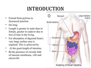
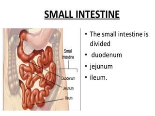
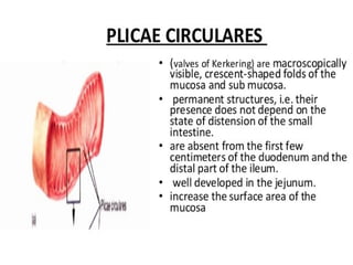
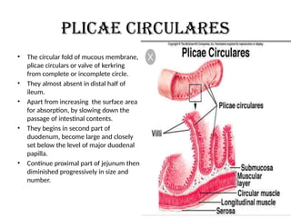
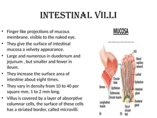
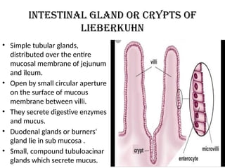
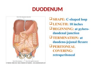
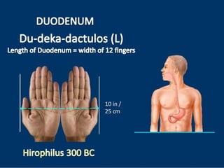
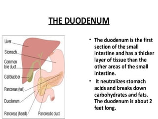
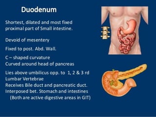
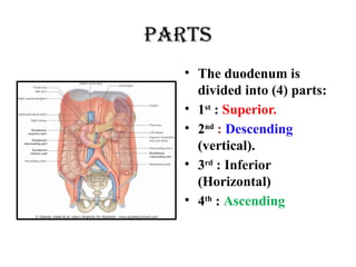
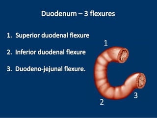
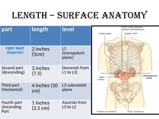
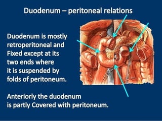
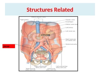
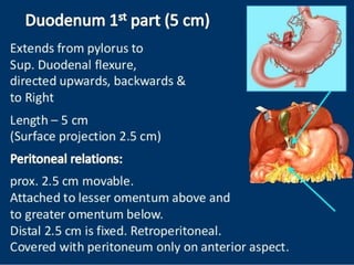
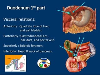
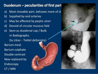
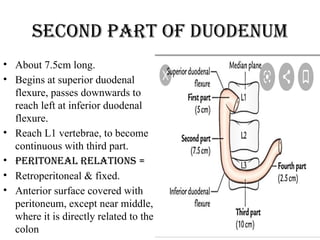
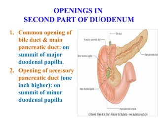
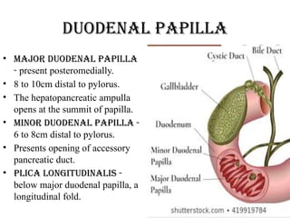
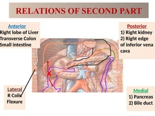
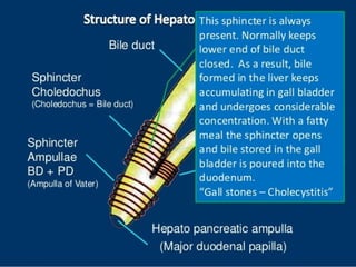
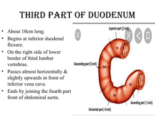
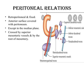
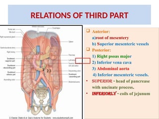
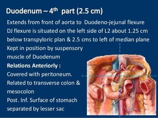
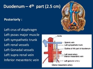
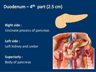
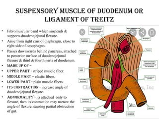
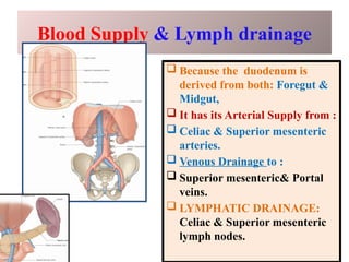
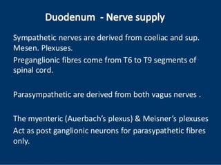
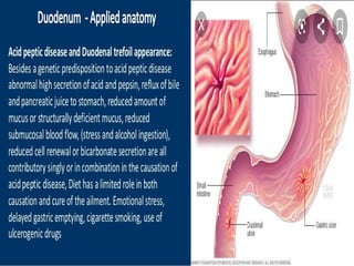
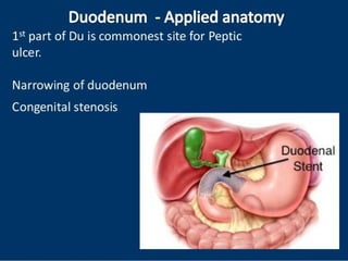
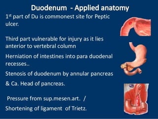
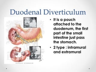
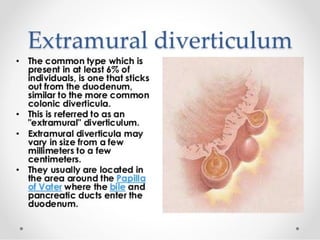
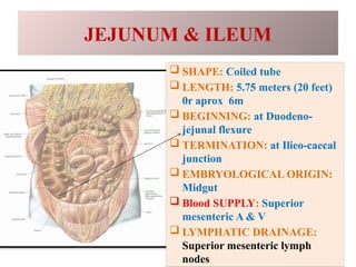
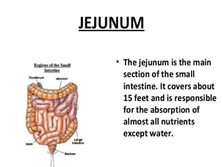
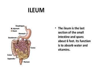
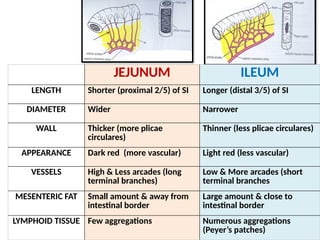
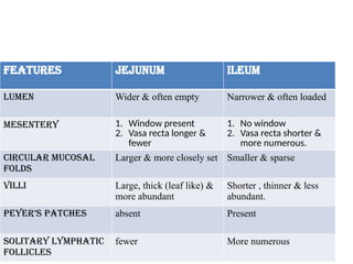
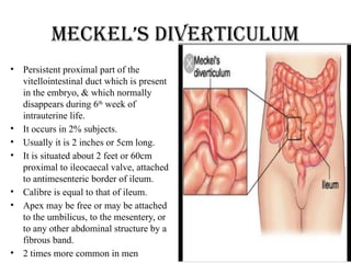
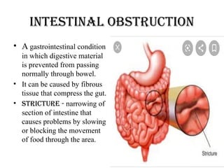
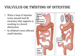
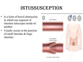
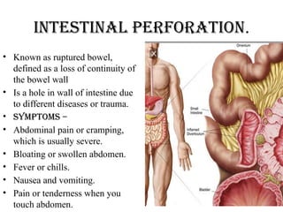
•ANATOMY OF THE
SMALL INTESTINE
{kqnzkUr
• ^^vl`t% ‘ys”e.k’pkfi ;% izlkn% ijkser% ra iP;ekua fiRrsu
ok;q’pkI;uq /kkofrAA
rrksvL;kU=kf.k tk;Urs xqna ofLr’p nsfgu%AA** ¼lq-’kk-
4@25&26½
• vkpk;Z lqJqr ds vuqlkj jDr vkSj dQ ds lkjre Hkkx esa fiRr
ds }kjk idrs le; mlesa ok;q Hkh vk tkrh gS] rc mlls vkU= dh
yEckbZ lk<s rhu O;ke ,oa fL=;ksa esa rhu O;ke crk;h gSA
• ^^ lk/kZf=O;kekU;U=kf.k iqalk] L=h.kke/kZO;keghukfu**AA
¼lq-’kk-5@8½
introduction
• Extend from pylorus to
ileocaecal junction.
• 6m long.
• Length is greater in male than in
female, greater in cadaver due to
loss of tone in the living.
• For absorption of digested food a
very large surface area is
required. This is achieved by
• A) the great length of intestine.
• B) the presence of circular fold
of mucous membrane, villi and
microvilli.
Plicae circulares
• The circular fold of mucous membrane,
plicae circulars or valve of kerkring
from complete or incomplete circle.
• They almost absent in distal half of
ileum.
• Apart from increasing the surface area
for absorption, by slowing down the
passage of intestinal contents.
• They begins in second part of
duodenum, become large and closely
set below the level of major duodenal
papilla.
• Continue proximal part of jejunum then
diminished progressively in size and
number.
Intestinal villi
• Finger like projections of mucous
membrane, visible to the naked eye.
• They give the surface of intestinal
mucosa a velvety appearance.
• Large and numerous in duodenum and
jejunum , but smaller and fewer in
ileum.
• They increase the surface area of
intestine about eight times.
• They vary in density from 10 to 40 per
square mm, 1 to 2 mm long.
• Villus is covered by a layer of absorptive
columnar cells, the surface of these cells
has a striated border, called microvilli.
Intestinal gland or crypts of
lieberkuhn
• Simple tubular glands,
distributed over the entire
mucosal membrane of jejunum
and ileum.
• Open by small circular aperture
on the surface of mucous
membrane between villi.
• They secrete digestive enzymes
and mucus.
• Duodenal glands or burners‘
gland lie in sub mucosa .
• Small, compound tubuloacinar
glands which secrete mucus.
Lymphatic follicles
• Contains two types of lymphatic follicles.
• Solitory lymphatic follicles - 1
or 2mm in diameter.
• Distributed throughout small & large
intestine. Aggregated lymphatic
follicles or peyer’s patches
form circular or oval patches,
• Length = 2 to 10cm & containing 10 to
over 200 follicles.
• Largest & most numerous in ileum, &
small, circular & fewer in distal jejunum.
• Placed lengthwise along the antimesenteric
border of intestine.
• Payer’s patches - most numerous at
puberty, diminished in size & number, may
persist up to old age, get ulcerated in
typhoid fever, oval ulcers.
xzg.kh
• ^^vXU;f/k”BkueUuL; xzg.kkn~ xzg.kh erkA
ukHks:ifj lkâfXu cyksiLrEHk c`afgrk**AA ¼p-fp-15@56½
• vkpk;Z pjd ds vuqlkj xzg.kh dks vfXu dk vkJ; gksus ,oa vUu dks xzg.k djus
ds dkj.k bls xzg.kh laKk nsrs gq, bldh fLFkfr ukfHk ds mij crk;h gSA
• ^^ iDokek’k;e/;LFkk xzg.kh lk izdhfrZrk**A ¼lq-m-40@169½
• lqJqr us Hkh vkek’k; ,oa iDok’k; ds e/; fLFkr vo;o dks xzg.kh laKk nh gSA
• ^^“k”Bh fiRr/kjk uke] ;k prqZfo/keUuikueqiHkqDrekEk’k;kr~ izP;qr
iDok’k;ksifLFkra /kkj;fr**AA ¼lq-’kk-4@17½
• vkpk;Z lqJqr us fiRr/kjk dyk dk LFkku Hkh vkek’k; ,oa iDok’k; ds e/; crk;k
gSA lsou fd;s prqfoZ/k vUuiku tks vkek’k; ls vkrk gS] mls xzg.k djus okyk
vo;o xzg.kh gSA xzg.kh esa fiRr ds }kjk vUu th.kZ gksdj iDok’k; dh vksj
ikpu ds fy, Hkst fn;k gSA
DUODENUM
SHAPE: C-shaped loop
LENGTH: 10 inches
BEGINNING: at pyloro-
duodenal junction
TERMINATION: at
duodeno-jejunal flexure
PERITONEAL
COVERING:
retroperitoneal
parts
• The duodenum is
divided into (4) parts:
• 1st
: Superior.
• 2nd
: Descending
(vertical).
• 3rd
: Inferior
(Horizontal)
• 4th
: Ascending
Length – surface anatomy
part length level
FIRST PART
(Superior)
2 inches
(5cm)
L1
(transpyloric
plane)
Second part
(descending)
3 inches
(7.5)
Descends from
L1 to L3)
Third part
(Horizontal)
4 inches (10
cm)
L3 subcoastal
plane
Fourth part
(Ascending
Part
1 inches
(2.5 cm)
Ascends from
L3 to L2
Structures Related
psoas
pancreas
Second part of duodenum
• About 7.5cm long.
• Begins at superior duodenal
flexure, passes downwards to
reach left at inferior duodenal
flexure.
• Reach L1 vertebrae, to become
continuous with third part.
• Peritoneal relations =
• Retroperitoneal & fixed.
• Anterior surface covered with
peritoneum, except near middle,
where it is directly related to the
colon
OPENINGS IN
SECOND PART OF DUODENUM
1. Common opening of
bile duct & main
pancreatic duct: on
summit of major
duodenal papilla.
2. Opening of accessory
pancreatic duct (one
inch higher): on
summit of minor
duodenal papilla
Duodenal papilla
• Major duodenal papilla
- present posteromedially.
• 8 to 10cm distal to pylorus.
• The hepatopancreatic ampulla
opens at the summit of papilla.
• Minor duodenal papilla -
6 to 8cm distal to pylorus.
• Presents opening of accessory
pancreatic duct.
• Plica longitudinalis -
below major duodenal papilla, a
longitudinal fold.
RELATIONS OF SECOND PART
X
Anterior
Right lobe of Liver
Transverse Colon
Small intestine
Posterior
1) Right kidney
2) Right edge
of inferior vena
cava
Lateral
R Colic
Flexure
Medial
1) Pancreas
2) Bile duct
Third part of duodenum
• About 10cm long.
• Begins at inferior duodenal
flexure.
• On the right side of lower
border of third lumbar
vertebrae.
• Passes almost horizontally &
slightly upwards in front of
inferior vena cava.
• Ends by joining the fourth part
front of abdominal aorta.
Peritoneal relations
• Retroperitoneal & fixed.
• Anterior surface covered
with peritoneum.
• Except in the median plane.
• Crossed by superior
mesenteric vessels & by the
root of mesentery.
RELATIONS OF THIRD PART
 Anterior:
a)root of mesentery
b) Superior mesenteric vessels
 Posterior:
1) Right psoas major
2) Inferior vena cava
3) Abdominal aorta
4) Inferior mesenteric vessels.
• Superior - head of pancrease
with uncinate process.
• Inferiorly - coils of jejunum
1 2 3
Suspensory muscle of duodenum or
ligament of treitz
• Fibromuscular band which suspends &
supports duodenojejunal flexure.
• Arise from right crus of diaphragm, close to
right side of oesophagus.
• Passes downwards behind pancreas, attached
to posterior surface of duodenojejunal
flexure & third & fourth parts of duodenum.
• Made up of –
• Upper part - striped muscle fiber.
• Middle part – elastic fibers.
• Lower part - plain muscle fibers.
• Its contraction - increase angle of
duodenojejunal flexure.
• Abnormality - its attached only to
flexure, then its contraction may narrow the
angle of flexure, causing partial obstruction
of gut.
Blood Supply & Lymph drainage
 Because the duodenum is
derived from both: Foregut &
Midgut,
 It has its Arterial Supply from :
 Celiac & Superior mesenteric
arteries.
 Venous Drainage to :
 Superior mesenteric& Portal
veins.
 LYMPHATIC DRAINAGE:
Celiac & Superior mesenteric
lymph nodes.
JEJUNUM & ILEUM
 SHAPE: Coiled tube
 LENGTH: 5.75 meters (20 feet)
0r aprox 6m
 BEGINNING: at Duodeno-
jejunal flexure
 TERMINATION: at Ilieo-caecal
junction
 EMBRYOLOGICAL ORIGIN:
Midgut
 Blood SUPPLY: Superior
mesenteric A & V
 LYMPHATIC DRAINAGE:
Superior mesenteric lymph
nodes
JEJUNUM ILEUM
LENGTH Shorter (proximal 2/5) of SI Longer (distal 3/5) of SI
DIAMETER Wider Narrower
WALL Thicker (more plicae
circulares)
Thinner (less plicae circulares)
APPEARANCE Dark red (more vascular) Light red (less vascular)
VESSELS High & Less arcades (long
terminal branches)
Low & More arcades (short
terminal branches
MESENTERIC FAT Small amount & away from
intestinal border
Large amount & close to
intestinal border
LYMPHOID TISSUE Few aggregations Numerous aggregations
(Peyer’s patches)
features jejunum ileum
Lumen Wider & often empty Narrower & often loaded
Mesentery 1. Window present
2. Vasa recta longer &
fewer
1. No window
2. Vasa recta shorter &
more numerous.
Circular mucosal
folds
Larger & more closely set Smaller & sparse
Villi Large, thick (leaf like) &
more abundant
Shorter , thinner & less
abundant.
Peyer’s patches absent Present
Solitary lymphatic
follicles
fewer More numerous
Meckel’s diverticulum
• Persistent proximal part of the
vitellointestinal duct which is present
in the embryo, & which normally
disappears during 6th
week of
intrauterine life.
• It occurs in 2% subjects.
• Usually it is 2 inches or 5cm long.
• It is situated about 2 feet or 60cm
proximal to ileocaecal valve, attached
to antimesenteric border of ileum.
• Calibre is equal to that of ileum.
• Apex may be free or may be attached
to the umbilicus, to the mesentery, or
to any other abdominal structure by a
fibrous band.
• 2 times more common in men
Intestinal obstruction
• A gastrointestinal condition
in which digestive material
is prevented from passing
normally through bowel.
• It can be caused by fibrous
tissue that compress the gut.
• Stricture - narrowing of
section of intestine that
causes problems by slowing
or blocking the movement
of food through the area.
Volvulus or twisting of intestine
• When a loop of intestine
twists around itself &
mesentery that supports it,
resulting in a bowel
obstruction.
• In children's most affected
small intestine.
Intussusception
• Is a form of bowel obstruction
in which one segment of
intestine telescopes inside of
another.
• Usually occurs at the junction
of small intestine & large
intestine
Intestinal perforation.
• Known as ruptured bowel,
defined as a loss of continuity of
the bowel wall
• Is a hole in wall of intestine due
to different diseases or trauma.
• Symptoms –
• Abdominal pain or cramping,
which is usually severe.
• Bloating or swollen abdomen.
• Fever or chills.
• Nausea and vomiting.
• Pain or tenderness when you
touch abdomen.
Intestinal hernia.
• A hernia occurs when an organ
pushes through an opening in
the muscle or tissue that holds it
in place.
• The intestine may break through
a weakened area in the
abdominal wall
THANK YOU

SMALL INTESTINE.pptx....................

  • 1.
  • 2.
    {kqnzkUr • ^^vl`t% ‘ys”e.k’pkfi;% izlkn% ijkser% ra iP;ekua fiRrsu ok;q’pkI;uq /kkofrAA rrksvL;kU=kf.k tk;Urs xqna ofLr’p nsfgu%AA** ¼lq-’kk- 4@25&26½ • vkpk;Z lqJqr ds vuqlkj jDr vkSj dQ ds lkjre Hkkx esa fiRr ds }kjk idrs le; mlesa ok;q Hkh vk tkrh gS] rc mlls vkU= dh yEckbZ lk<s rhu O;ke ,oa fL=;ksa esa rhu O;ke crk;h gSA • ^^ lk/kZf=O;kekU;U=kf.k iqalk] L=h.kke/kZO;keghukfu**AA ¼lq-’kk-5@8½
  • 3.
    introduction • Extend frompylorus to ileocaecal junction. • 6m long. • Length is greater in male than in female, greater in cadaver due to loss of tone in the living. • For absorption of digested food a very large surface area is required. This is achieved by • A) the great length of intestine. • B) the presence of circular fold of mucous membrane, villi and microvilli.
  • 6.
    Plicae circulares • Thecircular fold of mucous membrane, plicae circulars or valve of kerkring from complete or incomplete circle. • They almost absent in distal half of ileum. • Apart from increasing the surface area for absorption, by slowing down the passage of intestinal contents. • They begins in second part of duodenum, become large and closely set below the level of major duodenal papilla. • Continue proximal part of jejunum then diminished progressively in size and number.
  • 7.
    Intestinal villi • Fingerlike projections of mucous membrane, visible to the naked eye. • They give the surface of intestinal mucosa a velvety appearance. • Large and numerous in duodenum and jejunum , but smaller and fewer in ileum. • They increase the surface area of intestine about eight times. • They vary in density from 10 to 40 per square mm, 1 to 2 mm long. • Villus is covered by a layer of absorptive columnar cells, the surface of these cells has a striated border, called microvilli.
  • 8.
    Intestinal gland orcrypts of lieberkuhn • Simple tubular glands, distributed over the entire mucosal membrane of jejunum and ileum. • Open by small circular aperture on the surface of mucous membrane between villi. • They secrete digestive enzymes and mucus. • Duodenal glands or burners‘ gland lie in sub mucosa . • Small, compound tubuloacinar glands which secrete mucus.
  • 9.
    Lymphatic follicles • Containstwo types of lymphatic follicles. • Solitory lymphatic follicles - 1 or 2mm in diameter. • Distributed throughout small & large intestine. Aggregated lymphatic follicles or peyer’s patches form circular or oval patches, • Length = 2 to 10cm & containing 10 to over 200 follicles. • Largest & most numerous in ileum, & small, circular & fewer in distal jejunum. • Placed lengthwise along the antimesenteric border of intestine. • Payer’s patches - most numerous at puberty, diminished in size & number, may persist up to old age, get ulcerated in typhoid fever, oval ulcers.
  • 10.
    xzg.kh • ^^vXU;f/k”BkueUuL; xzg.kkn~xzg.kh erkA ukHks:ifj lkâfXu cyksiLrEHk c`afgrk**AA ¼p-fp-15@56½ • vkpk;Z pjd ds vuqlkj xzg.kh dks vfXu dk vkJ; gksus ,oa vUu dks xzg.k djus ds dkj.k bls xzg.kh laKk nsrs gq, bldh fLFkfr ukfHk ds mij crk;h gSA • ^^ iDokek’k;e/;LFkk xzg.kh lk izdhfrZrk**A ¼lq-m-40@169½ • lqJqr us Hkh vkek’k; ,oa iDok’k; ds e/; fLFkr vo;o dks xzg.kh laKk nh gSA • ^^“k”Bh fiRr/kjk uke] ;k prqZfo/keUuikueqiHkqDrekEk’k;kr~ izP;qr iDok’k;ksifLFkra /kkj;fr**AA ¼lq-’kk-4@17½ • vkpk;Z lqJqr us fiRr/kjk dyk dk LFkku Hkh vkek’k; ,oa iDok’k; ds e/; crk;k gSA lsou fd;s prqfoZ/k vUuiku tks vkek’k; ls vkrk gS] mls xzg.k djus okyk vo;o xzg.kh gSA xzg.kh esa fiRr ds }kjk vUu th.kZ gksdj iDok’k; dh vksj ikpu ds fy, Hkst fn;k gSA
  • 11.
    DUODENUM SHAPE: C-shaped loop LENGTH:10 inches BEGINNING: at pyloro- duodenal junction TERMINATION: at duodeno-jejunal flexure PERITONEAL COVERING: retroperitoneal
  • 15.
    parts • The duodenumis divided into (4) parts: • 1st : Superior. • 2nd : Descending (vertical). • 3rd : Inferior (Horizontal) • 4th : Ascending
  • 17.
    Length – surfaceanatomy part length level FIRST PART (Superior) 2 inches (5cm) L1 (transpyloric plane) Second part (descending) 3 inches (7.5) Descends from L1 to L3) Third part (Horizontal) 4 inches (10 cm) L3 subcoastal plane Fourth part (Ascending Part 1 inches (2.5 cm) Ascends from L3 to L2
  • 19.
  • 23.
    Second part ofduodenum • About 7.5cm long. • Begins at superior duodenal flexure, passes downwards to reach left at inferior duodenal flexure. • Reach L1 vertebrae, to become continuous with third part. • Peritoneal relations = • Retroperitoneal & fixed. • Anterior surface covered with peritoneum, except near middle, where it is directly related to the colon
  • 24.
    OPENINGS IN SECOND PARTOF DUODENUM 1. Common opening of bile duct & main pancreatic duct: on summit of major duodenal papilla. 2. Opening of accessory pancreatic duct (one inch higher): on summit of minor duodenal papilla
  • 25.
    Duodenal papilla • Majorduodenal papilla - present posteromedially. • 8 to 10cm distal to pylorus. • The hepatopancreatic ampulla opens at the summit of papilla. • Minor duodenal papilla - 6 to 8cm distal to pylorus. • Presents opening of accessory pancreatic duct. • Plica longitudinalis - below major duodenal papilla, a longitudinal fold.
  • 26.
    RELATIONS OF SECONDPART X Anterior Right lobe of Liver Transverse Colon Small intestine Posterior 1) Right kidney 2) Right edge of inferior vena cava Lateral R Colic Flexure Medial 1) Pancreas 2) Bile duct
  • 28.
    Third part ofduodenum • About 10cm long. • Begins at inferior duodenal flexure. • On the right side of lower border of third lumbar vertebrae. • Passes almost horizontally & slightly upwards in front of inferior vena cava. • Ends by joining the fourth part front of abdominal aorta.
  • 29.
    Peritoneal relations • Retroperitoneal& fixed. • Anterior surface covered with peritoneum. • Except in the median plane. • Crossed by superior mesenteric vessels & by the root of mesentery.
  • 30.
    RELATIONS OF THIRDPART  Anterior: a)root of mesentery b) Superior mesenteric vessels  Posterior: 1) Right psoas major 2) Inferior vena cava 3) Abdominal aorta 4) Inferior mesenteric vessels. • Superior - head of pancrease with uncinate process. • Inferiorly - coils of jejunum 1 2 3
  • 34.
    Suspensory muscle ofduodenum or ligament of treitz • Fibromuscular band which suspends & supports duodenojejunal flexure. • Arise from right crus of diaphragm, close to right side of oesophagus. • Passes downwards behind pancreas, attached to posterior surface of duodenojejunal flexure & third & fourth parts of duodenum. • Made up of – • Upper part - striped muscle fiber. • Middle part – elastic fibers. • Lower part - plain muscle fibers. • Its contraction - increase angle of duodenojejunal flexure. • Abnormality - its attached only to flexure, then its contraction may narrow the angle of flexure, causing partial obstruction of gut.
  • 35.
    Blood Supply &Lymph drainage  Because the duodenum is derived from both: Foregut & Midgut,  It has its Arterial Supply from :  Celiac & Superior mesenteric arteries.  Venous Drainage to :  Superior mesenteric& Portal veins.  LYMPHATIC DRAINAGE: Celiac & Superior mesenteric lymph nodes.
  • 43.
    JEJUNUM & ILEUM SHAPE: Coiled tube  LENGTH: 5.75 meters (20 feet) 0r aprox 6m  BEGINNING: at Duodeno- jejunal flexure  TERMINATION: at Ilieo-caecal junction  EMBRYOLOGICAL ORIGIN: Midgut  Blood SUPPLY: Superior mesenteric A & V  LYMPHATIC DRAINAGE: Superior mesenteric lymph nodes
  • 46.
    JEJUNUM ILEUM LENGTH Shorter(proximal 2/5) of SI Longer (distal 3/5) of SI DIAMETER Wider Narrower WALL Thicker (more plicae circulares) Thinner (less plicae circulares) APPEARANCE Dark red (more vascular) Light red (less vascular) VESSELS High & Less arcades (long terminal branches) Low & More arcades (short terminal branches MESENTERIC FAT Small amount & away from intestinal border Large amount & close to intestinal border LYMPHOID TISSUE Few aggregations Numerous aggregations (Peyer’s patches)
  • 47.
    features jejunum ileum LumenWider & often empty Narrower & often loaded Mesentery 1. Window present 2. Vasa recta longer & fewer 1. No window 2. Vasa recta shorter & more numerous. Circular mucosal folds Larger & more closely set Smaller & sparse Villi Large, thick (leaf like) & more abundant Shorter , thinner & less abundant. Peyer’s patches absent Present Solitary lymphatic follicles fewer More numerous
  • 48.
    Meckel’s diverticulum • Persistentproximal part of the vitellointestinal duct which is present in the embryo, & which normally disappears during 6th week of intrauterine life. • It occurs in 2% subjects. • Usually it is 2 inches or 5cm long. • It is situated about 2 feet or 60cm proximal to ileocaecal valve, attached to antimesenteric border of ileum. • Calibre is equal to that of ileum. • Apex may be free or may be attached to the umbilicus, to the mesentery, or to any other abdominal structure by a fibrous band. • 2 times more common in men
  • 50.
    Intestinal obstruction • Agastrointestinal condition in which digestive material is prevented from passing normally through bowel. • It can be caused by fibrous tissue that compress the gut. • Stricture - narrowing of section of intestine that causes problems by slowing or blocking the movement of food through the area.
  • 51.
    Volvulus or twistingof intestine • When a loop of intestine twists around itself & mesentery that supports it, resulting in a bowel obstruction. • In children's most affected small intestine.
  • 52.
    Intussusception • Is aform of bowel obstruction in which one segment of intestine telescopes inside of another. • Usually occurs at the junction of small intestine & large intestine
  • 53.
    Intestinal perforation. • Knownas ruptured bowel, defined as a loss of continuity of the bowel wall • Is a hole in wall of intestine due to different diseases or trauma. • Symptoms – • Abdominal pain or cramping, which is usually severe. • Bloating or swollen abdomen. • Fever or chills. • Nausea and vomiting. • Pain or tenderness when you touch abdomen.
  • 54.
    Intestinal hernia. • Ahernia occurs when an organ pushes through an opening in the muscle or tissue that holds it in place. • The intestine may break through a weakened area in the abdominal wall
  • 55.