Downloaded 330 times
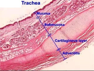
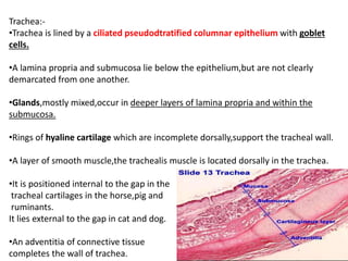
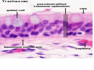
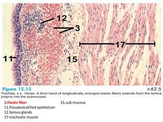
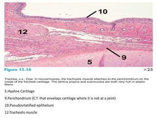
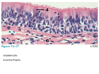
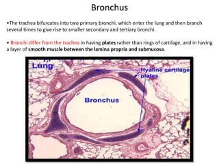
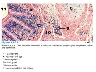
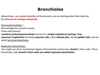
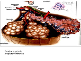
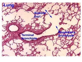
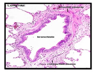
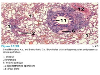
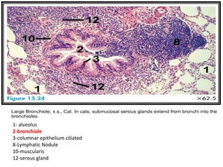
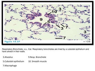
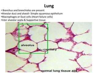
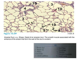
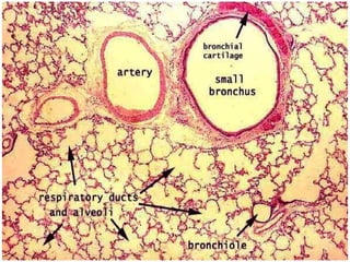


The document summarizes the histology of the respiratory system. It describes the structure of the trachea, bronchi, bronchioles, and lungs. The trachea has ciliated pseudostratified epithelium and incomplete hyaline cartilage rings. The bronchi have plates of cartilage instead of rings and a smooth muscle layer between the lamina propria and submucosa. Bronchioles lack cartilage and glands. Terminal bronchioles have Clara cells and cuboidal epithelium. Respiratory bronchioles have alveoli in their walls. The lungs contain alveolar ducts, alveoli lined with simple squamous epithelium, and macrophages in the interalveolar septa.


![CASE_PRESENTATION_ON_subdural_hematoma(SDH)[1 FINAL PPT]-1.pptx](https://cdn.slidesharecdn.com/ss_thumbnails/casepresentationonsubduralhematomasdh1finalppt-1-260129172522-d405d375-thumbnail.jpg?width=640&height=640&fit=bounds)


