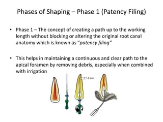
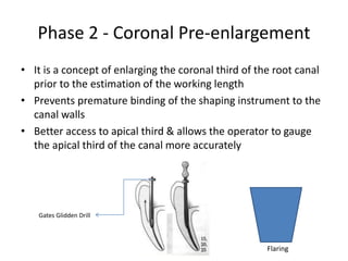
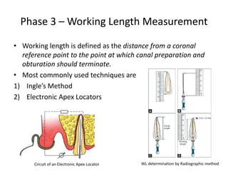
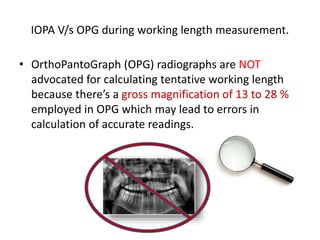
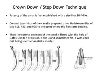
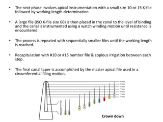
The second phase of a root canal treatment.
This presentation covers the most basic techniques of root canal shaping.
provides the reader with a concise overview of the big picture.
2. Introduction
⢠In an endodontic treatment, the root canal
system is our road map to success
⢠In 1852 Arthur used small files for root canal
enlargement.
⢠Ingle and LeVine (1958) listed standardization
for instruments.
⢠Ingle described conventional technique/
standardized taper technique of root canal
preparation.
⢠Almost 50 years ago, Herbert Schilder
introduced the concept of âcleaning and
shaping."
5. Cleaning
⢠Cleaning refers to removal of all contents of root
canal system before and during shaping that is
organic substrates, micro-flora, bacterial byproducts,
food debris, caries, denticles, pulp-stones, previous
root canal filling materials and dentinal shavings
6. Shaping
⢠The final preparation of the canal should be an exact
replica of the original canal configuration in shape,
taper, and flow only larger
7. Phases of Shaping â Phase 1 (Patency Filing)
⢠Phase 1 â The concept of creating a path up to the working
length without blocking or altering the original root canal
anatomy which is known as âpatency filingâ
⢠This helps in maintaining a continuous and clear path to the
apical foramen by removing debris, especially when combined
with irrigation
8. Phase 2 - Coronal Pre-enlargement
⢠It is a concept of enlarging the coronal third of the root canal
prior to the estimation of the working length
⢠Prevents premature binding of the shaping instrument to the
canal walls
⢠Better access to apical third & allows the operator to gauge
the apical third of the canal more accurately
Gates Glidden Drill
Flaring
9. Phase 3 â Working Length Measurement
⢠Working length is defined as the distance from a coronal
reference point to the point at which canal preparation and
obturation should terminate.
⢠Most commonly used techniques are
1) Ingleâs Method
2) Electronic Apex Locators
Circuit of an Electronic Apex Locator WL determination by Radiographic method
10. IOPA V/s OPG during working length measurement.
⢠OrthoPantoGraph (OPG) radiographs are NOT
advocated for calculating tentative working length
because thereâs a gross magnification of 13 to 28 %
employed in OPG which may lead to errors in
calculation of accurate readings.
11. Phase 4 â Canal Shaping Techniques
⢠The endodontic hand instruments can be employed in any
one of the functional motions :
1. Step-Back Technique
(a) Conventional Step-Back
(b) Passive Step-Back
2. Crown-down (Step-Down) Technique
and its modifications :
(a) Crown-Down Pressure-less
(b) Double Flare
(c) Balanced Force
Apical to Coronal Coronal to Apical
12. Phase 5 â Working Width
⢠Most canals are oval in cross-section. Oval canals
have two diameters, a minor (smaller) and a major
(larger) diameter.
⢠The quality of cleaning is dependant on
instrumenting to the larger diameter
⢠Working Width is best understood by studying
cross-sections of apical canals
⢠If the greater diameter of the original canal is
measured, the correct WW is an instrument size,
slightly larger than that dimension.
13. Some basic terminologies
⢠Anatomic Apex: It is defined as the tip or the end
of the root determined morphologically.
⢠Radiographic Apex: It is defined as the tip or the
end of the root determined radio-graphically.
⢠Apical Foramen (Major Diameter): It is the main
apical opening of the root canal. It is frequently
eccentrically located away from the anatomic or
radiographic apex.
⢠Apical Constriction (Minor Diameter): It is the
apical portion of the root canal having the
narrowest diameter.
⢠Cementodentinal Junction: It is the region where
the dentin and cementum are united.
14. Step Back / Telescopic / Serial Root Canal Preparation
⢠Schilder suggested a âserial preparationâ that
included enlarging to a file size #30 or #35 up to
working length and then serially reducing WL for
the following instruments.
⢠To overcome instrument transportation in the
apical third of root canal, Mullaney in 1960
divided the step-back preparation into two
phases â
1) Phase I - Apical preparation starting at the
apical constriction.
2) Phase II - Preparation of the remainder of the
root canal, gradually stepping back while
increasing in size.
15. Phase 1 - Apical preparation starting at the
apical constriction
⢠Establish WL, then insert the first #10 K-File
into the canal with watch winding motion.
⢠Remove the file and irrigate the canal.
⢠Lubricate the instrument for use in apical
area.
⢠Place the next larger size files to the WL in
similar manner and again irrigate the canal.
⢠Repeat the process until a #25 K-file reaches
the WL (MAF) as minimum.
⢠Recapitulation should be done between the
files by placing previous smaller number file
to the WL. This breaks up apical debris which
are washed away with the irrigant.
16. Phase 2 - Preparation of the remainder of the root canal,
gradually stepping back while increasing in size
⢠Place next file in the series to a length 1 mm
short of WL.
⢠Insert the file into the canal with watch winding
motion, remove it after circumferential filing,
irrigate and recapitulate by #10 file.
⢠Repeat the same procedure with successively
larger files at length 1mm lesser from the
previous length of previous file.
⢠Similarly mid canal area and coronal part of the
canal is prepared and shaped with larger number
files.
⢠Finally refining of the root canal is done by
master apical file with push-pull strokes to
achieve a smooth taper form of the root canal.
17. Phase 2 contd.
⢠Gates Glidden drills number
â 2, 3, 4 are used to create
coronal and middle
preparations.
18. Phase 2 contd.
⢠Patency of the apical canal segment, which
has been enlarged to size 25, must be ensured
by continued use of this file after each step
back (RECAPITULATION).
⢠Circumferential filing using the master apical
file can be used to smooth out and further
refine the steps created by the step back. This
preparation allows an adequate amount of
coronal space in the root canal for lateral
compaction.
⢠Instrumentation is finished when the walls are
smooth and clean and when the preparation
shows continuous taper in an apical direction
19. Advantages
⢠This technique creates small apical preparation with larger
instruments used at successively decreasing lengths to create
a taper
⢠Taper of canal preparation can be altered by changing the
interval between the consecutive instruments, for example,
taper of prepared canal can be increased by reducing the
intervals between each successive file from 1 to 0.5 mm.
20. Disadvantages
⢠Difficult to irrigate apical region.
⢠More chances of pushing debris peri-apically.
⢠Time consuming.
⢠It has a tendency to straighten the curved canal.
⢠Increased chances of iatrogenic errors; ledge formation, instrument
separation, zipping of the apical area, apical blockage, etc.
⢠Since, curvature of the canal is reduced during mid-root flaring, there will
be a loss in the working length.
⢠Difficult to insert instruments in canal.
21. Passive Step Back Technique
⢠This technique was introduced by Dr. Mahmoud Torabinejad.
⢠It involves the insertion of progressively larger hand
instruments as deep as they can be passively placed
⢠Subsequently, Gates-Glidden drills are used for additional
coronal enlargement followed by apical instrumentation using
the step-back technique.
22. Advantages
⢠Removal of debris and minor canal obstructions.
⢠Knowledge of the canal morphology.
⢠Gradual passive enlargement of the canal in an apical to
coronal direction.
⢠This technique can also be used with ultrasonic
instruments.
⢠Decreased incidence of procedural errors like
transportation of the canal, ledge or zip formation.
23. Crown Down / Step Down Technique
⢠Patency of the canal is first established with a size 8 or 10 K-file.
⢠Coronal two-thirds of the canal is prepared using Hedstroem files of
size #15, #20, and #25 to the point where the file starts binding.
⢠Then the coronal segment of the canal is flared with the help of
Gates-Glidden drills Nos. 2 and 3 and sometimes No. 4 with each
drill being used sequentially shorter.
24. ⢠The next phase involves apical instrumentation with a small size 10 or 15 K-file
followed by working length determination
⢠A large file (ISO K-file size 60) is then placed in the canal to the level of binding
and the canal is instrumented using a watch winding motion until resistance is
encountered
⢠The process is repeated with sequentially smaller files until the working length
is reached.
⢠Recapitulation with #10 or #15 number file & copious irrigation between each
step.
⢠The final canal taper is accomplished by the master apical file used in a
circumferential filing motion.
25. Advantages
⢠Permits straighter access to the apical region.
⢠Eliminates coronal interferences which allows better determination of
apical canal sizes
⢠Removes bulk of the tissue and microorganisms before apical shaping.
⢠Allows deeper penetration of irrigants.
⢠Eliminates the amount of necrotic debris that could be extruded
through the apical foramen during instrumentation.
⢠Less chance of zipping near the apical constriction.
⢠It provides a coronal escape-way that reduces the âpiston in a cylinder
effectâ responsible for debris extrusion from the apex.
26. Crown Down Pressure-less Technique
⢠Suggested by Marshall and Pappin.
⢠Early coronal flaring with Gates-Glidden drills is followed by
an incremental removal of dentin from coronal to apical
direction, and hence called âcrown-downâ technique.
⢠Straight K-files are then used in a large to small sequence with
a reaming motion and no apical pressureâhence called
âpressure-lessâ technique
27. Double Flare Technique
⢠Introduced by Fava in 1983
⢠In this, canal is explored using a small file (#8/#10)
⢠Then the canal is prepared in a crown down manner
using K files in decreasing sizes.
⢠Followed by Step Back method with K-File in
ascending sizes.
28. Double Flare Technique
⢠Indications
- Straight root canals
- Straight portions of curved canals of
mature teeth
⢠Contraindications
- Calcified canals
- Young permanent teeth
- Teeth with open apex
Calcified Canal Blunderbuss Apex
Straight Canal
29. Advantages
⢠Greater taper in the cervical and middle third such that
removal of canal contents is more effective and root canal is
better cleaned.
⢠Elected technique in cases of necrotic or gangrenous teeth.
⢠Improved quality of root canal filling when compared to
conventional technique.
⢠The flared technique maintains the root canal shape and
produces neither the hour glass appearance nor the apical zip.
30. Balanced Force Technique
⢠This technique was developed by Roane and Sabala in 1985. It involves the
use of instrument with non- cutting tip (ex: Flex-R).
⢠Triangular cross-sectioned instruments are used. The decreased mass of
the instrument and deeper cutting flutes improves the flexibility of
instrument and decrease the restoring force of the instrument when
placed in curved canals.
⢠This technique can be described as positioning and preloading an
instrument through clockwise rotation and then shaping the canal with
counter-clockwise rotation.
31. ⢠Coronal third of the canal is prepared using the crown-down technique
⢠After pressure-less insertion of the Flex-R file the instrument is rotated
clockwise (90°), using only light apical pressure.
⢠The instrument is then rotated counter-clockwise (180°â270°), and
sufficient apical pressure is used to keep the file at the same insertion
depth during this step. In this step, dentinal shavings are removed with a
characteristic clicking sound.
⢠After two or three cycles, the file loaded with dentinal shavings is
removed from the canal with a prolonged clockwise rotation. This action
loads the debris into the flutes
⢠Sequential files are used in a crown-down fashion before preparing the
apical third to the appropriate master apical file size.
32. Advantages
⢠Lesser chances of canal transportation
⢠File manipulation can be done at any point without
creating a ledge or blockage
⢠File cutting only occurs at the apical extent.
⢠Extrusion of materials less than other techniques.