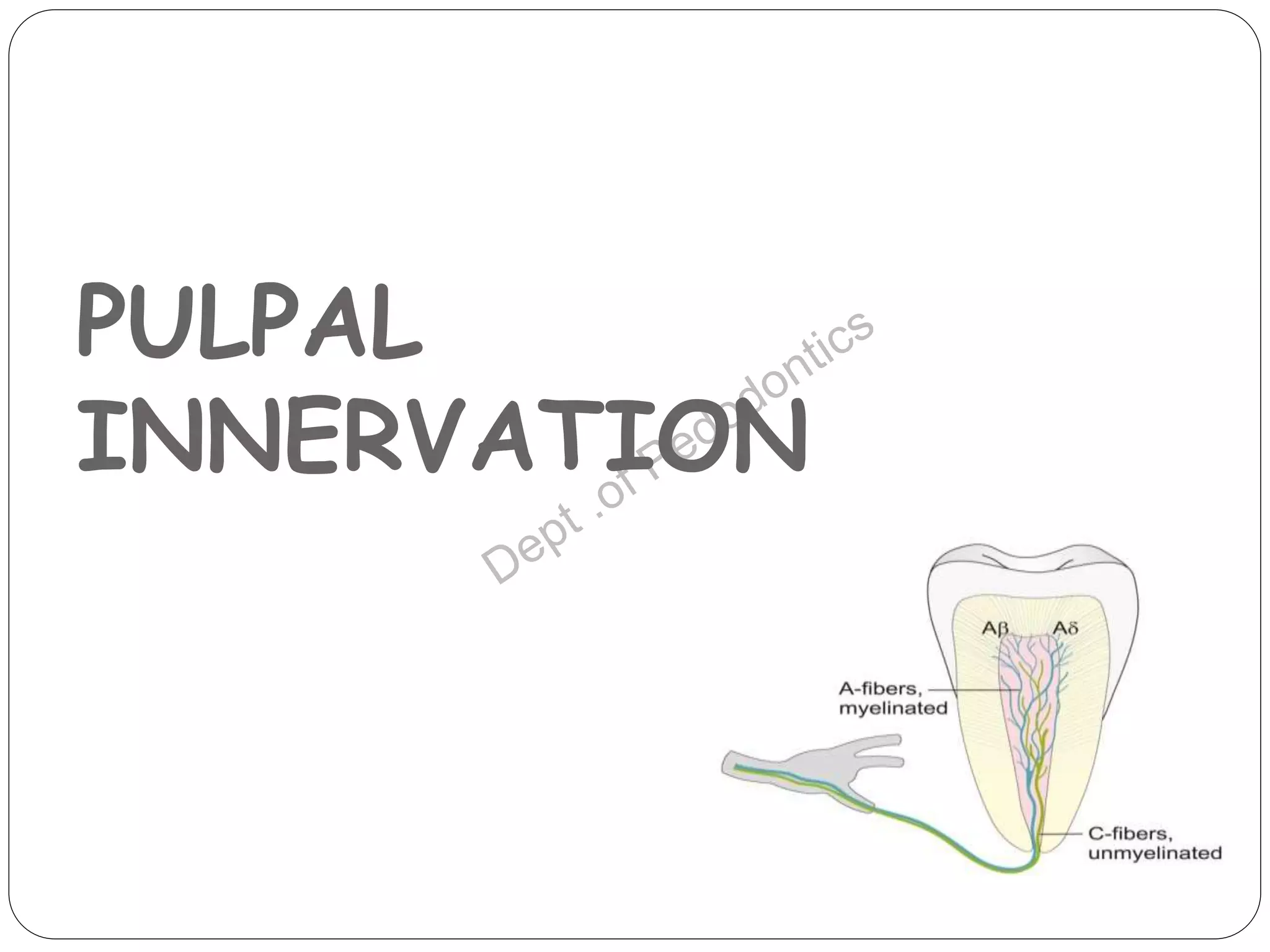
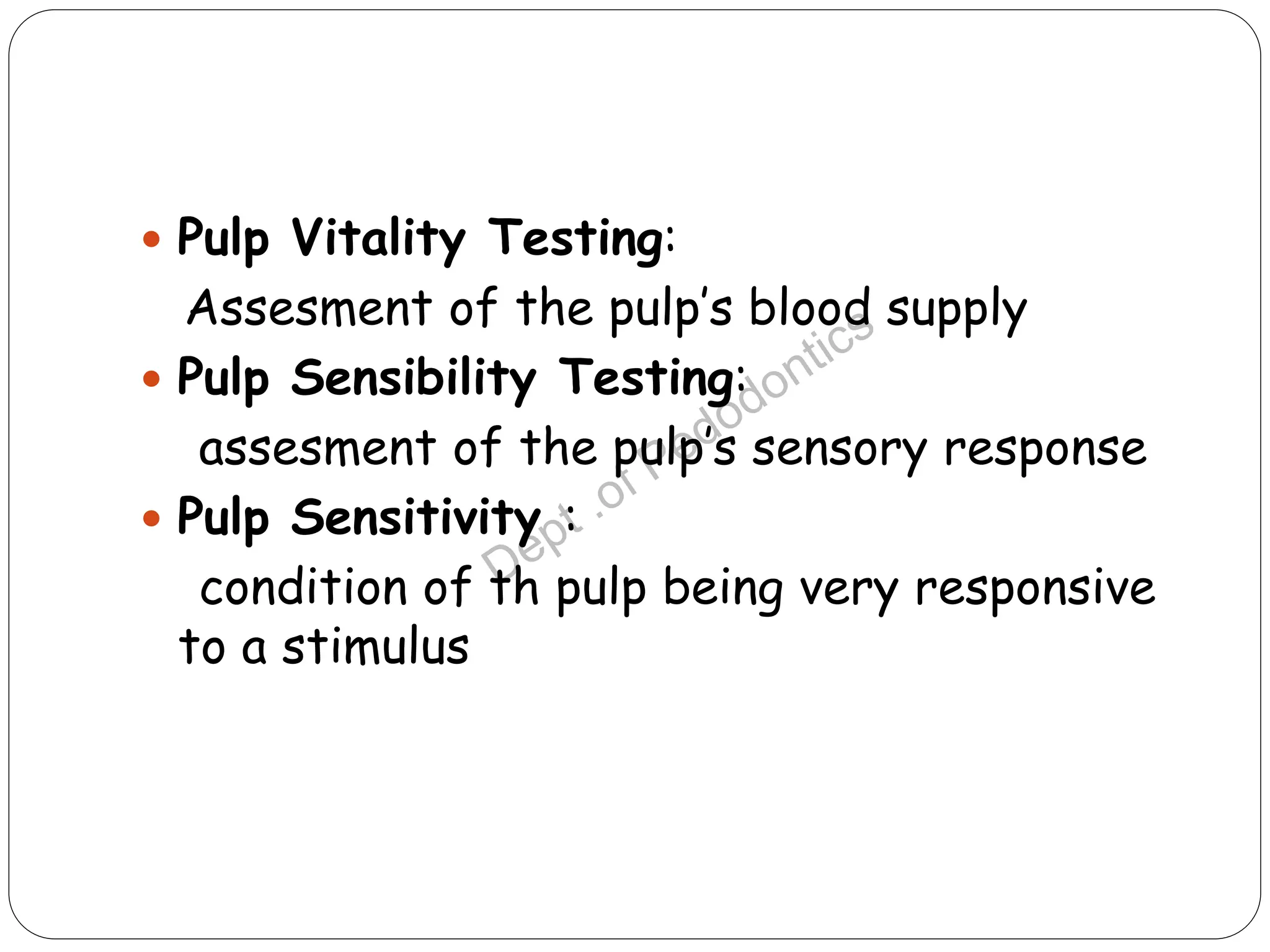
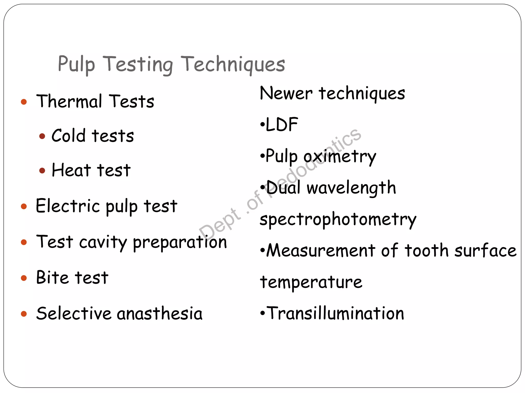
2. 2
Introduction
Uses
Ideal requirements
Pulpal innervation
Pulp vitality tests
Various conditions and their
responses to various tests
Conclusion
Refrences
CONTENTS
3. INTRODUCTION
3
Diagnosis in dentistry may be defined as ‘the process
whereby the data obtained from questioning,
examining and testing are combined by the dentist to
identify deviations from the normal.
The diagnosis of dental pulp status -history, clinical
examination, special tests, and radiological
examination-not as the outcome of any one specific
test.
4. 4
Vitality testing -important aid -diagnosis of pulp
disease
One of the greatest diagnostic challenges in clinical
practice -accurate assessment of pulp status.
This may be further complicated in paediatric
dentistry -developing dentition, traumatized teeth, or
young children who have a limited ability to recall a
pain history for the tooth in question.
5. USES
5
Prior to operative procedures : for selected teeth prior to
restorative or orthodontic interventions, where pulp health
may be in question.
Diagnosis of pain: identifying pulpal pain from other
conditions such as myofascial pain dysfunction syndrome and
referred pain . Normal response to pulp testing -eliminate
pulpal pathology.
Investigation of radiolucent areas: Radiolucent areas -
periapical extension of pulpal pathology, due to other
pathological processes, normal structures. Pulpal pathology is
not responsible -normal response to vitality testing.
6. 6
Post-trauma assessment : important part of the
examination and review of traumatized teeth.
Assessment of anesthesia: Grossman -pulp testers to
assess -a tooth is completely anaesthetized -local
anesthetic prior to operative procedures
Assessment of teeth which have been pulp capped or
required deep restoration : prognosis of teeth -
clinically assessed on the basis of reported symptoms;
clinical palpation; and percussion, radiographs, and pulp
testing.
9. 9
Dental pulp- sensory & autonomic nerve fibers.
Nerve fibers + blood vessels- apical foramen.
After entering pulp- nerve bundles run coronally-
divide into smaller branches- single axon –pulp dentin
margin- plexus of Raschkow.
Individual axons- numerous terminal filaments-
dentinal tubules.
2 types sensory fibers- A fibers (myelinated) C fibers
(unmyelinated).
10. 10
A Fiber
Large
Myelinated
High conduction velocity (6-
30 m/sec)
Innervate pulp dentin border
Subgroups - A β fibres & A δ
fibres ( according to diameter
and conduction velocities).A β
more sensitive to stimulation
than A δ
C Fiber
Small
Unmyelinated
Slow conduction velocities
(0.5- 2 m/sec)
Innervate pulp core
11. 11
A δ FIBRES
Costitute 90% of A fiber
Large
Increased conduction velocity
Acute, sharp, momentary pain
Pain quiclky disappears on
stimulus removal
Excited by hydro mechanical
events in dentinal tubules e.g.
drilling ,air drying
C FIBRES
Small
Decreased conduction velocities
Dull, burning, poorly located pain
Associated with pain caused by tissue
injury & modulated by inflammatory
mediators, vascular changes in blood flow
and volume
Pain increase with pressure
Activation- stimuli reaching pulp- intense
heating or cooling of tooth crown.
Have high threshold
12. 12
As the intensity of the stimulus increases, more
sensory nerves are activated, and this results in a
progressive increase in the sensory response.
The response to a given stimulus will be greatest
where neural density is the highest.
14. 14
Pulp Vitality Testing:
Assesment of the pulp’s blood supply
Pulp Sensibility Testing:
assesment of the pulp’s sensory response
Pulp Sensitivity :
condition of th pulp being very responsive
to a stimulus
15. Pulp Testing Techniques
15
Thermal Tests
Cold tests
Heat test
Electric pulp test
Test cavity preparation
Bite test
Selective anasthesia
Newer techniques
•LDF
•Pulp oximetry
•Dual wavelength
spectrophotometry
•Measurement of tooth surface
temperature
•Transillumination
16. THERMAL TESTING
16
Application of cold and heat stimuli to a tooth, to
determine sensitivity to thermal changes.
Hot and cold -tests of sensitivity, -conducted for
different diagnostic reasons.
A response to cold -vital pulp, regardless of whether
that pulp is normal or abnormal.
Increased response to heat -pulpal or periapical
pathology -endodontic intervention .
17. Cold test
17
Cold -causes contraction the dentinal tubules-rapid
outward flow of fluid within the patent tubules.
Rapid movement dentinal fluid -‘hydrodynamic forces’
acting on the A δ nerve fibres within the pulp–dentine
complex - sharp sensation lasting for the duration of
the thermal test.
A variety of cold tests may be employed,
Major difference -the degree of cold applied to the
tooth.
18. 18
The cold test -differentiate between reversible and
irreversible pulpitis.
Stimulus application -lingering effect or if the pain
subsides immediately on removal of the stimulus.
Lingering pain-even after the removal of cold
stimulus- irreversible pulpitis.
Pain subsides immediately after stimulus removal-
reversible pulpitis.
19. 19
Cold testing + electric pulp tester -the results from
one test verify the findings of the other test.
If a mature, non-traumatized tooth does not respond
either to EPT or cold-considered non-vital .
When testing multi-rooted teeth-respond positively to
cold-only one root actually contains vital pulp tissue.
20. Methods of application
20
Wrap - SLIVER OF ICE in wet gauze -place it against the
buccal surface, comparing the reaction between -test tooth &
control tooth.
PENCILS OF ICE -filling a plastic straw with water –freezing
upright position.
ETHYL CHLORIDE (boiling point – 41 ° C) sprayed onto a
cotton pledget-formation of ice crystals, prior to application
to the tooth.
DICHLORODIflUOROMETHANE (DDM) (boiling point – 0 ° C)
-compressed refrigerant spray-sprayed onto a cotton pledget.
21. 21
Frozen carbon dioxide (CO 2 ) ‘dry ice’ or ‘carbon dioxide
snow’ (boiling point – 72 ° C). Solid stick of CO 2 gas
prepared – deliver CO 2 gas into custom-made plastic
cylinder -the stick is applied to buccal surface.
Ozone-friendly NON-CHLOROflUOROCARBON sprays -
introduced in certain countries.
cold stimulus -begin with the most posterior tooth -advance
towards the anterior teeth.
Such a sequence will prevent any melted ice water dripping
in a posterior direction -cause stimulation of other teeth -
a false response.
22. 22
Dachi etal 1967 recommended use of ¼” ice cone
placed against the tooth for 5 sec to quantify cold
test.
23. Heat test
23
A stick of heated gutta-percha or hot water.
A gutta percha stick-heated with a naked flame or an
electric heater -soft and glistens –applied vaseline-coated
surface of the test tooth.
Tooth surface temperature -150 ° C - gutta-percha softens
at 65 ° C -heated in delivery devices up to 200 ° C.
Prolonged heat application -bi-phasic stimulation of A δ
fibres initially, following C fibres . Activation of C fibres -
lingering pain-applied for not more than 5 s.
24. Disadvantages :
24
Difficult to use on posterior teeth because of limited
access.
Excessive heating may result in pulp damage .
A light layer of lubricant should be placed onto the tooth surface prior to
applying the heated material to prevent the hot gutta purcha (78 C TO
150 C ) or compound from adhering to the dry tooth surface. ( ZACH
ET AL ,1998)
26. Electric pulp test
26
Objective :to stimulate intact A δ nerves in the pulp–dentine complex
by electric current. an ionic shift in the dentinal fluid local
depolarization generation of an action potential from intact A δ
nerves positive result.
The electric pulp tester -battery-operated instrument-connected to
a probe -applied to the tooth under investigation.
Produce a pulsating electrical stimulus, the initial intensity very low
value -prevent excessive stimulation and discomfort.
Intensity of the electric stimulus is then increased steadily at a pre-
selected rate, - note read-out on the digital display when the patient
acknowledges a warm or tingling sensation.
28. 28
Response threshold is reached -adequate number of nerve
terminals are activated -summation effect. An area of high
neural density -fast and strong response-least electric
current.
Desirable area of assessment in incisor teeth -incisal edge,
where the enamel is thinnest or absent.
The tester -tooth surface adjacent to a pulp horn, -highest
nerve density within the pulp.
This position equates to the incisal third region of anterior
teeth and the mid-third region of posterior teeth.
(Assessment of pulp vitality : a review V. Gopikrishna etal 2009 @ IJPD)
29. 29
The threshold for response may be influenced by the
thickness of the enamel and dentine overlying the pulp.
Thus, the response threshold for healthy teeth may be
lowest in incisors, slightly greater in premolars, and
greatest in molar teeth.
A recent study has revealed that the optimum site for
tester electrode placement on molars is on the tip of the
mesiobuccal cusp.
Appropriatelectrode placement site for electric pulp testing .Lin J et al 2007@journal of endodontics
31. Bite test
•Identifying cracked tooth or fractured cusp.
•It also helpful in diagnosing cases where in pulpal
pathosis is extended to periradicular region causing
apical periodontitis
Patient biting on cotton swab
31
32. 32
Devices –cotton swab , tooth picks, orange wood
sticks, rubber polishing wheels.
Commercially available device- tooth slooth-small
concave area on top- contact cusp to be tested.
Adjacent teeth -controls - patient is aware of the
normal response to these test.
33. 33
Biting pressure -applied slowly until full closure achieved.
Firm pressure -applied for few seconds, the patient is
then asked to release the pressure quickly.
Each individual pressure on a tooth can be tested like
this manner.
The practitioner should note if the pain is elicited during
the pressure phase or upon quick release of pressure.
Pain present on biting- apical periodontitis
Pain on release of biting force- cracked tooth
34. Staining and transillumination
34
The presence of crack in the surface of the tooth-stain to the
area is often of great assistance.
Shining a bright light on the surface of the tooth.
Transillumination is the detection method -easily and
graphically-presence of crack or not.
Based on a law of physics-a beam of light will continue to
penetrate through a substance until is meets a space, after
which the light beam is reflected.
This results in a light and a dark area of the tooth separated
by the fracture line.
35. 35
Sources of light other than from the
transilluminator (i.e., overhead lighting and
operatory lighting)- not to use.
A dental mirror -a fracture is present.
TRANSILLUMINATION: THE “LIGHT DETECTOR”@ ENDODONTICS: Colleagues for
Excellence Summer 2008
36. Selective anesthesia
36
Restricted to the patient who are in pain at the
time of the test -usual tests have failed to
identify the tooth.
Objective -anesthetize one tooth at a time until
the pain disappears and is localized to a specific
tooth .
37. 37
Technique
Using either infiltration or intra ligamentary injection,
inject the most posterior tooth in the area suspected
of being the cause of pain.
If the pain persist anesthetize the next tooth mesial
to it and continue to do so until the pain disappears.
If the pain cannot be identified as from maxillary or
mandibular origin ,an inferior alveolar block is given.
Cessation of pain naturally indicates involvement of
mandibular tooth.
38. 38
Advantage
This test is obviously a last resort and has an
advantage over the ‘test cavity’ during which
iatrogenic damage is possible.
39. TEST CAVITY
39
Only when all other test methods - deemed impossible
or the result of the other tests - inconclusive.
The test cavity- drilling through the enamel-dentin
junction of an un-anesthetized tooth .
Drilling - high speed round bur with appropriate
coolant and air
Sensitivity or the pain felt- an indication of pulp
vitality- procedure terminated- restore prepared
cavity.
If no pain- cavity preparation till pulp chamber-
enododontic treatment
40. 40
FALSE POSITIVES
Anxious patients
Liquefaction necrosis
Contact with metal restorations
Vital tissue still present in partially
necrotic root canal system
FALSE NEGATIVES
Incomplete root
development
Recently traumatized
teeth
Sclerosed canals
Recent orthodontic
activation
Patients with psychotic
disorders
RESPONSES FROM PULP TESTING
41. THERMOGRAPHY/ TEMPERATURE
MEASUREMENT
41
As a diagnostic procedure for human teeth-use of
thermistors, infrared thermography, and liquid
crystals.
Cholesteric liquid crystals-different colours when
heated-previously employed.
Underlying principle -teeth with an intact pulp blood
supply (vital/ healthy pulp tissue) had a warmer tooth
surface temperature compared with teeth that had no
blood supply.
42. 42
Surface temperature of teeth - measured over a
period of time at 15 s intervals using an electric
thermometer attached to a surface probe, placed in
contact with the tooth.
The study showed that, following cooling, only vital
teeth showed a subsequent rise in surface
temperature.
Diagnosis of tooth vitality by crown surface temperature measurement: a clinical
43. 43
Disadvantages
Teeth must be isolated with rubber dam, after
which a period of acclimatization is necessary prior
to imaging.
The technique is complex and requires the
subjects to be at rest for 1 h prior to testing.
45. •Developed- Tenland (1982)) later- Holloway (1983)
•1st in dentistry by GAZELIUS ET AL IN 1986
•a non-invasive method of assessing and measuring the
blood flow of pulp tissue.
•Laser light wavelengths 633 and 780 nm is directed onto
the tooth -by securing a fiber-optic probe against the
tooth surface.
LASER DOPPLER FLOWMETRY (LDF)
45
46. 46
The laser light from the probe -passes along the
enamel prisms to the enamel–dentine junction and the
S-shaped dentinal tubules, which act as light guides,
to the pulp .
Light absorbed by red blood cells in the capillary
plexus is scattered and undergoes a shift in frequency
according to the Doppler principle; light absorbed by
stationary objects does not undergo a shift in
frequency.
47. 47
A signal is produced which measures the influx of the blood
cells (number of red blood cells times mean velocity).
The proportion of Doppler-shifted light is detected by a
photo detector. The detected signal is weak and therefore
highly amplified.
A trace of signals from vital and non- vital teeth. Fourier
analysis of the traces has revealed a heart beat frequency in
the vital tooth, but not in the non-vital tooth and is
therefore an effective discriminator
49. A LDF probe showing laser light
guides.
A LDF probe applied to a sectioned
tooth
showing the passage of light via the
enamel prisms and
dentinal tubules to the pulp.
A LDF trace showing signals from two teeth; the
upper is from a vital tooth while the lower is from a non-
vital tooth.
49
50. PULSE OXIMETRY
50
Invented by Aoyagi in the early 1970.
Non-invasive method
Entirely objective test -no subjective response from
the patient.
Consists of two light-emitting diodes, one to transmit
red light (640 nm) and the other to transmit infrared
light (940 nm), and a photodetector on the opposite
side of the vascular bed.
The light emitting diode transmits light through a
vascular bed such as the finger or ear.
Oxygenated haemoglobin and deoxygenated
haemoglobin absorb different amounts of
red/infrared light.
51. 51
The pulsatile change in the blood volume causes periodic
changes in the amount of red/ infrared light absorbed
by the vascular bed before reaching the photo
detector.
The relationship between the pulsatile change in the
absorption of red light and the pulsatile change in the
absorption of infrared light is analysed by the pulse
oximeter to determine the saturation of arterial blood.
Schnettler and Wallace reported a correlation between
pulpal and systemic oxygen saturation readings using a
modified ear pulse oximeter probe on a tooth. They
recommended its use as a definitive pulp vitality tester.
(Schnettler JM, Wallace JA. Pulse oximeter as a diagnostic tool of pulp vitality.@ J
Endod 1991.)
52. 52
To be accurate-a normal arterial blood flow is required.
Arterial pulsatile blood flow is low, pulse oximeter measurements
are unobtainable.
This may occur during hypovolaemia, hypothermia, or intense
peripheral vasoconstriction.
Lower specificity in cases where the coronal pulp is undergoing
calcific changes.
This may occur following trauma, placement of deep restorations,
or with physiological ageing.
In such cases, a radicular vital pulp with coronal calcification could
potentially cause a false negative response.
53. 53
SPECTROPHOTOMETRY
Also called Dual Wavelength Spectrophotometry (DWLS)
Method independent of pulsatile circulation.
Measures oxygen changes in capillary bed .
Advantages :
Avulsed or replanted teeth with open apices- blood supply
regained in 1st 20 days but nerve supply lags behind- repeated
readings 40 days- healing process.
Non – invasive.
Objective test.
Instrument- small, portable, inexpensive.
55. Reversible pulpitis / Pulpal hyperemia
•Mild to moderate inflammatory condition of the pulp -
noxious stimuli
•Symptomatic reversible pulpitis -sharp pain lasting for a
moment- disappears when the stimulus is removed.
•Pain may become chronic.
55
56. 56
The pulp is sensitive to temperature changes,
particularly cold, application of cold is an excellent
method of locating and diagnosing the involved
tooth.
The electric pulp test, using less current than on a
control tooth, is an excellent corroborating test.
57. Irreversible pulpitis
•Irreversible pulpitis is a persistant inflammatory
condition of the pulp, symptomatic or asymptomatic
caused by a noxious stimulus.
•A tooth with symptomatic irreversible pulpitis exhibit
intermittent or spontaneous pain, whereby rapid exposure
to dramatic temperature changes (especially to cold
stimuli) will elicit heightened and prolong episodes of pain
even after the source of the pain is removed.
•The pain may be sharp or dull localised or referred.
57
58. 58
In the early stages the thermal test may elicit pain
that persists after removal of the thermal stimulus.
In the late stages when the pulp is exposed ,it may
respond normally to the thermal stimulus.
The electric pulp test induces a response with a
marked variation in current form of normal.
Result of examination for mobility and perussion and
palpation tests are negative.
59. Chronic Hyperplasic Pulpitis
•Chronic hyperplastic pulpitis or ”pulp polyp” is a
productive pulpal inflammation due to an extensive
carious of a young pulp.
•Mechanical irritation from chewing and bacterial
infection often provide the stimulus.
•The tooth may respond feebly or not at all to the
thermal test, unless one use extreme cold, as from an
methyl chloride spray.
•More current than normal may be required to elicit a
response by means of the electric pulp tester.
59
60. Internal resorption
•Internal resorption is an idiopathic slow or fast
progressive resorptive process occurring in the dentin of
the pulp chamber or root canals of the teeth
•Internal resorption in the root of a tooth is asymptomatic.
•In the crown of the tooth ,internal resorption may be
manifested as a reddish area called “pink spot”.
•The defect is usually is recognized by means of a
radiograph.
60
61. Necrosis of pulp
•Necrosis is death of the pulp.
• Partial or total, depending on whether part or the entire pulp is
removed.
•It can be caused by any noxious insult to the pulp , such as bacteria,
trauma ,and chemical irritation.
•A normal tooth with a nerotic pulp cause no pain
•Radiograph is non diagnostic.
•The teeth with partial necrosis -respond to thermal changes -presence
of vital nerve fibers in the adjacent inflamed tissue.
•Tooth with necrotic pulp does not respond to cold ,the electric pulp
test ,or the test cavity
61
62. CONCLUSION
62
An accurate assessment of tooth vitality is of paramount
importance in clinical practice.
Although sensitivity testing is commonly employed by
the majority of clinicians, it has acknowledged
limitations.
Rapid advances in knowledge and applied technology
relating to pulpal blood flow may lead the way for a more
objective, accurate, and predictable means of pulp
vitality assessment.
63. References
1. Text of Grossman’s Endodontic practice 12th edition
2. Samraj RV Recent advances in pulp vitality testing
3. Stephen cohen Kenneth M. HargreaveText book of Pathways of the pulp
9th edition.
4. Nisha garg. Textbook of endodontics- 3rd edition.
5. Velayutham GopikrishnArticle on Evaluation of Efficacy of a New Custom-
Made Pulse Oximeter
6. Diagnosis of tooth vitality by crown surface temperature measurement: a
clinical evaluation.Fanibunda KB. @J Dent 1986
7. TRANSILLUMINATION: THE “LIGHT DETECTOR”@ ENDODONTICS:
Colleagues for Excellence Summer 2008
8. Assessment of pulp vitality : a review V. Gopikrishna etal 2009 @ IJPD
63