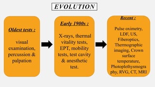
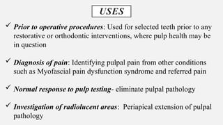
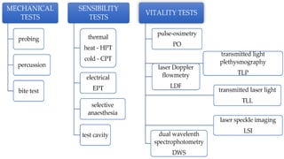
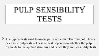
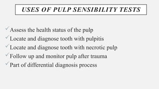
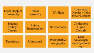
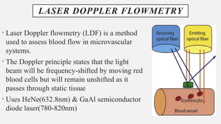
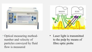
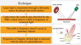
ENDODONTIC
DIAGNOSIS- PART 2
Dr. Sushmita Rane
Seminar- 7
6/02/2023
INTRODUCTION
• STEDMAN’S MEDICAL DICTIONARY ‘clinical diagnosis is the
determination of the nature of the disease made from a study of the
signs and symptoms of the disease.’
Latin
“Diagignoskein”
Dia : apart
Gignoskein : to learn
The patient tells the
clinician why the
patient is seeking
advice.
The clinician
questions the patient
about the symptoms
and history that led
to the visit.
The clinician
performs the
objective testings.
The clinician correlates the
objective findings with the
subjective details and creates a
tentative differential diagnosis.
The clinician
formulate a definitive
diagnosis.
EVOLUTION
Oldest tests :
visual
examination,
percussion &
palpation
Early 1900s :
X-rays, thermal
vitality tests,
EPT, mobility
tests, test cavity
& anesthetic
test.
Recent :
Pulse oximetry,
LDF, US,
Fiberoptics,
Thermographic
imaging, Crown
surface
temperature,
Photoplethysmogra
phy, RVG, CT, MRI
USES
 Prior to operative procedures: Used for selected teeth prior to any
restorative or orthodontic interventions, where pulp health may be
in question
 Diagnosis of pain: Identifying pulpal pain from other conditions
such as Myofascial pain dysfunction syndrome and referred pain
 Normal response to pulp testing- eliminate pulpal pathology
 Investigation of radiolucent areas: Periapical extension of pulpal
pathology
PULP SENSIBILITY
TESTS
 The typical tests used to assess pulps are either Thermal(cold, heat)
or electric pulp tests – These all test depends on whether the pulp
responds to the applied stimulus and hence they are Sensibility Tests
USES OF PULP SENSIBILITY TESTS
Assess the health status of the pulp
Locate and diagnose tooth with pulpitis
Locate and diagnose tooth with necrotic pulp
Follow up and monitor pulp after trauma
Part of differential diagnosis process
Necrotic pulp
Clinically normal/ Pulpitis
Pulp Response to the stimulus
Response Responds
Nature of the response
Normal
(compared to other
teeth)
Exaggerated
(Reproduces the pain that the
patient has been experiencing-
Pulpitis)
What type of Pulpitis- History,
duration, nature
THERMAL TESTS
• Application of heat / cold to a tooth to determine sensitivity to
thermal changes.
• A response indicates a vital pulp, regardless of whether the pulp is
normal / abnormal.
Cold testing
Heat testing
MECHANISM OF COLD SENSIBILITY PULP TEST
contraction the dentinal tubules
Rapid outward flow of fluid within the patent tubules.
act on the A δ nerve fibres producing sharp sensation lasting
for the duration of the thermal test.
Also, the nerve fibres will feel this change in temperature if
they are functioning normally or if there is inflammation
Methods of
application
PENCILS OF ICE
WRAPPING ICE IN WET GAUZE
FROZEN CARBON DIOXIDE
‘DRY ICE’ (boiling point – 72 ° C)
ETHYL CHLORIDE(boiling point –
41 ° C)
TETRAFLUOROETHANE (DDM)
(boiling point – 26.2 ° C)
COLD PULP SENSIBILITY TESTS
 Dry ice
 Chen and abbott reported that Dry ice sticks were the most reliable cold test
 formed in the device called Odontotest
This device uses pressurized liquid
CO2 that is forced through a small
orifice. It becomes Dry ice when it
comes under atmospheric pressure
This produces stick of dry ice.
Its temperature is -56 °C when
applied to the tooth
Refrigerant spray
• Endo ice- 1,1,1,2 tetrafluoroethane
• It has a temperature of about -26 °C when first sprayed but this
reduces to about-18.5 °C in the mouth
• Application- sprayed on cotton pellet then applied to the tooth
• Disadvantage- becomes liquid quite rapidly in the warm
environment of the mouth. The liquid can then flow in the
gingival region where it may be felt and misinterpretated as
being from pulp of the tooth
Ice
Used by freezing water in the local anesthetic catridge and then
applying the extruded portion to the tooth
HEAT PULP SENSIBILITY TEST
• Teeth with clinically normal pulp do
not respond to heat- they only respond
to heat when they are inflamed
When do we use Heat test?
patient’s chief complaint is intense
pain on having hot liquid/food.
METHODS
Ball burnisher Hot GP sticks
Dry rubber polishing wheel Hot water
Isolate the area of tooth to be tested
Warm air is directed to exposed surface of tooth and patient’s response is noted
Heat is applied to the occlusal-buccal third of exposed crown
If no response occurs, the hot substance can be moved to the central portion of
the crown or close to the tooth cervical margin
When response occurs, heat should be removed immediately
Care to be taken to avoid using excessive heat or prolonged application of heat to
tooth
Technique
ELECTRIC PULP TESTING
• Uses nerve stimulation.
• Objective is to stimulate a pulpal response by subjecting
the tooth to an increasing degree of electric current.
• Electric Pulp Tester (EPT) scale used : 0-64 units
• Healthy vital pulp : 11-16 units
Sequence of testing :1st contralateral tooth
2nd opposing tooth
3rd adjacent tooth
Last suspicious tooth
Isolate the area of tooth to be tested
Apply an electrolyte (toothpaste) on tooth and place electrode against the dried
enamel of crown’s occlusobuccal / incisolabial surface.
Turn the rheostat slowly to introduce minimal current into the tooth & increase
the current slowly. Ask the pt to indicate when tingling sensation occurs
Record the result according to the numeric scale on EPT
Repeat the procedure for each tooth to be tested
Technique
• In case of complete coverage crown the tip of endodontic explorer is
coated with toothpaste and placed in contact with the tooth structure
• The tip on the electronic pulp tester is placed on the explorer and
checked for response
Response Interpretation
Positive Vital
Negative Non vital
False negative Immature teeth, recent trauma, atrophied
pulp/calcification, poor contact with
tooth, inadequate electrolyte.
False positive Patient anxiety, multirooted teeth, contact
with metallic restoration
Delayed respose (>30units) Partial necrosis
Early/Exaggerated(10 or <) Hyperemia
Responses of EPT
Contraindications :
 cardiac pacemaker &
insulin pumps
 Teeth with full coverage
restorations
Disadvantages :
 Gives no indication of
state of vascular supply.
 Doesn’t measure the
degree of health /disease
of pulp.
• A study conducted by Peterson et al compared the ability
of thermal and electric pulp testing methods in detection of
pulp. The sensitivity of cold test was 0.83, 0.86 for heat
testing and 0.72 for EPT.
• Same study evaluated the specificity of these tests. 93% of
teeth were correctly identified by cold and EPT whereas
only 41% teeth were identified by heat tests
• Results showed accuracy of cold test to be 86%,EPT
81%,heat test 71%
BITE TEST
• Useful in identifying a cracked tooth or fractured cusp, when pressure
is applied in a certain direction to one cusp or section of the tooth.
• Methods used : Biting on
Orangewood stick Wet cotton roll Burlew rubber disk
• Many methods have been described to identify crack undermining
the cusp. But the most common and generally used are
Commercially available devices : Tooth slooth & FracFinder
• The test is repeated on each cusp and on several teeth in the region
of patients complaint
How to distinguish between different conditions?
Symptomatic Apical
periodontitis
Apical Abcess
Lateral periodontitis
(periodontal pocket,
swelling and tenderness on
palpation on lateral aspect)
Cracked tooth or cusp
flexure
Pain on biting
Pain on
release of
biting force
DYE / STAIN TEST
• Can be done individually or along with other tests.
• Used to determine the presence of crack in the surface of a tooth
& caries detection
Crack detection dyes : Methylene blue / India ink
Caries detecting dyes : 1% acid red in propylene
glycol (stains infected dentin)
• A small amount of dye is placed on a
small cotton pellet & crown/root is
coated with it.
• Any dye on surface is washed away &
if fracture present , dye remains in the
crack & can be observed via loupes
Technique
FIBEROPTIC TRANSILLUMINATION
• An FOTI device can be used to confirm the
presence of cracks underneath any restoration &
to determine the extent & depth of the cracks
• Shining a bright light on the buccal / lingual
surface of tooth.
• For a tooth that is not cracked, light will shine
through the opposite surface without any
interruption.
• Friedman & Marcus reported its usefulness for
detection of proximal caries.
ANESTHETIC TEST
• Restricted to patients who are in pain at the time of the test when
the usual tests have failed to identify the tooth.
Using either infiltration / intraligamentary injection,
inject the most posterior tooth in the suspected area.
If pain persist when the tooth has been fully
anesthetized, anesthetize the next tooth mesial to it &
continue to do so until the pain disappears
TEST CAVITY
• Involves drilling through enamel-dentin junction of
an unanesthetized tooth using high speed
handpiece.
• Sensitivity/ pain felt by the patient indicates vital
pulp – no endodontic treatment indicated cavity is
restored.
• No pain felt : cavity preparation continued until
pulp chamber is reached.
OCCLUSAL ANALYSIS
• Occlusion should be checked to
determine if the offending tooth is in
traumatic occlusion.
• Excessive occlusal forces can be the
cause of pulpal inflammation.
GP POINT TRACING
• Can localize Endodontic lesion to a specific tooth
• Aids in the differential diagnosis between a periodontal and endodontic
lesion
• Place a gutta percha point through the sinus/ Fistula tract and take a
radiograph
ADVANCED DIAGNOSTIC
AIDS
Laser Doppler
flowmetry
Pulse
oximetry
UV light
Ultrasound
Doppler / Color
Power Doppler
Hughes
Probeye
Camera
Infrared
Thermography
Thermocouple
Cholesteric
Liquid
Crystals
Thermistor Ultrasound
Photoplethys
m-ography
Dual
wavelength
spectrophotome
try
LASER DOPPLER FLOWMETRY
• Laser Doppler flowmetry (LDF) is a method
used to assess blood flow in microvascular
systems.
• The Doppler principle states that the light
beam will be frequency-shifted by moving red
blood cells but will remain unshifted as it
passes through static tissue
• Uses HeNe(632.8nm) & GaAl semiconductor
diode laser(780-820nm)
• Optical measuring method-
number and velocity of
particles conveyed by fluid
flow is measured
• Laser light is transmitted
to the pulp by means of
fibre optic probe
Laser light is transmitted through a fiberoptic
source & placed onto tooth surface
Light enters the tooth & gets absorbed by the
RBCs which lead to a shift in frequency of
scattered light
This shift in frequency doesn’t occur in
stationary objects
Proportion of Doppler shifted light is detected
with the help of a photodetector
Technique
a) Painless
b) Useful in traumatized
tooth
c) Used in patients who
have undergone
orthognathic surgery
d) No noxious stimuli
e) Can be used in both
adults & children
a) Technique sensitive
b) Expensive
c) Affected by
antihypertensive drugs &
nicotine
d) Not useful in teeth with
crowns/large restorations
f) Detects only coronal
blood flow of pulp
Ani Belcheva et al(2021)
 two groups: test group (44
traumatised teeth) and control
group (44 sound and healthy
teeth). The measurement of
Pulpal Blood Flow was
performed, using a LDF monitor
 The analysis of the LDF
outcomes in function of
diagnosis indicated that the
measurements of the traumatised
teeth were significantly higher
than those of non-traumatised
 This study concluded that LDF application provides dentists with
fundamental benefits in terms of an early and precise investigation of Pulpal
Blood Flow .
 In addition, LDF is a useful monitoring tool for revascularization of
traumatised teeth and reliable objective diagnostic indicator of pulp vitality.
PULSE OXIMETRY
• Noninvasive method to measure oxygen saturation
levels.
Sensor
2 LEDs
Red light (660nm) Infrared light (940nm)
• A pulse oximeter works by transmitting light
through translucent portion of body(eg. Finger, ear,
• The amount absorbed depends on the ratio
of oxygenated to deoxygenated hemoglobin
in the blood.
• On the basis of difference between the light
emitted and light received a microprocessor
calculates the pulse rate and oxygen
concentration
• It Transmits light through
translucent portion of body
• Some of this light is
absorbed as it passes through
the tissue
Indications:
 Recent Trauma
 Primary and immature
permanent teeth
 Patient monitoring:
sedation
Limitation:
 Intrinsic interference:
Venous blood and tissue
constituents, acidity, CO2
 Extrinsic interference
 Well adapting sensor
 Hb bound to other gases
 Extensive Restorations
 Environmental-ambient
light interference
MEASUREMENT OF SURFACE TEMPERATURE
THERMISTOR THERMOCOUPLE
CHOLESTERIC
LIQUID CRYSTALS
INFRARED
THERMOGRAPHY
HUGHES
PROBEYE
CAMERA
THERMISTOR
• The thermistor is a small, resistance thermometer with very high
sensitivity and low heat capacity.
• Researchers have stated that a vital tooth and a nonvital teeth if cooled
below room temperature it is seen that vital teeth rewarm back to the
room temperature faster than the nonvital tooth
• 2 Types
Measuring Thermistor
Reference Thermistor
• Banes & Hammond – evaluated accuracy of thermistor in
teeth with vital pulps & previous RCT both with & without full
gold crowns. Surface temp of RC treated teeth without full
gold crowns were significantly lower than those of with vital
pulps
CHOLESTERIC LIQUID CRYSTALS
• Howell et al employed color change
of cholesteric liquid crystals in
detecting pulp vitality.
• Used to show difference in tooth
temp between teeth with vital
(hotter) pulp & necrotic (cooler)
pulp.
The teeth in question were isolated with a rubber
dam or cotton rolls and coated with a very thin
layer of petroleum jelly.
Thin black plastic strips, approximately 3 by 8
mm, were coated on one surface with the liquid
crystals, and the uncoated surface was placed in
contact with the tooth to be studied.
The resultant colours were recorded and
correlated with the findings of cold and
electrical stimulation.
Technique
THERMOCOUPLE
• Thermocouples are used for measuring the tooth surface temperature.
• Brown and Goldberg et al using thermocouple found out that surface
temperature decreased in an anterior direction.
• Labial surface had lesser temperature when compared to palatal
surface.
• Aidin Farugi et al(2021)
• This clinical study was
designed to compare cold pulp
testing (CPT), heat pulp
testing (HPT) and electric pulp
test (EPT) with pulse oximetry
(PO), flowmetry(FM) and
thermometry (TM) in terms of
sensitivity and specificity
Methodology
 20 Premolar teeth in need of Root canal treatment age between 20 and 50 years
 All tests (CPT, HPT, EPT,,FM, PO and TM) were performed by a single clinician. No
response within 7 s was considered negative for both tests. The sequence of tests was EPT,
CPT and finally HPT.
 Oxygen saturation percentage was measured for determining vitality; An infrared
transmitter circuit was used for Flowmetry which detects the variations in blood volume.
 For Thermometry, temperature of two teeth was measured for each patient: the test and the
control
 HPT was the most accurate of all in diagnosing pulp status without
any false results
 PO was the most accurate of all vitality tests
 This study suggests that Pulp sensibility tests may be more accurate
than vitality tests and are reliable for pulp vitality assessment.
INFRARED THERMOGRAPHY
• Computer controlled infrared imaging system
is a noninvasive sensitive method of
measuring body temperature
• Vital pulp higher surface temp than necrotic
pulp
• Components : IR sensor, control unit, thermal
image computer, software, color monitor &
printer
Teeth in question are isolated with rubber dam and
cooled with stream of cold air
Symmetrical cooling of teeth of about 22°C is done
Then teeth are rewarmed to their former temperature.
Vital teeth will rewarm in 5 sec whereas non-vital teeth
take up 15 sec to rewarm.
Technique
Sara Mendes et al in this study(2020)
 For the study group, 33 non-vital teeth and 33
vital upper central incisors were assessed. The
inclusion criteria were: age between 20-65 years
old; negative response to the vitality tests, teeth
suspected to be in a necrotic pulpal state
confirmed by periapical X-ray.
 They concluded that the use of IRT to assess
humans anterior teeth temperature with different
pulp states may present different temperatures.
Thus, the results suggested that the use of IRT to
assess a tooth’s vitality may be an aiding tool
during a diagnostic exam.
HUGHES PROBEYE CAMERA
• It can detect temperature changes as small as
0.10°C.
• It can also measure pulp vitality, by
measuring blood flow in pulp.
• This consists of thermal video system with a
silicon close up lens with a resolvable spot
size of 0.023 inch.
UV LIGHT
• Some objects possess an unusual feature of
being able to emit light of higher
wavelength when illuminated with UV light.
This feature is called ‘Fluorescence’
• Foreman et al (1983) reported that teeth with
necrotic pulps & teeth with RCT did not
fluoresce when exposed to UV light, while
teeth with vital pulp fluoresced normally.
ULTRASOUND
• Ultrasound (US) is a non-invasive, inexpensive and painless
imaging method.
• Tremendous potential to compliment conventional
radiography as an imaging technique.
• High resolution : 3D images of inner macrostructures of
tooth.
• Based on the reflection (echoes) of ultrasonic waves at the
interface b/w tissues having different acoustic properties
Use of Ultrasound
Healing
of
periapical
lesion
Detect the
pulpal
blood flow
in vital
tooth.
Detects
cracks
Detects
Vertical
root
fractures
• The ultrasound apparatus incorporates
Coupling
agent
Ultrasound
probe/
Transducer
Software
with
electronic
and digital
signal
processing
units.
High
resolution :
3D images
of inner
macrostruct
ures of
tooth.
Device uses a transducer (crystal containing probe) (Yoon MJ et
al,2010)
US waves generated when an alternating current (3-10 MHz) is applied
to crystal as a consequence of piezoelectric effect
When the probe is moved. Ultrasound has the ability to penetrate
biological tissues and can detect the discontinuities and pathosis
Once the sound waves hits on the biological tissues it gets scattered and
reflected back which is called the echo.
The echo signals are converted to a light image of various grey shades
Technique
An area of tissue that causes
a considerable reflection of
ultrasound is described as
Hyperechoic
Solid lesion
Biopsy
An area where there is no
reflection of echoes,
typically within
homogeneous liquids is
Anechoic
Cystic lesion
Enucleation
Elisabetta Cotti et al (2020)
 Ultrasound real-time examination
is a promising tool to detect and
trace noninvasively sinus tracts of
endodontic origin.
 It produces a direct image rather
than one obtained with a gutta-
percha cone inserted into the tract.
The vascular reaction within and
around the tract can be also
observed by the 3D rendering and
the color and power Doppler
implementation.
 10 patients who had a lesion of apical periodontitis (AP) and
Sinus tract and 10 patients in the control group with AP without
an Sinus tract recruited in 2 endodontic practices.
 They also traced the pattern of the Sinus tracts with a computer
program.
 The images were then submitted to 2 calibrated and blinded
observers who were asked to describe the presence of AP and
Sinus tract and to trace it with the same program.
 The data obtained were compared with the clinical and
radiographic diagnosis of Sinus tract.
Methology
• The main application of ultrasound in
dentistry in the past years has always been
the diagnosis of pathologies of the major
salivary glands, and sialolithiasis
• This examination, due to its reduced
invasiveness and absence of radiation
• This major salivary glands may present with
symptoms such as dry mouth, dysphagia and
obstruction of duct, inflammation, or
swelling
ULTRASOUND DOPPLER / COLOR POWER
DOPPLER
• Ultrasound Doppler allows for the detection of the rate and direction
of the blood flow.
• Intensity of Doppler signal is represented by changes in real time on a
graph (Doppler) & also shown in form of color spots on gray scale
image (color).
Positive doppler shifts : blood moving toward transducer = RED
Negative doppler shifts : blood moving in opposite direction = BLUE
Vital tooth : US doppler reveals a
pulsating waveform & sound
characteristic.
Non vital tooth : Linear non-
pulsed waveform without
pulsating sound.
In this study, 30 patients having
inflammatory periapical lesions of
the maxillary or mandibular anterior
teeth and requiring endodontic
surgery were selected
They used conventional periapical
radiographs as well as USG with
color Doppler and power Doppler
for the diagnosis of these lesions.
 Their diagnostic performances were
compared against histopathologic
examination.
Methodology
Figure 1 (A-C): Periapical cyst.
A) Intraoral periapical radiograph
shows a well-circumscribed radiolucent
periapical lesion (arrow) with a partial
sclerotic border, measuring more than 1
cm in diameter.
B) USG with color Doppler and power
Doppler shows a hypoechoic,well-
contoured cavity (arrow) with no
evidence of internal vasculature
on color and power Doppler
examinations.
C)H and E stained section shows the
presence of an epithelial lining (arrow)
with underlying dense
fibrocellular connective tissue stroma
Figure 2 (A-C): Periapical granuloma.
A)Intraoral periapical radiograph shows
a well-circumscribed radiolucent
periapical lesion (arrow) without a
sclerotic border, measuring less than 1
cm in size.
B)USG with color Doppler and power
Doppler shows a poorly defined
hypoechoic lesion (arrow) exhibiting a
rich vascular supply on color and
power Doppler examinations.
C)H&E stained section shows
fibrocellular connective tissue stroma
(arrow) consisting of chronic
inflammatory cell infiltrate and
endothelium-lined blood capillaries with
red blood cells, fibroblasts, and
collagen fibers
o The difference
between the accuracy
of radiographs and
USG for the detection
of periapical cyst and
periapical granuloma
proved to be highly
significant ( P<0.001)
USG with color Doppler and power Doppler is superior to
conventional intraoral radiographic methods for diagnosing the
nature of periapical lesions in the anterior jaws.
DUAL WAVELENGTH SPECTROPHOTOMETRY
• This method measures oxygenation changes in
the capillary bed rather than in the supply vessels.
• Independent of pulsatile circulation
• Detects presence or absence of oxygenated blood
at 760nm & 850nm
• It is useful method for testing vitality
PHOTOPLETHYSMOGRAPHY
• Optical measurement technique : used to detect
blood volume changes in microvascular bed of
tissue.
• Can also be used to investigate circulatory
changes in pulp.
• Hb absorbs certain wavelengths of light, while
remaining light passes through the tooth & is
detected by a receptor.
Vital pulp : Pulsatile
waveforms
Non-Vital pulp : loss of
pulsatile
(Miwa et al,2002)
RECENT ADVANCES IN MAGNIFICATION
• ENDOSCOPE The use of the endoscope in
endodontics was first described in 1979 as an aid
in the diagnosis of root fracture of a maxillary
central incisor
• It captures images of the subgingival environment
on a real-time and has fiber optic–delivered camera
• LOUPES are magnification devices that are widely
used in dental procedures that allow easy focus on
the surgical field and that have been used to enhance
visualization of tissues and to facilitate optimal
instrument placement
MICROSCOPE It has also been used as a diagnostic
tool in the detection of small orifices, in vertical root
fractures and as a complementary tool in procedures
such as repair of perforations and removal of
obstacles inside the root canal.
ORASCOPY involves using the ORASCOPE, a
modified medical endoscope, for treatment in the oral
cavity. It uses fiber optics, making the instrument
lightweight and flexible.
CONCLUSION
• Diagnosis forms the basis of treatment. The techniques of
endodontic diagnosis are ever evolving. Careful attention to
diagnostic aids & an understanding of both their usefulness &
limitations is essential.
• The newer techniques gives more accurate and clear diagnosis
about status of the dental disease. Few systems are in their
infancy and many are based solely in laboratories, however such
technologies may prove useful in future.
REFERENCES
• COHEN-Pathways of the Pulp (11th Edition)
• INGLE-Endodontjcs (7th Edition).
• WEINE-Endodontic Therapy (Fifth Edition)
• Benjamin Salmon; Dominique Le Denmat (2012). Intraoral ultrasonography:
development of a specific high-frequency probe and clinical pilot study.
• Dr.Divya Pandey,Dr Pooja Chaturvedi(2020)Recent Diagnostic Aids in
Endodontics .
• Recent Advances in Endodontics Exploring theTrends in Diagnosis.(2020).
International Journal of Innovative Science and ResearchTechnology
THANK YOU

7. DIAGNOSTIC PROCEDURES IN ENDODONTICS.pptx

  • 1.
    ENDODONTIC DIAGNOSIS- PART 2 Dr.Sushmita Rane Seminar- 7 6/02/2023
  • 2.
    INTRODUCTION • STEDMAN’S MEDICALDICTIONARY ‘clinical diagnosis is the determination of the nature of the disease made from a study of the signs and symptoms of the disease.’ Latin “Diagignoskein” Dia : apart Gignoskein : to learn
  • 3.
    The patient tellsthe clinician why the patient is seeking advice. The clinician questions the patient about the symptoms and history that led to the visit. The clinician performs the objective testings. The clinician correlates the objective findings with the subjective details and creates a tentative differential diagnosis. The clinician formulate a definitive diagnosis.
  • 4.
    EVOLUTION Oldest tests : visual examination, percussion& palpation Early 1900s : X-rays, thermal vitality tests, EPT, mobility tests, test cavity & anesthetic test. Recent : Pulse oximetry, LDF, US, Fiberoptics, Thermographic imaging, Crown surface temperature, Photoplethysmogra phy, RVG, CT, MRI
  • 5.
    USES  Prior tooperative procedures: Used for selected teeth prior to any restorative or orthodontic interventions, where pulp health may be in question  Diagnosis of pain: Identifying pulpal pain from other conditions such as Myofascial pain dysfunction syndrome and referred pain  Normal response to pulp testing- eliminate pulpal pathology  Investigation of radiolucent areas: Periapical extension of pulpal pathology
  • 7.
    PULP SENSIBILITY TESTS  Thetypical tests used to assess pulps are either Thermal(cold, heat) or electric pulp tests – These all test depends on whether the pulp responds to the applied stimulus and hence they are Sensibility Tests
  • 8.
    USES OF PULPSENSIBILITY TESTS Assess the health status of the pulp Locate and diagnose tooth with pulpitis Locate and diagnose tooth with necrotic pulp Follow up and monitor pulp after trauma Part of differential diagnosis process
  • 9.
    Necrotic pulp Clinically normal/Pulpitis Pulp Response to the stimulus Response Responds Nature of the response Normal (compared to other teeth) Exaggerated (Reproduces the pain that the patient has been experiencing- Pulpitis) What type of Pulpitis- History, duration, nature
  • 10.
    THERMAL TESTS • Applicationof heat / cold to a tooth to determine sensitivity to thermal changes. • A response indicates a vital pulp, regardless of whether the pulp is normal / abnormal. Cold testing Heat testing
  • 11.
    MECHANISM OF COLDSENSIBILITY PULP TEST contraction the dentinal tubules Rapid outward flow of fluid within the patent tubules. act on the A δ nerve fibres producing sharp sensation lasting for the duration of the thermal test. Also, the nerve fibres will feel this change in temperature if they are functioning normally or if there is inflammation
  • 12.
    Methods of application PENCILS OFICE WRAPPING ICE IN WET GAUZE FROZEN CARBON DIOXIDE ‘DRY ICE’ (boiling point – 72 ° C) ETHYL CHLORIDE(boiling point – 41 ° C) TETRAFLUOROETHANE (DDM) (boiling point – 26.2 ° C) COLD PULP SENSIBILITY TESTS
  • 13.
     Dry ice Chen and abbott reported that Dry ice sticks were the most reliable cold test  formed in the device called Odontotest This device uses pressurized liquid CO2 that is forced through a small orifice. It becomes Dry ice when it comes under atmospheric pressure This produces stick of dry ice. Its temperature is -56 °C when applied to the tooth
  • 14.
    Refrigerant spray • Endoice- 1,1,1,2 tetrafluoroethane • It has a temperature of about -26 °C when first sprayed but this reduces to about-18.5 °C in the mouth • Application- sprayed on cotton pellet then applied to the tooth • Disadvantage- becomes liquid quite rapidly in the warm environment of the mouth. The liquid can then flow in the gingival region where it may be felt and misinterpretated as being from pulp of the tooth Ice Used by freezing water in the local anesthetic catridge and then applying the extruded portion to the tooth
  • 15.
    HEAT PULP SENSIBILITYTEST • Teeth with clinically normal pulp do not respond to heat- they only respond to heat when they are inflamed When do we use Heat test? patient’s chief complaint is intense pain on having hot liquid/food.
  • 16.
    METHODS Ball burnisher HotGP sticks Dry rubber polishing wheel Hot water
  • 17.
    Isolate the areaof tooth to be tested Warm air is directed to exposed surface of tooth and patient’s response is noted Heat is applied to the occlusal-buccal third of exposed crown If no response occurs, the hot substance can be moved to the central portion of the crown or close to the tooth cervical margin When response occurs, heat should be removed immediately Care to be taken to avoid using excessive heat or prolonged application of heat to tooth Technique
  • 18.
    ELECTRIC PULP TESTING •Uses nerve stimulation. • Objective is to stimulate a pulpal response by subjecting the tooth to an increasing degree of electric current. • Electric Pulp Tester (EPT) scale used : 0-64 units • Healthy vital pulp : 11-16 units Sequence of testing :1st contralateral tooth 2nd opposing tooth 3rd adjacent tooth Last suspicious tooth
  • 19.
    Isolate the areaof tooth to be tested Apply an electrolyte (toothpaste) on tooth and place electrode against the dried enamel of crown’s occlusobuccal / incisolabial surface. Turn the rheostat slowly to introduce minimal current into the tooth & increase the current slowly. Ask the pt to indicate when tingling sensation occurs Record the result according to the numeric scale on EPT Repeat the procedure for each tooth to be tested Technique
  • 20.
    • In caseof complete coverage crown the tip of endodontic explorer is coated with toothpaste and placed in contact with the tooth structure • The tip on the electronic pulp tester is placed on the explorer and checked for response
  • 21.
    Response Interpretation Positive Vital NegativeNon vital False negative Immature teeth, recent trauma, atrophied pulp/calcification, poor contact with tooth, inadequate electrolyte. False positive Patient anxiety, multirooted teeth, contact with metallic restoration Delayed respose (>30units) Partial necrosis Early/Exaggerated(10 or <) Hyperemia Responses of EPT
  • 22.
    Contraindications :  cardiacpacemaker & insulin pumps  Teeth with full coverage restorations Disadvantages :  Gives no indication of state of vascular supply.  Doesn’t measure the degree of health /disease of pulp.
  • 23.
    • A studyconducted by Peterson et al compared the ability of thermal and electric pulp testing methods in detection of pulp. The sensitivity of cold test was 0.83, 0.86 for heat testing and 0.72 for EPT. • Same study evaluated the specificity of these tests. 93% of teeth were correctly identified by cold and EPT whereas only 41% teeth were identified by heat tests • Results showed accuracy of cold test to be 86%,EPT 81%,heat test 71%
  • 24.
    BITE TEST • Usefulin identifying a cracked tooth or fractured cusp, when pressure is applied in a certain direction to one cusp or section of the tooth. • Methods used : Biting on Orangewood stick Wet cotton roll Burlew rubber disk
  • 25.
    • Many methodshave been described to identify crack undermining the cusp. But the most common and generally used are Commercially available devices : Tooth slooth & FracFinder • The test is repeated on each cusp and on several teeth in the region of patients complaint
  • 26.
    How to distinguishbetween different conditions? Symptomatic Apical periodontitis Apical Abcess Lateral periodontitis (periodontal pocket, swelling and tenderness on palpation on lateral aspect) Cracked tooth or cusp flexure Pain on biting Pain on release of biting force
  • 27.
    DYE / STAINTEST • Can be done individually or along with other tests. • Used to determine the presence of crack in the surface of a tooth & caries detection Crack detection dyes : Methylene blue / India ink Caries detecting dyes : 1% acid red in propylene glycol (stains infected dentin)
  • 28.
    • A smallamount of dye is placed on a small cotton pellet & crown/root is coated with it. • Any dye on surface is washed away & if fracture present , dye remains in the crack & can be observed via loupes Technique
  • 29.
    FIBEROPTIC TRANSILLUMINATION • AnFOTI device can be used to confirm the presence of cracks underneath any restoration & to determine the extent & depth of the cracks • Shining a bright light on the buccal / lingual surface of tooth. • For a tooth that is not cracked, light will shine through the opposite surface without any interruption. • Friedman & Marcus reported its usefulness for detection of proximal caries.
  • 30.
    ANESTHETIC TEST • Restrictedto patients who are in pain at the time of the test when the usual tests have failed to identify the tooth.
  • 31.
    Using either infiltration/ intraligamentary injection, inject the most posterior tooth in the suspected area. If pain persist when the tooth has been fully anesthetized, anesthetize the next tooth mesial to it & continue to do so until the pain disappears
  • 32.
    TEST CAVITY • Involvesdrilling through enamel-dentin junction of an unanesthetized tooth using high speed handpiece. • Sensitivity/ pain felt by the patient indicates vital pulp – no endodontic treatment indicated cavity is restored. • No pain felt : cavity preparation continued until pulp chamber is reached.
  • 33.
    OCCLUSAL ANALYSIS • Occlusionshould be checked to determine if the offending tooth is in traumatic occlusion. • Excessive occlusal forces can be the cause of pulpal inflammation.
  • 34.
    GP POINT TRACING •Can localize Endodontic lesion to a specific tooth • Aids in the differential diagnosis between a periodontal and endodontic lesion • Place a gutta percha point through the sinus/ Fistula tract and take a radiograph
  • 35.
  • 36.
    Laser Doppler flowmetry Pulse oximetry UV light Ultrasound Doppler/ Color Power Doppler Hughes Probeye Camera Infrared Thermography Thermocouple Cholesteric Liquid Crystals Thermistor Ultrasound Photoplethys m-ography Dual wavelength spectrophotome try
  • 37.
    LASER DOPPLER FLOWMETRY •Laser Doppler flowmetry (LDF) is a method used to assess blood flow in microvascular systems. • The Doppler principle states that the light beam will be frequency-shifted by moving red blood cells but will remain unshifted as it passes through static tissue • Uses HeNe(632.8nm) & GaAl semiconductor diode laser(780-820nm)
  • 38.
    • Optical measuringmethod- number and velocity of particles conveyed by fluid flow is measured • Laser light is transmitted to the pulp by means of fibre optic probe
  • 39.
    Laser light istransmitted through a fiberoptic source & placed onto tooth surface Light enters the tooth & gets absorbed by the RBCs which lead to a shift in frequency of scattered light This shift in frequency doesn’t occur in stationary objects Proportion of Doppler shifted light is detected with the help of a photodetector Technique
  • 41.
    a) Painless b) Usefulin traumatized tooth c) Used in patients who have undergone orthognathic surgery d) No noxious stimuli e) Can be used in both adults & children a) Technique sensitive b) Expensive c) Affected by antihypertensive drugs & nicotine d) Not useful in teeth with crowns/large restorations f) Detects only coronal blood flow of pulp
  • 42.
    Ani Belcheva etal(2021)  two groups: test group (44 traumatised teeth) and control group (44 sound and healthy teeth). The measurement of Pulpal Blood Flow was performed, using a LDF monitor  The analysis of the LDF outcomes in function of diagnosis indicated that the measurements of the traumatised teeth were significantly higher than those of non-traumatised
  • 43.
     This studyconcluded that LDF application provides dentists with fundamental benefits in terms of an early and precise investigation of Pulpal Blood Flow .  In addition, LDF is a useful monitoring tool for revascularization of traumatised teeth and reliable objective diagnostic indicator of pulp vitality.
  • 44.
    PULSE OXIMETRY • Noninvasivemethod to measure oxygen saturation levels. Sensor 2 LEDs Red light (660nm) Infrared light (940nm) • A pulse oximeter works by transmitting light through translucent portion of body(eg. Finger, ear,
  • 45.
    • The amountabsorbed depends on the ratio of oxygenated to deoxygenated hemoglobin in the blood. • On the basis of difference between the light emitted and light received a microprocessor calculates the pulse rate and oxygen concentration • It Transmits light through translucent portion of body • Some of this light is absorbed as it passes through the tissue
  • 46.
    Indications:  Recent Trauma Primary and immature permanent teeth  Patient monitoring: sedation Limitation:  Intrinsic interference: Venous blood and tissue constituents, acidity, CO2  Extrinsic interference  Well adapting sensor  Hb bound to other gases  Extensive Restorations  Environmental-ambient light interference
  • 47.
    MEASUREMENT OF SURFACETEMPERATURE THERMISTOR THERMOCOUPLE CHOLESTERIC LIQUID CRYSTALS INFRARED THERMOGRAPHY HUGHES PROBEYE CAMERA
  • 48.
    THERMISTOR • The thermistoris a small, resistance thermometer with very high sensitivity and low heat capacity. • Researchers have stated that a vital tooth and a nonvital teeth if cooled below room temperature it is seen that vital teeth rewarm back to the room temperature faster than the nonvital tooth • 2 Types Measuring Thermistor Reference Thermistor
  • 49.
    • Banes &Hammond – evaluated accuracy of thermistor in teeth with vital pulps & previous RCT both with & without full gold crowns. Surface temp of RC treated teeth without full gold crowns were significantly lower than those of with vital pulps
  • 50.
    CHOLESTERIC LIQUID CRYSTALS •Howell et al employed color change of cholesteric liquid crystals in detecting pulp vitality. • Used to show difference in tooth temp between teeth with vital (hotter) pulp & necrotic (cooler) pulp.
  • 51.
    The teeth inquestion were isolated with a rubber dam or cotton rolls and coated with a very thin layer of petroleum jelly. Thin black plastic strips, approximately 3 by 8 mm, were coated on one surface with the liquid crystals, and the uncoated surface was placed in contact with the tooth to be studied. The resultant colours were recorded and correlated with the findings of cold and electrical stimulation. Technique
  • 52.
    THERMOCOUPLE • Thermocouples areused for measuring the tooth surface temperature. • Brown and Goldberg et al using thermocouple found out that surface temperature decreased in an anterior direction. • Labial surface had lesser temperature when compared to palatal surface.
  • 53.
    • Aidin Farugiet al(2021) • This clinical study was designed to compare cold pulp testing (CPT), heat pulp testing (HPT) and electric pulp test (EPT) with pulse oximetry (PO), flowmetry(FM) and thermometry (TM) in terms of sensitivity and specificity
  • 54.
    Methodology  20 Premolarteeth in need of Root canal treatment age between 20 and 50 years  All tests (CPT, HPT, EPT,,FM, PO and TM) were performed by a single clinician. No response within 7 s was considered negative for both tests. The sequence of tests was EPT, CPT and finally HPT.  Oxygen saturation percentage was measured for determining vitality; An infrared transmitter circuit was used for Flowmetry which detects the variations in blood volume.  For Thermometry, temperature of two teeth was measured for each patient: the test and the control
  • 55.
     HPT wasthe most accurate of all in diagnosing pulp status without any false results  PO was the most accurate of all vitality tests  This study suggests that Pulp sensibility tests may be more accurate than vitality tests and are reliable for pulp vitality assessment.
  • 56.
    INFRARED THERMOGRAPHY • Computercontrolled infrared imaging system is a noninvasive sensitive method of measuring body temperature • Vital pulp higher surface temp than necrotic pulp • Components : IR sensor, control unit, thermal image computer, software, color monitor & printer
  • 57.
    Teeth in questionare isolated with rubber dam and cooled with stream of cold air Symmetrical cooling of teeth of about 22°C is done Then teeth are rewarmed to their former temperature. Vital teeth will rewarm in 5 sec whereas non-vital teeth take up 15 sec to rewarm. Technique
  • 58.
    Sara Mendes etal in this study(2020)  For the study group, 33 non-vital teeth and 33 vital upper central incisors were assessed. The inclusion criteria were: age between 20-65 years old; negative response to the vitality tests, teeth suspected to be in a necrotic pulpal state confirmed by periapical X-ray.  They concluded that the use of IRT to assess humans anterior teeth temperature with different pulp states may present different temperatures. Thus, the results suggested that the use of IRT to assess a tooth’s vitality may be an aiding tool during a diagnostic exam.
  • 59.
    HUGHES PROBEYE CAMERA •It can detect temperature changes as small as 0.10°C. • It can also measure pulp vitality, by measuring blood flow in pulp. • This consists of thermal video system with a silicon close up lens with a resolvable spot size of 0.023 inch.
  • 60.
    UV LIGHT • Someobjects possess an unusual feature of being able to emit light of higher wavelength when illuminated with UV light. This feature is called ‘Fluorescence’ • Foreman et al (1983) reported that teeth with necrotic pulps & teeth with RCT did not fluoresce when exposed to UV light, while teeth with vital pulp fluoresced normally.
  • 61.
    ULTRASOUND • Ultrasound (US)is a non-invasive, inexpensive and painless imaging method. • Tremendous potential to compliment conventional radiography as an imaging technique. • High resolution : 3D images of inner macrostructures of tooth. • Based on the reflection (echoes) of ultrasonic waves at the interface b/w tissues having different acoustic properties
  • 62.
    Use of Ultrasound Healing of periapical lesion Detectthe pulpal blood flow in vital tooth. Detects cracks Detects Vertical root fractures
  • 63.
    • The ultrasoundapparatus incorporates Coupling agent Ultrasound probe/ Transducer Software with electronic and digital signal processing units. High resolution : 3D images of inner macrostruct ures of tooth.
  • 64.
    Device uses atransducer (crystal containing probe) (Yoon MJ et al,2010) US waves generated when an alternating current (3-10 MHz) is applied to crystal as a consequence of piezoelectric effect When the probe is moved. Ultrasound has the ability to penetrate biological tissues and can detect the discontinuities and pathosis Once the sound waves hits on the biological tissues it gets scattered and reflected back which is called the echo. The echo signals are converted to a light image of various grey shades Technique
  • 66.
    An area oftissue that causes a considerable reflection of ultrasound is described as Hyperechoic Solid lesion Biopsy An area where there is no reflection of echoes, typically within homogeneous liquids is Anechoic Cystic lesion Enucleation
  • 67.
    Elisabetta Cotti etal (2020)  Ultrasound real-time examination is a promising tool to detect and trace noninvasively sinus tracts of endodontic origin.  It produces a direct image rather than one obtained with a gutta- percha cone inserted into the tract. The vascular reaction within and around the tract can be also observed by the 3D rendering and the color and power Doppler implementation.
  • 68.
     10 patientswho had a lesion of apical periodontitis (AP) and Sinus tract and 10 patients in the control group with AP without an Sinus tract recruited in 2 endodontic practices.  They also traced the pattern of the Sinus tracts with a computer program.  The images were then submitted to 2 calibrated and blinded observers who were asked to describe the presence of AP and Sinus tract and to trace it with the same program.  The data obtained were compared with the clinical and radiographic diagnosis of Sinus tract. Methology
  • 71.
    • The mainapplication of ultrasound in dentistry in the past years has always been the diagnosis of pathologies of the major salivary glands, and sialolithiasis • This examination, due to its reduced invasiveness and absence of radiation • This major salivary glands may present with symptoms such as dry mouth, dysphagia and obstruction of duct, inflammation, or swelling
  • 72.
    ULTRASOUND DOPPLER /COLOR POWER DOPPLER • Ultrasound Doppler allows for the detection of the rate and direction of the blood flow. • Intensity of Doppler signal is represented by changes in real time on a graph (Doppler) & also shown in form of color spots on gray scale image (color). Positive doppler shifts : blood moving toward transducer = RED Negative doppler shifts : blood moving in opposite direction = BLUE
  • 73.
    Vital tooth :US doppler reveals a pulsating waveform & sound characteristic. Non vital tooth : Linear non- pulsed waveform without pulsating sound.
  • 74.
    In this study,30 patients having inflammatory periapical lesions of the maxillary or mandibular anterior teeth and requiring endodontic surgery were selected They used conventional periapical radiographs as well as USG with color Doppler and power Doppler for the diagnosis of these lesions.  Their diagnostic performances were compared against histopathologic examination. Methodology
  • 75.
    Figure 1 (A-C):Periapical cyst. A) Intraoral periapical radiograph shows a well-circumscribed radiolucent periapical lesion (arrow) with a partial sclerotic border, measuring more than 1 cm in diameter. B) USG with color Doppler and power Doppler shows a hypoechoic,well- contoured cavity (arrow) with no evidence of internal vasculature on color and power Doppler examinations. C)H and E stained section shows the presence of an epithelial lining (arrow) with underlying dense fibrocellular connective tissue stroma
  • 76.
    Figure 2 (A-C):Periapical granuloma. A)Intraoral periapical radiograph shows a well-circumscribed radiolucent periapical lesion (arrow) without a sclerotic border, measuring less than 1 cm in size. B)USG with color Doppler and power Doppler shows a poorly defined hypoechoic lesion (arrow) exhibiting a rich vascular supply on color and power Doppler examinations. C)H&E stained section shows fibrocellular connective tissue stroma (arrow) consisting of chronic inflammatory cell infiltrate and endothelium-lined blood capillaries with red blood cells, fibroblasts, and collagen fibers
  • 77.
    o The difference betweenthe accuracy of radiographs and USG for the detection of periapical cyst and periapical granuloma proved to be highly significant ( P<0.001) USG with color Doppler and power Doppler is superior to conventional intraoral radiographic methods for diagnosing the nature of periapical lesions in the anterior jaws.
  • 78.
    DUAL WAVELENGTH SPECTROPHOTOMETRY •This method measures oxygenation changes in the capillary bed rather than in the supply vessels. • Independent of pulsatile circulation • Detects presence or absence of oxygenated blood at 760nm & 850nm • It is useful method for testing vitality
  • 79.
    PHOTOPLETHYSMOGRAPHY • Optical measurementtechnique : used to detect blood volume changes in microvascular bed of tissue. • Can also be used to investigate circulatory changes in pulp. • Hb absorbs certain wavelengths of light, while remaining light passes through the tooth & is detected by a receptor.
  • 80.
    Vital pulp :Pulsatile waveforms Non-Vital pulp : loss of pulsatile (Miwa et al,2002)
  • 81.
    RECENT ADVANCES INMAGNIFICATION • ENDOSCOPE The use of the endoscope in endodontics was first described in 1979 as an aid in the diagnosis of root fracture of a maxillary central incisor • It captures images of the subgingival environment on a real-time and has fiber optic–delivered camera • LOUPES are magnification devices that are widely used in dental procedures that allow easy focus on the surgical field and that have been used to enhance visualization of tissues and to facilitate optimal instrument placement
  • 82.
    MICROSCOPE It hasalso been used as a diagnostic tool in the detection of small orifices, in vertical root fractures and as a complementary tool in procedures such as repair of perforations and removal of obstacles inside the root canal. ORASCOPY involves using the ORASCOPE, a modified medical endoscope, for treatment in the oral cavity. It uses fiber optics, making the instrument lightweight and flexible.
  • 85.
    CONCLUSION • Diagnosis formsthe basis of treatment. The techniques of endodontic diagnosis are ever evolving. Careful attention to diagnostic aids & an understanding of both their usefulness & limitations is essential. • The newer techniques gives more accurate and clear diagnosis about status of the dental disease. Few systems are in their infancy and many are based solely in laboratories, however such technologies may prove useful in future.
  • 86.
    REFERENCES • COHEN-Pathways ofthe Pulp (11th Edition) • INGLE-Endodontjcs (7th Edition). • WEINE-Endodontic Therapy (Fifth Edition) • Benjamin Salmon; Dominique Le Denmat (2012). Intraoral ultrasonography: development of a specific high-frequency probe and clinical pilot study. • Dr.Divya Pandey,Dr Pooja Chaturvedi(2020)Recent Diagnostic Aids in Endodontics . • Recent Advances in Endodontics Exploring theTrends in Diagnosis.(2020). International Journal of Innovative Science and ResearchTechnology
  • 87.

Editor's Notes

  • #1 Last week we saw general aspects pertaining to Diagnosis In todays seminar we will be going to discuss exclusively on clinical methods of endodontic diagnosis
  • #2 An accurate diagnosis is cornerstone of any endodontic therapy
  • #3 The process of making the diagnosis can be divided into five stages:
  • #4 Recently, there are advances in diagnostic aids like
  • #5 So what are uses of all these tests
  • #6 So in further slides we will see each one of these tests in detail
  • #7 It is Performed to help with the diagnosis of the condition of the pulp.\Traditionally they have been called vitality test. It is an incorrect term to be used. Sensibility is defined as the ability to respond to stimulus whereas Vitality defined as capacity to live grow and develop. These tests attempt to determine the responsiveness of pulpal sensory neurons Specific tests are available for testing blood flow. Like pulse oximetry
  • #8 Prior to Restorative, Endodontic, Orthodontic, Periodontal Or surgical procedures Used to differentiate between reversible and irreversible pulpitis by observing patient’s response Tooth has become pulpless and infected After trauma As a part of differential diagnostic process such as to exclude periapical pathosis of pulpal origin
  • #9 2 general aspects that should be considered while performing tests 1) Pulpal response 2) Nature of the response
  • #11 When there is cold application
  • #12 There are several methods for performing cold sensibility test. These include pencil:filling a plastic straw with water –freezing upright position
  • #13 Cheapest and quickest method Can be used in Full coverage restoration as temperature of dry ice is -76°C
  • #14 As they are gases they will evaporate as their temperature increases in the mouth. Therefore, cotton pellet must be resprayed with the gas A study conducted by Rutsatz el al in 2012 found that periodontal loss and gingival recession may influence the pain response with cold stimuli
  • #15 Hence heat test cannot be used to differentiate a normal pulp from a necrotic pulp or a tooth with pulpess or infected root canal system
  • #16 Ball burnisher- must not touch the tooth. It should be red hot and held 1-2 mm away from the tooth so the heat can radiate onto the tooth Rubber polishing cup is rotated as fast as possible in a low speed handpiece while in contact with the tooth surface, heat will be generated by friction Gutta percha sticks can be heated and in the molten state they can be applied For Hot water test , operator should isolate one tooth at a time with Rubber Dam. Hot water is loaded into syringe and applied . Ideally 1or 2 teeth that are expected to have normal pulps should be tested first.
  • #18 The assumption is that if patient feels this sensation then the pulp has viable nerve fibres
  • #19 Avoid contact with any rest in the tooth or adjacent gingival tissue The scales used are not indicative of any particular amount of electric current being applied apart from indicating an increase from the lowest to the highest level
  • #23 Sensitivity-ability of test to identify the teeth that are diseased. Specificity-ability of test to identify teeth without disease
  • #24 A clinician should differentiate between periodntiits and cracked tooth
  • #25 These instrument has pyramidal shape on one side and flat or serrated other side This allows biting force to be placed on one cusp at a time inorder to identify the cusp with the crack The pt is asked to bite down hard and to maintain the pressure on it while the operator manually moves it around in various direction to try and simulate the biting forces
  • #26 The only symptom that different cracked tooth is pain on release of biting force
  • #29 Decayed tooth scatters light more strongly & has lower index of light transmission than sound tooth.
  • #30 It is used as a last resort
  • #32 Most reliable test, but invasive Used only when all other tests are deemed impossible / results are inconclusive.
  • #35 The tests commonly used depend on the patient’s perception to the thermal, electric stimulation and/or direct stimulation (cavity test) which activate the A. fibres of the pulp Tooth complex, reflects a sensitive result, with no indication of vascular or histological supply, thus representing a lack of scientific evidence for the determination of the pulp Condition. Therefore there are various advances in diagnostic aids which will help to formulate more accurately the definitive diagnosis
  • #37 at a power of 1-2mW
  • #40 A. Laser Doppler flowmeter unit. Mold made of quick-set putty with two laser Doppler probes embedded. D and E, Laser Doppler probes for assessment of blood flow in anterior teeth (D) and posterior teeth (E). F, Quick-set putty with dental probes in position in the mouth.
  • #45 So how it works A is light emitting diode emitting red light at 660nm, B is Light emitting diode emitting infrared light at 940nm C is photodetector, D is pulse oximeter monitor, E is pulse oximeter sensor, F is custom made pulse oximeter holder Probe is placed on labial surface & sensor on palatal surface – ideal middle third. Oxygen saturation percentage; values more than 86% were considered to be related to vital pulp as opposed to less than 86% values, which signified partial or total necrosis.
  • #47 Measurement of surface temperature also tells us about vitality of pulp However,even the most sensitive temp measurement device may not be able to distinguish whether a vital pulp is reversible or irreversibly involved.
  • #48 a measuring and a reference thermistor. Measuring thermistor is kept in contact with the surface enamel and reference thermistor measures the change in atmospheric temperature
  • #51 Vital tooth : Blue-green , red-green , green Non vital tooth : Red, yellow, yellow-red
  • #52 Periodontium is the heat source which is conducted through the pulpal tissues. A vital pulp is capable of circulation and thereby a source of temperature.
  • #53 One article published by
  • #55 This study showed that the
  • #57 How the test is performed
  • #58 Sara Mendes et al in this study(2020) Quantified and compared the temperature of vital and non vital anterior teeth by using infrared thermography However, future studies are needed in this area to support the present findings, namely to test its specificity and sensibility.
  • #60 Loss of fluorescence is due to increase in iron content around dentin. This can be sequalae to diapedesis which happens during inflammation
  • #62 Ultra sound can be used for both hard and soft tissue detection Unlike X-rays, it does not cause harmful ionizing radiation
  • #64 ultrasound may not establish the definitive diagnosis, it can facilitate the differential diagnosis between cystic and solid granulomatous lesions.
  • #65 Schematic representation of ultrasound waves into biologic tissues. Where Operator moves the probe in the examination area …. The waves penetrate biological tissues ….. gets scattered and reflected back which produces echo., ….. Real time 3D image is produced
  • #66 Is indicative of Ultrasonography was first used for diagnosis of periapical lesions by Cotti et al. (2002). They reported the US findings of periapical cysts as hypoechoic lesions without internal vascularity, whereas periapical granulomas were seen either as hyperechoic or a mixture of hypo- and hyperechoic lesions with rich vascular supply.
  • #67 In one of the article which was published by Elisabetta Cotti et al in the 2020 , they said that
  • #68 In this study
  • #69 Figure 1 showing ultrasound showing image of AP showing an intact cortical plate (arrows)..and a periapical radiograph depicting same lesion
  • #70 Showing sinus tract interrupting the cortical bone plate in ultrasound Showing Sinus tract with periapical radiograph clinical image ,E, F is tracing of another sinus tract
  • #71 In another article published by Rodolfo Reda et al in the year 2021….they suggested that
  • #74 This article was published by Sumit Goel et al All data were compared and statistically analyzed.
  • #76 In this way we can differentiate granuloma from cyst
  • #78 The presence of arterioles rather than arteries in the pulp and its rigid encapsulation by surrounding dentine and enamel make it difficult to detect a pulse in the pulp space DWLS is of a great use in the replantation cases where the healing period can be monitored more accurately. DWLS use the visible light thus minimizing the hazards caused by other mode like the laser beam. It is an inexpensive, accurate device which is portable and can be used like a pulp tester.
  • #81  LED lighting illuminates the working area, allowing a clear image to display on either a flat-screen monitor or tablet. The image is magnified at 50x or up to 100x with new technology. However, dental loupes have limitations, such as a fixed magnification, which may not allow for proper visualization in all surgical steps. Loupes with integrated light sources could be clinically useful; however, it has been reported that they do not provide any measurable acuity inside the root canal and are dependent on the operator’s experience
  • #82 In the past, fiber-optic imaging provided superior ergonomics but suffered from poor image quality. But the Orascope uses a unique lens design combined with a digital image processing system in the camera, allowing the fiber optics to surpass the image quality of the medical rod lens.