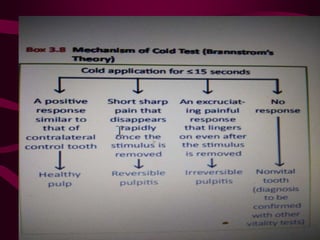
CLINICAL DIAGNOSTIC
METHODS
BY- DR. SONAL BANSAL
DEFINITION
• Diagnosis is the correct determination,
discriminative estimation, and logical
appraisal of conditions found during
examination as evidenced by distinctive
signs, marks, and symptoms.
• Diagnosis is also defined as the art of
distinguishing one disease from another.
HISTORY AND RECORD
• Case history is defined as the data
concerning an individual and his or her
family and environment, including the
individual medical history that may be
useful in analyzing and diagnosing his or
her case or for instructional purposes.
CHIEF COMPLAINT
• Reasons for consulting the clinician.
• Actual expressed complaint – own words.
• Determine the chronology of events that led
to the complaint
• Any other relevant problems
MEDICAL HISTORY
• Thorough & complete medical history.
• Baseline BP & pulse measurement
Evaluate medical conditions that will alter
the dental treatment
 Evaluate medical conditions that have oral
manifestations.
• CVS
• PULMONARY
• GI & RENAL
• HEAMTOLOGIC
• NEUROLOGIC
• ALLERGIES
• CURRENT MEDICATIONS & SIDE EFFECTS
DENTAL HISTORY
• Chronology of events that led up to the
chief complaints is dental history.
• Past and present symptoms
• Trauma & previous dental procedures
• S.O.A.P
SYMPTOMS
• Symptoms are the units of information sought in
clinical diagnosis. They are defined as phenomena
or signs of a departure from the normal state and
are indicative of illness.
Symptoms can be classified as follows:
• Subjective symptoms: Those experienced and
reported to the clinician by the patient
• Objective symptoms: Those ascertained by the
clinician through various tests
PAIN
• Kind of pain
• (a) Sharp, piercing, and lancinating: This type of
painful response is consistent with those usually
associated with excitation of the “Aδ” nerve fibers
in the pulp.
• (b) Dull, boring, gnawing, and excruciating: This
kind of painful response is consistent with those
resulting from excitation and slower rate of
transmission of the “C” nerve fibers in the pulp.
Determining the category of the pain is important
in suggesting the next group of questions to be
Location: The ability to localize the pain is obviously
important.
• (a) Localized pain: Pain is localized when the
patient can point to a specific tooth or site with
assurance and speed when asked to do so. Sharp,
piercing, lancinating pain in a tooth usually
responds promptly to cold and is easy to localize.
Symptoms from such teeth are rarely referred to
other sites.
• (b) Diffuse pain: When the pain is diffuse, however,
the patient describes an area of discomfort rather
than a specific site. When the patient is asked to
point to the most painful spot, the patient’s finger
moves along the dental arch or between the maxilla
and the mandible. This diffuseness is diagnostic
because the inability to localize the pain frequently
relates to dental pain that is dull, boring, and
gnawing, from a tooth that responds abnormally to
heat more than to cold and with symptoms that can
be referred to other sites. The cause of diffuse pain
is because of the fact that proprioceptive fibers are
not located in the pulp
Duration of pain: The duration of the pain is also
diagnostic.
(a) Short and specific to stimuli: At times, pulpal pain
lasts only as long as an irritant is present. Acute
reversible pulpitis (hyperemia) is characterized by
pain of short duration, caused by a specific irritant,
which disappears as soon as the irritant is
removed. The pain is usually localized and is more
responsive to cold than to heat. The pain may
either be intermittent or constant. Clinical
experience has shown that a tooth with fleeting
pulpal pain that disappears on removal of the
irritant has an excellent chance of recovery
• (b) Persistent and lingering: If the pain persists and
if it lasts for minutes to hours after the removal of
the stimuli, the pulpitis will usually be irreversible
and the patient will require endodontic therapy.
• (c) Spontaneous pain: Spontaneous pain is one that
occurs without any apparent cause and is usually a
pain of long duration which is a symptom of
irreversible pulpitis.
• (d) Nocturnal pain: Pain that occurs on changing
the position of the head awakens the patient from
sleep and usually is a symptom of irreversible
pulpitis.
Dentinal Hypersensitivity
• According to Holland et al., dentin hypersensitivity
is characterized by short, sharp pain arising from
exposed dentin in response to stimuli typically
thermal, evaporative, tactile, osmotic or chemical
and which cannot be ascribed to any other form of
dental defect or pathology
Brannstrom’s Hydrodynamic Theory
Stimulus
Fluid flow in exposed open dentinal tubules
Stimulation of Aδ nerve fibers
Pain
Objective Symptoms
• Objective symptoms are determined by tests
and observations performed by the
clinician.
Diagnostic Methods in
Endodontics
1.Visual and tactile inspection
(a) Hard tissues
(b) Soft tissues
(c) Gingiva
(ii) Periodontium
2. percussion
3. palpation
4. mobility and depressibility
5. bite test
6. Magnification
7. radiography (a) Intraoral periapical radiographs (b) Cone beam
computed tomography (CBCT)
• Assessment of pulp vitality
(a) Neural sensibility tests
(i) Thermal tests
(ii) Heat testing
(iii) Cold testing
(iv) Electric pulp tester (EPT)
(v) Anesthetic test
(vi) Test cavity
(b) Pulp vascularity tests
(i) Pulse oximetry
(ii) Laser Doppler flowmetry
(iii) Recent technologies
Dual-wavelength spectrophotometry
Thermography
Crown surface temperature
Transmitted light photoplethysmography
Visual & tactile inspection
• 3 C’s – color, contour , consistency
• Tactile – fingers & explorer
• Dry & well illuminated field
• Soft tissue & periodontal assessment
PERCUSSION
• Tenderness upon percussion indicates some degree of
inflammation in periodontal ligament.
• Not a test of vitality.
• Clinican should randomly and gently tap teeth in
suspected quadrant on all the quadrants.
• Low intensity – finger
• Increasing intensity – mirror handle
• Random percussion to eliminate bias
• However, in chronic periapical inflammation, percussion
yields negative result.
• Horizontal and vertical percussion.
PALPATION
• Uses digital pressure to check for tenderness.
• To check for fluctuation and induration of the soft
tissues.
• Changes in underlying bony architecture.
• Tenderness indicates inflammation in the
periodontal ligament surrounding affected tooth
has spread to the periosteum overlying the
jawbone.
MOBILITY &
DEPRESSIBILITY
• Integrity of attachment apparatus – MOBILITY
• Lateral movement
• DEPRESSIBILITY – vertical movement
• 1st degree – noticeable movement
• 2nd degree - < 1 mm
• 3rd degree - > 1mm
• To differentiate between mobility due to acute
alveolar abscess and periodontal abscess, vitality
tests must be performed.
• In acute alveolar abscess, the tooth will be non
vital.
• Palpation, percussion, mobility, and depressibility
test the integrity of the attachment apparatus, i.e.,
periodontal ligament and bone, and are not
diagnostic when the disease is confined within the
pulp cavity of a tooth.
Bite Test
• The bite test is useful in identifying a cracked
tooth or fractured cusp when pressure is applied
in a certain direction to one cusp or section of the
tooth. The bite test is also helpful in diagnosing
cases wherein the pulpal pathosis has extended
into the periradicular region causing
• Pain on biting → Symptomatic apical
periodontitis
• Pain on release of biting force → Cracked tooth
MAGNIFICATION
• Magnification is an essential requisite in current
precision-based endodontic practice. Initially,
devices that enhanced vision were restricted to
magnification loupes. More recently, the use of
dental operating microscopes has gained
momentum and its applications in endodontic
diagnosis include:
• Locating hidden canals obstructed by calcifications
• Detection of cracks and fractures
TRANSILLUMINATION
• Used to diagnose vertical fracture and cracks in
the crown.
• Uses a fibreoptic illuminating device horizontally
at gingival level.
• If a fracture exists, light will illuminate the side of
the crown that it contacts but the crown on the
opposite side remains dark.
RADIOGRAPHY
• The radiograph is one of the most important
clinical tools in making a diagnosis. It
permits visual examination of the oral
structures that would otherwise be unseen
by the naked eye. Without it, diagnosis,
case selection, treatment, and evaluation of
healing would be impossible.
Intraoral Periapical Radiographs
• To produce an excellent radiograph, one must
master the necessary skills:
• Proper placement of the film in the patient’s
mouth
• Correct angulation of the cone in relation to the
film and oral structures to prevent distortion of the
anatomical images
• Correct exposure time, so that images are recorded
with identifiable contrasts
• Proper developing technique to ensure a clear,
permanent record that can be retained and stored
Cone Beam Computed
Tomography (CBCT)
• Limitations in conventional radiography such as
providing two-dimensional representations of
three-dimensional objects, image distortion, and
superimposition of structures led to the
development of three-dimensional imaging
systems. The introduction of cone beam computed
tomography (CBCT) or cone beam volumetric
tomography (CBVT) imaging facilitated the
transition from 2D to a 3D approach in image
acquisition and interpretation
• Steps in Processing
• According to Scarfe et al., the following are
the steps involved in CBCT image
processing:
• 1. Acquisition configuration
• 2. Image detection
• 3. Image reconstruction
• 4. Image display
Applications in Endodontics
Diagnosis of periradicular lesions, canal
visualization, assessment of internal and external
resorption, detection of root fractures and other
dentoalveolar trauma, preparation for endodontic
surgery, and detection of calcific metamorphosis.
Advantages
• Rapid scan time, beam limitation, image accuracy,
reduced patient radiation dose, interactive display
modes applicable to maxillofacial imaging,
multiplanar reformation, and three-dimensional
volume rendering.
Limitations
• Artifacts (X-ray beam related, patient related,
conebeam related, and scanner related), image
noise, poor soft-tissue contrast.
ASSESSMENT OF PULP
VITALITY
• Most common methods clinically employed assess
neural sensitivity, while the recent research and
developments are trying to find a clinical method
of vascularity assessment.
NEURAL SENSIBILITY TESTS
• These are the tests that indirectly tell us about the
vitality status of the pulp. They work on the
principle of stimulating the neural fibers present in
the pulp
THERMAL PULP TESTS
• Involves application of cold and heat to a tooth.
• Used to :
1. Isolate the offending tooth.
2. To determine whether the tooth is vital or
nonvital.
 The cold test is used in differentiating between
reversible and irreversible pulpitis.
 A response to cold indicates a vital pulp,
regardless of whether it is normal or abnormal.
• An abnormal response to heat usually indicates the
presence of a pulpal or periodontal disorder
requiring endodontic treatment.
• Neither of the tests is totally reliable but can
provide useful information in cases of pulpal
involvement.
• Cold test :
• Uses ice sticks, compressed gases, carbon di oxide
snow.
• Carbon dioxide snow is the most effective method
of eliciting response in vital teeth.
• Isolating and bathing each tooth with ice water is
will elicit the most accurate response because it
simultaneously cools all the surfaces of the tooth.
• When a reaction to cold occurs, the patient can
quickly point to the offending tooth.
• Heat test:
• Commonly used methods are warm sticks
of gutta percha and hot water bath.
• The heat response can be localized or
diffuse or referred to a different site.
• Four possible responses include:
1. No response – non vital tooth or false negative due to
calcifications, immature apex, recent trauma or
premedication.
2. Mild to moderate degree of awareness that subsides
within 1-2 second after stimulus has been removed -
within normal limits.
3. Strong momentary painful response that subsides
within 1-2 sec after stimulus has been removed –
reversible pulpitis.
4. Moderate to strong painful response that lingers for
several seconds or longer after stimulus has been
removed – irreversible pulpitis.
Heat application for ≤5 seconds
Vasodilatation NO RESPONSE
Increased intrapulpal pressure
Reduced neural excitation threshold
Immediate excruciating painful response which
is significantly different from the contralateral
control tooth OR A painful response that lingers
on even after the removal of the heat stimulus
Irreversible pulpitis
Positive response similar to
contralateral control tooth Healthy
state of the pulp
Nonvital tooth (diagnosis to
be confirmed with other
vitality tests)
Electric Pulp Test (EPT)
• The electric pulp test is one of the tests used to
determine pulp vitality. The electric pulp tester ,
when testing for pulp vitality, uses nerve
stimulation.
• The objective is to stimulate a pulpal response by
subjecting the tooth to an increasing degree of
electric current. A positive response is an
indication of vitality and helps in determining the
normality or abnormality of that pulp. No response
to the electrical stimulus can be an indication of
pulp necrosis
• The procedure for electrical pulp testing can be
performed in the following simple steps: Describe
the test to the patient in a way that will reduce
anxiety and will eliminate a biased response.
• Isolate the area of control tooth and the tooth to be
tested with cotton rolls and a saliva ejector, and air-
dry all the teeth. Check the electric pulp tester for
function and determine that current is passing
through the electrode.
• The test is always performed on a control tooth
prior to testing the tooth in question. Apply an
electrolyte (prophy paste or toothpaste) on the
tooth electrode and place it against the dried
enamel of the crown’s occlusobuccal or
incisolabial surface. It is important to avoid
contacting any restorations in the tooth or the
adjacent gingival tissue with the electrolyte or the
electrode; this would cause a false and misleading
response.
• Location of probe tip: The placement of the tester is critical to ensure
accurate response from the tooth. –
• Anterior teeth incisal third
• Posterior teeth mid-third of the mesiobuccal cusp of molars and
buccal cusp of premolars
• Completion of the circuit:
• Retract the patient’s cheek away from the tooth electrode and the
electrical circuit is completed by either: – A ground wire (lip clip) is
placed over the patient’s lip in contact with the oral mucosa – Or, the
clinician instructs the patient to rest a finger on the metal sheath of the
pulp tester Turn the rheostat slowly to introduce minimal current into
the tooth and increase the current slowly. Ask the patient to indicate
when sensation occurs by using such words as “tingling” or
“warmth.” Record the result according to the numeric scale on the
pulp tester. Repeat the foregoing for each tooth to be tested.
Normal response: A positive response is a response
that occurs at the same neural excitation threshold
as the control tooth. For example, during the
assessment of a maxillary left lateral incisor, the
control tooth would be the maxillary right lateral
incisor. The test tooth would be considered normal
and vital if both the teeth exhibit a positive
response at a similar numerical value of the EPT.
Negative response: This denotes a nonvital tooth,
which fails to respond even when the tester is set to
the highest electrical excitation value.
• Early response: This denotes a diseased
state of pulp as the tooth responds to a
threshold which is less than that of the
control tooth.
• Delayed response: This also denotes a
diseased state of the pulp wherein the tooth
responds at a significantly higher electrical
excitation level than compared to the
control tooth.
• A false positive can occur when moist gangrenous
pulp is present, patient anxiety, saliva conducting
stimulus, metallic restorations conducting stimulus
to adjacent teeth.
• In multirooted tooth pulp can be partially necrotic
leading confusion.
• A false negative test can occur in extensive
calcification , teeth with extensive restoration,
patients with high pain threshold, recently
traumatized teeth, incomplete root formation.
• EPT is an imperfect though useful way to
determine pulpal status of tooth.
• In case of a periapical radiolucency, EPT
determines whether pulp is vital.
• When used with thermal and periodontal testing,
EPT can help differentiate pulpal disease from
periodontal or nonodontogenic causes.
SELECTIVE ANAESTHESIA
TEST
• Intraligamentary anaesthesia is used when the
clinician has determined through prior testing, the
offending tooth, and the patient reports severe
lingering residual pain as a result of thermal testing.
• Use of this test breaks the cycle of pain for several
mins and reconfirms through elimination of pain the
diagnosis.
• If the pain still continues, the cause may be
heterotropic pain and non odnotogenic causes should
be considered.
TEST CAVITY
• Occasionally the clinician will encounter mixed
responses to pulp testing, where the most accurate
technique to discover a pulp is vital is the test cavity.
• A preparation is made without anesthetizing and if a
vital response is elicited cavity should be restored
• When No response is seen, endodontic therapy is
completed.
• Although damage can be repaired, this is not a
reversible procedure.
• Hence is reserved as a last resort to diagnosis.
LASER DOPPLER
FLOWMETRY
• Uses a laser beam of known wavelength that is
directed through the crown of the tooth to the blood
vessels in the pulp.
• Moving blood vessels causes the laser beam to be
doppler shifted and reflected which is detected by a
photocell, output of which is proportional to the
number and velocity of the blood vessels..
• It is an objective measurement of the pulp vitality
unlike other tests.
• Limitations include equipment costs, use of multiple
probes etc.
• The position on the crown and the location of the
pulp within the tooth cause variation in pulpal
blood flow measurement.
• Anti hypertensive medication and nicotine may
affect bolld flow to the pulp producing inaccurate
results.
PULSE OXIMETRY
• Widely used technique for recording blood oxygen
saturation levels.
• Increased acidity metabolic rate produced by
inflammation cause deoxygenation of hemoglobin
and change the oxygen saturation of blood.
• Can detect pulpal inflammation or partial necrosis
in teeth that are still vital.
Thank you

CLINICAL DIAGNOSTIC METHOD aids and advantages

  • 1.
  • 2.
    DEFINITION • Diagnosis isthe correct determination, discriminative estimation, and logical appraisal of conditions found during examination as evidenced by distinctive signs, marks, and symptoms. • Diagnosis is also defined as the art of distinguishing one disease from another.
  • 3.
    HISTORY AND RECORD •Case history is defined as the data concerning an individual and his or her family and environment, including the individual medical history that may be useful in analyzing and diagnosing his or her case or for instructional purposes.
  • 4.
    CHIEF COMPLAINT • Reasonsfor consulting the clinician. • Actual expressed complaint – own words. • Determine the chronology of events that led to the complaint • Any other relevant problems
  • 5.
    MEDICAL HISTORY • Thorough& complete medical history. • Baseline BP & pulse measurement Evaluate medical conditions that will alter the dental treatment  Evaluate medical conditions that have oral manifestations.
  • 6.
    • CVS • PULMONARY •GI & RENAL • HEAMTOLOGIC • NEUROLOGIC • ALLERGIES • CURRENT MEDICATIONS & SIDE EFFECTS
  • 7.
    DENTAL HISTORY • Chronologyof events that led up to the chief complaints is dental history. • Past and present symptoms • Trauma & previous dental procedures • S.O.A.P
  • 8.
    SYMPTOMS • Symptoms arethe units of information sought in clinical diagnosis. They are defined as phenomena or signs of a departure from the normal state and are indicative of illness. Symptoms can be classified as follows: • Subjective symptoms: Those experienced and reported to the clinician by the patient • Objective symptoms: Those ascertained by the clinician through various tests
  • 9.
    PAIN • Kind ofpain • (a) Sharp, piercing, and lancinating: This type of painful response is consistent with those usually associated with excitation of the “Aδ” nerve fibers in the pulp. • (b) Dull, boring, gnawing, and excruciating: This kind of painful response is consistent with those resulting from excitation and slower rate of transmission of the “C” nerve fibers in the pulp. Determining the category of the pain is important in suggesting the next group of questions to be
  • 10.
    Location: The abilityto localize the pain is obviously important. • (a) Localized pain: Pain is localized when the patient can point to a specific tooth or site with assurance and speed when asked to do so. Sharp, piercing, lancinating pain in a tooth usually responds promptly to cold and is easy to localize. Symptoms from such teeth are rarely referred to other sites.
  • 11.
    • (b) Diffusepain: When the pain is diffuse, however, the patient describes an area of discomfort rather than a specific site. When the patient is asked to point to the most painful spot, the patient’s finger moves along the dental arch or between the maxilla and the mandible. This diffuseness is diagnostic because the inability to localize the pain frequently relates to dental pain that is dull, boring, and gnawing, from a tooth that responds abnormally to heat more than to cold and with symptoms that can be referred to other sites. The cause of diffuse pain is because of the fact that proprioceptive fibers are not located in the pulp
  • 12.
    Duration of pain:The duration of the pain is also diagnostic. (a) Short and specific to stimuli: At times, pulpal pain lasts only as long as an irritant is present. Acute reversible pulpitis (hyperemia) is characterized by pain of short duration, caused by a specific irritant, which disappears as soon as the irritant is removed. The pain is usually localized and is more responsive to cold than to heat. The pain may either be intermittent or constant. Clinical experience has shown that a tooth with fleeting pulpal pain that disappears on removal of the irritant has an excellent chance of recovery
  • 13.
    • (b) Persistentand lingering: If the pain persists and if it lasts for minutes to hours after the removal of the stimuli, the pulpitis will usually be irreversible and the patient will require endodontic therapy. • (c) Spontaneous pain: Spontaneous pain is one that occurs without any apparent cause and is usually a pain of long duration which is a symptom of irreversible pulpitis. • (d) Nocturnal pain: Pain that occurs on changing the position of the head awakens the patient from sleep and usually is a symptom of irreversible pulpitis.
  • 14.
    Dentinal Hypersensitivity • Accordingto Holland et al., dentin hypersensitivity is characterized by short, sharp pain arising from exposed dentin in response to stimuli typically thermal, evaporative, tactile, osmotic or chemical and which cannot be ascribed to any other form of dental defect or pathology
  • 16.
    Brannstrom’s Hydrodynamic Theory Stimulus Fluidflow in exposed open dentinal tubules Stimulation of Aδ nerve fibers Pain
  • 17.
    Objective Symptoms • Objectivesymptoms are determined by tests and observations performed by the clinician.
  • 18.
    Diagnostic Methods in Endodontics 1.Visualand tactile inspection (a) Hard tissues (b) Soft tissues (c) Gingiva (ii) Periodontium 2. percussion 3. palpation 4. mobility and depressibility 5. bite test 6. Magnification 7. radiography (a) Intraoral periapical radiographs (b) Cone beam computed tomography (CBCT)
  • 19.
    • Assessment ofpulp vitality (a) Neural sensibility tests (i) Thermal tests (ii) Heat testing (iii) Cold testing (iv) Electric pulp tester (EPT) (v) Anesthetic test (vi) Test cavity (b) Pulp vascularity tests (i) Pulse oximetry (ii) Laser Doppler flowmetry (iii) Recent technologies Dual-wavelength spectrophotometry Thermography Crown surface temperature Transmitted light photoplethysmography
  • 20.
    Visual & tactileinspection • 3 C’s – color, contour , consistency • Tactile – fingers & explorer • Dry & well illuminated field • Soft tissue & periodontal assessment
  • 21.
    PERCUSSION • Tenderness uponpercussion indicates some degree of inflammation in periodontal ligament. • Not a test of vitality. • Clinican should randomly and gently tap teeth in suspected quadrant on all the quadrants. • Low intensity – finger • Increasing intensity – mirror handle • Random percussion to eliminate bias • However, in chronic periapical inflammation, percussion yields negative result. • Horizontal and vertical percussion.
  • 23.
    PALPATION • Uses digitalpressure to check for tenderness. • To check for fluctuation and induration of the soft tissues. • Changes in underlying bony architecture. • Tenderness indicates inflammation in the periodontal ligament surrounding affected tooth has spread to the periosteum overlying the jawbone.
  • 25.
    MOBILITY & DEPRESSIBILITY • Integrityof attachment apparatus – MOBILITY • Lateral movement • DEPRESSIBILITY – vertical movement • 1st degree – noticeable movement • 2nd degree - < 1 mm • 3rd degree - > 1mm
  • 27.
    • To differentiatebetween mobility due to acute alveolar abscess and periodontal abscess, vitality tests must be performed. • In acute alveolar abscess, the tooth will be non vital. • Palpation, percussion, mobility, and depressibility test the integrity of the attachment apparatus, i.e., periodontal ligament and bone, and are not diagnostic when the disease is confined within the pulp cavity of a tooth.
  • 28.
    Bite Test • Thebite test is useful in identifying a cracked tooth or fractured cusp when pressure is applied in a certain direction to one cusp or section of the tooth. The bite test is also helpful in diagnosing cases wherein the pulpal pathosis has extended into the periradicular region causing • Pain on biting → Symptomatic apical periodontitis • Pain on release of biting force → Cracked tooth
  • 30.
    MAGNIFICATION • Magnification isan essential requisite in current precision-based endodontic practice. Initially, devices that enhanced vision were restricted to magnification loupes. More recently, the use of dental operating microscopes has gained momentum and its applications in endodontic diagnosis include: • Locating hidden canals obstructed by calcifications • Detection of cracks and fractures
  • 31.
    TRANSILLUMINATION • Used todiagnose vertical fracture and cracks in the crown. • Uses a fibreoptic illuminating device horizontally at gingival level. • If a fracture exists, light will illuminate the side of the crown that it contacts but the crown on the opposite side remains dark.
  • 32.
    RADIOGRAPHY • The radiographis one of the most important clinical tools in making a diagnosis. It permits visual examination of the oral structures that would otherwise be unseen by the naked eye. Without it, diagnosis, case selection, treatment, and evaluation of healing would be impossible.
  • 33.
    Intraoral Periapical Radiographs •To produce an excellent radiograph, one must master the necessary skills: • Proper placement of the film in the patient’s mouth • Correct angulation of the cone in relation to the film and oral structures to prevent distortion of the anatomical images • Correct exposure time, so that images are recorded with identifiable contrasts • Proper developing technique to ensure a clear, permanent record that can be retained and stored
  • 34.
    Cone Beam Computed Tomography(CBCT) • Limitations in conventional radiography such as providing two-dimensional representations of three-dimensional objects, image distortion, and superimposition of structures led to the development of three-dimensional imaging systems. The introduction of cone beam computed tomography (CBCT) or cone beam volumetric tomography (CBVT) imaging facilitated the transition from 2D to a 3D approach in image acquisition and interpretation
  • 35.
    • Steps inProcessing • According to Scarfe et al., the following are the steps involved in CBCT image processing: • 1. Acquisition configuration • 2. Image detection • 3. Image reconstruction • 4. Image display
  • 36.
    Applications in Endodontics Diagnosisof periradicular lesions, canal visualization, assessment of internal and external resorption, detection of root fractures and other dentoalveolar trauma, preparation for endodontic surgery, and detection of calcific metamorphosis.
  • 37.
    Advantages • Rapid scantime, beam limitation, image accuracy, reduced patient radiation dose, interactive display modes applicable to maxillofacial imaging, multiplanar reformation, and three-dimensional volume rendering. Limitations • Artifacts (X-ray beam related, patient related, conebeam related, and scanner related), image noise, poor soft-tissue contrast.
  • 38.
    ASSESSMENT OF PULP VITALITY •Most common methods clinically employed assess neural sensitivity, while the recent research and developments are trying to find a clinical method of vascularity assessment. NEURAL SENSIBILITY TESTS • These are the tests that indirectly tell us about the vitality status of the pulp. They work on the principle of stimulating the neural fibers present in the pulp
  • 39.
    THERMAL PULP TESTS •Involves application of cold and heat to a tooth. • Used to : 1. Isolate the offending tooth. 2. To determine whether the tooth is vital or nonvital.  The cold test is used in differentiating between reversible and irreversible pulpitis.  A response to cold indicates a vital pulp, regardless of whether it is normal or abnormal.
  • 40.
    • An abnormalresponse to heat usually indicates the presence of a pulpal or periodontal disorder requiring endodontic treatment. • Neither of the tests is totally reliable but can provide useful information in cases of pulpal involvement. • Cold test : • Uses ice sticks, compressed gases, carbon di oxide snow. • Carbon dioxide snow is the most effective method of eliciting response in vital teeth.
  • 41.
    • Isolating andbathing each tooth with ice water is will elicit the most accurate response because it simultaneously cools all the surfaces of the tooth. • When a reaction to cold occurs, the patient can quickly point to the offending tooth.
  • 43.
    • Heat test: •Commonly used methods are warm sticks of gutta percha and hot water bath. • The heat response can be localized or diffuse or referred to a different site.
  • 46.
    • Four possibleresponses include: 1. No response – non vital tooth or false negative due to calcifications, immature apex, recent trauma or premedication. 2. Mild to moderate degree of awareness that subsides within 1-2 second after stimulus has been removed - within normal limits. 3. Strong momentary painful response that subsides within 1-2 sec after stimulus has been removed – reversible pulpitis. 4. Moderate to strong painful response that lingers for several seconds or longer after stimulus has been removed – irreversible pulpitis.
  • 47.
    Heat application for≤5 seconds Vasodilatation NO RESPONSE Increased intrapulpal pressure Reduced neural excitation threshold Immediate excruciating painful response which is significantly different from the contralateral control tooth OR A painful response that lingers on even after the removal of the heat stimulus Irreversible pulpitis Positive response similar to contralateral control tooth Healthy state of the pulp Nonvital tooth (diagnosis to be confirmed with other vitality tests)
  • 48.
    Electric Pulp Test(EPT) • The electric pulp test is one of the tests used to determine pulp vitality. The electric pulp tester , when testing for pulp vitality, uses nerve stimulation. • The objective is to stimulate a pulpal response by subjecting the tooth to an increasing degree of electric current. A positive response is an indication of vitality and helps in determining the normality or abnormality of that pulp. No response to the electrical stimulus can be an indication of pulp necrosis
  • 49.
    • The procedurefor electrical pulp testing can be performed in the following simple steps: Describe the test to the patient in a way that will reduce anxiety and will eliminate a biased response. • Isolate the area of control tooth and the tooth to be tested with cotton rolls and a saliva ejector, and air- dry all the teeth. Check the electric pulp tester for function and determine that current is passing through the electrode.
  • 50.
    • The testis always performed on a control tooth prior to testing the tooth in question. Apply an electrolyte (prophy paste or toothpaste) on the tooth electrode and place it against the dried enamel of the crown’s occlusobuccal or incisolabial surface. It is important to avoid contacting any restorations in the tooth or the adjacent gingival tissue with the electrolyte or the electrode; this would cause a false and misleading response.
  • 51.
    • Location ofprobe tip: The placement of the tester is critical to ensure accurate response from the tooth. – • Anterior teeth incisal third • Posterior teeth mid-third of the mesiobuccal cusp of molars and buccal cusp of premolars • Completion of the circuit: • Retract the patient’s cheek away from the tooth electrode and the electrical circuit is completed by either: – A ground wire (lip clip) is placed over the patient’s lip in contact with the oral mucosa – Or, the clinician instructs the patient to rest a finger on the metal sheath of the pulp tester Turn the rheostat slowly to introduce minimal current into the tooth and increase the current slowly. Ask the patient to indicate when sensation occurs by using such words as “tingling” or “warmth.” Record the result according to the numeric scale on the pulp tester. Repeat the foregoing for each tooth to be tested.
  • 53.
    Normal response: Apositive response is a response that occurs at the same neural excitation threshold as the control tooth. For example, during the assessment of a maxillary left lateral incisor, the control tooth would be the maxillary right lateral incisor. The test tooth would be considered normal and vital if both the teeth exhibit a positive response at a similar numerical value of the EPT. Negative response: This denotes a nonvital tooth, which fails to respond even when the tester is set to the highest electrical excitation value.
  • 54.
    • Early response:This denotes a diseased state of pulp as the tooth responds to a threshold which is less than that of the control tooth. • Delayed response: This also denotes a diseased state of the pulp wherein the tooth responds at a significantly higher electrical excitation level than compared to the control tooth.
  • 55.
    • A falsepositive can occur when moist gangrenous pulp is present, patient anxiety, saliva conducting stimulus, metallic restorations conducting stimulus to adjacent teeth. • In multirooted tooth pulp can be partially necrotic leading confusion. • A false negative test can occur in extensive calcification , teeth with extensive restoration, patients with high pain threshold, recently traumatized teeth, incomplete root formation.
  • 56.
    • EPT isan imperfect though useful way to determine pulpal status of tooth. • In case of a periapical radiolucency, EPT determines whether pulp is vital. • When used with thermal and periodontal testing, EPT can help differentiate pulpal disease from periodontal or nonodontogenic causes.
  • 57.
    SELECTIVE ANAESTHESIA TEST • Intraligamentaryanaesthesia is used when the clinician has determined through prior testing, the offending tooth, and the patient reports severe lingering residual pain as a result of thermal testing. • Use of this test breaks the cycle of pain for several mins and reconfirms through elimination of pain the diagnosis. • If the pain still continues, the cause may be heterotropic pain and non odnotogenic causes should be considered.
  • 58.
    TEST CAVITY • Occasionallythe clinician will encounter mixed responses to pulp testing, where the most accurate technique to discover a pulp is vital is the test cavity. • A preparation is made without anesthetizing and if a vital response is elicited cavity should be restored • When No response is seen, endodontic therapy is completed. • Although damage can be repaired, this is not a reversible procedure. • Hence is reserved as a last resort to diagnosis.
  • 59.
    LASER DOPPLER FLOWMETRY • Usesa laser beam of known wavelength that is directed through the crown of the tooth to the blood vessels in the pulp. • Moving blood vessels causes the laser beam to be doppler shifted and reflected which is detected by a photocell, output of which is proportional to the number and velocity of the blood vessels.. • It is an objective measurement of the pulp vitality unlike other tests. • Limitations include equipment costs, use of multiple probes etc.
  • 60.
    • The positionon the crown and the location of the pulp within the tooth cause variation in pulpal blood flow measurement. • Anti hypertensive medication and nicotine may affect bolld flow to the pulp producing inaccurate results.
  • 61.
    PULSE OXIMETRY • Widelyused technique for recording blood oxygen saturation levels. • Increased acidity metabolic rate produced by inflammation cause deoxygenation of hemoglobin and change the oxygen saturation of blood. • Can detect pulpal inflammation or partial necrosis in teeth that are still vital.
  • 62.