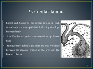
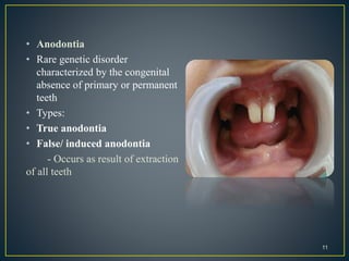
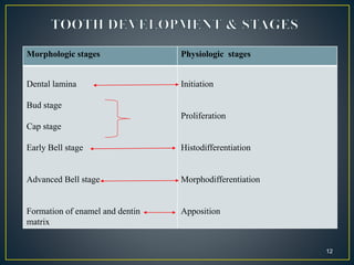
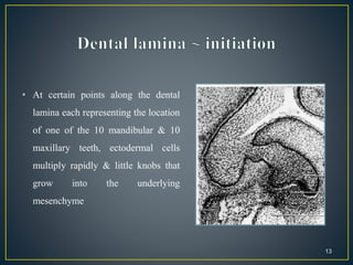
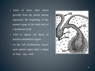
This document summarizes the process of tooth development from the dental lamina through the various stages to clinical considerations. It begins with the dental lamina and vestibular lamina developing from the oral ectoderm. Tooth buds then develop from the dental lamina, going through the bud, cap and bell stages as the enamel organ, dental papilla and follicle form. Root development occurs after crown formation. The stages and common anomalies in tooth development are described to provide dentists with an understanding of this complex process.
Introduction
Initiation of tooth development
Development of tooth
Stages of tooth development
Histophysiology
Development of enamel
Development of dentin
Development of pulp
Development of cementum
Development of root
Nerve and vascular supply during early development
Formation of the permanent dentition
Formation of supporting tissues
Tooth eruption
Theories of tooth eruption.
Developmental anomalies of tooth
References
For first year dental student, i got this from the internet.. Hope this can help u guys understand more about the tooth development.. Btw, good luck for minitest OB tomorrow.. No sacrifice, no victory!
Indian Dental Academy: will be one of the most relevant and exciting training center with best faculty and flexible training programs for dental professionals who wish to advance in their dental practice,Offers certified courses in Dental implants,Orthodontics,Endodontics,Cosmetic Dentistry, Prosthetic Dentistry, Periodontics and General Dentistry.
Growth & development of tooth & tongue/ dental crown & bridge coursesIndian dental academy
The Indian Dental Academy is the Leader in continuing dental education , training dentists in all aspects of dentistry and
offering a wide range of dental certified courses in different formats.for more details please visit
www.indiandentalacademy.com
BE UPDATE TO IT,, AS IT IS 3 years back from 2017
Kindly mail me if you feel, needy of this presentation
you can find my mail id @ slide share,,, if not mail me @
sukesh3567@gmail.com.
Good luck
Tooth development 3 /certified fixed orthodontic courses by Indian denta...Indian dental academy
The Indian Dental Academy is the Leader in
continuing dental education , training dentists
in all aspects of dentistry and offering a wide
range of dental certified courses in different
formats.
Indian dental academy provides dental crown &
Bridge,rotary endodontics,fixed orthodontics,
Dental implants courses.for details pls visit
www.indiandentalacademy.com ,or call
0091-9248678078
The Indian Dental Academy is the Leader in continuing dental education , training dentists in all aspects of dentistry and
offering a wide range of dental certified courses in different formats.for more details please visit
www.indiandentalacademy.com
The Indian Dental Academy is the Leader in continuing dental education , training dentists in all aspects of dentistry and
offering a wide range of dental certified courses in different formats.for more details please visit
www.indiandentalacademy.com
it describes tooth development and anomalies associated with tooth bud development along with other developmental stages of tooth development and some important terminologies.
The development of dentition is an interesting and a crucial topic for dentists. This presentation explains this topic in detail along with providing information on pathologies associated with each stage.
The Indian Dental Academy is the Leader in continuing dental education , training dentists in all aspects of dentistry and
offering a wide range of dental certified courses in different formats.
Indian Dental Academy: will be one of the most relevant and exciting training center with best faculty and flexible training programs for dental professionals who wish to advance in their dental practice,Offers certified courses in Dental implants,Orthodontics,Endodontics,Cosmetic Dentistry, Prosthetic Dentistry, Periodontics and General Dentistry.
Title: Sense of Smell
Presenter: Dr. Faiza, Assistant Professor of Physiology
Qualifications:
MBBS (Best Graduate, AIMC Lahore)
FCPS Physiology
ICMT, CHPE, DHPE (STMU)
MPH (GC University, Faisalabad)
MBA (Virtual University of Pakistan)
Learning Objectives:
Describe the primary categories of smells and the concept of odor blindness.
Explain the structure and location of the olfactory membrane and mucosa, including the types and roles of cells involved in olfaction.
Describe the pathway and mechanisms of olfactory signal transmission from the olfactory receptors to the brain.
Illustrate the biochemical cascade triggered by odorant binding to olfactory receptors, including the role of G-proteins and second messengers in generating an action potential.
Identify different types of olfactory disorders such as anosmia, hyposmia, hyperosmia, and dysosmia, including their potential causes.
Key Topics:
Olfactory Genes:
3% of the human genome accounts for olfactory genes.
400 genes for odorant receptors.
Olfactory Membrane:
Located in the superior part of the nasal cavity.
Medially: Folds downward along the superior septum.
Laterally: Folds over the superior turbinate and upper surface of the middle turbinate.
Total surface area: 5-10 square centimeters.
Olfactory Mucosa:
Olfactory Cells: Bipolar nerve cells derived from the CNS (100 million), with 4-25 olfactory cilia per cell.
Sustentacular Cells: Produce mucus and maintain ionic and molecular environment.
Basal Cells: Replace worn-out olfactory cells with an average lifespan of 1-2 months.
Bowman’s Gland: Secretes mucus.
Stimulation of Olfactory Cells:
Odorant dissolves in mucus and attaches to receptors on olfactory cilia.
Involves a cascade effect through G-proteins and second messengers, leading to depolarization and action potential generation in the olfactory nerve.
Quality of a Good Odorant:
Small (3-20 Carbon atoms), volatile, water-soluble, and lipid-soluble.
Facilitated by odorant-binding proteins in mucus.
Membrane Potential and Action Potential:
Resting membrane potential: -55mV.
Action potential frequency in the olfactory nerve increases with odorant strength.
Adaptation Towards the Sense of Smell:
Rapid adaptation within the first second, with further slow adaptation.
Psychological adaptation greater than receptor adaptation, involving feedback inhibition from the central nervous system.
Primary Sensations of Smell:
Camphoraceous, Musky, Floral, Pepperminty, Ethereal, Pungent, Putrid.
Odor Detection Threshold:
Examples: Hydrogen sulfide (0.0005 ppm), Methyl-mercaptan (0.002 ppm).
Some toxic substances are odorless at lethal concentrations.
Characteristics of Smell:
Odor blindness for single substances due to lack of appropriate receptor protein.
Behavioral and emotional influences of smell.
Transmission of Olfactory Signals:
From olfactory cells to glomeruli in the olfactory bulb, involving lateral inhibition.
Primitive, less old, and new olfactory systems with different path
MANAGEMENT OF ATRIOVENTRICULAR CONDUCTION BLOCK.pdfJim Jacob Roy
Cardiac conduction defects can occur due to various causes.
Atrioventricular conduction blocks ( AV blocks ) are classified into 3 types.
This document describes the acute management of AV block.
Couples presenting to the infertility clinic- Do they really have infertility...Sujoy Dasgupta
Dr Sujoy Dasgupta presented the study on "Couples presenting to the infertility clinic- Do they really have infertility? – The unexplored stories of non-consummation" in the 13th Congress of the Asia Pacific Initiative on Reproduction (ASPIRE 2024) at Manila on 24 May, 2024.
Pulmonary Thromboembolism - etilogy, types, medical- Surgical and nursing man...VarunMahajani
Disruption of blood supply to lung alveoli due to blockage of one or more pulmonary blood vessels is called as Pulmonary thromboembolism. In this presentation we will discuss its causes, types and its management in depth.
micro teaching on communication m.sc nursing.pdfAnurag Sharma
Microteaching is a unique model of practice teaching. It is a viable instrument for the. desired change in the teaching behavior or the behavior potential which, in specified types of real. classroom situations, tends to facilitate the achievement of specified types of objectives.
Explore natural remedies for syphilis treatment in Singapore. Discover alternative therapies, herbal remedies, and lifestyle changes that may complement conventional treatments. Learn about holistic approaches to managing syphilis symptoms and supporting overall health.
- Video recording of this lecture in English language: https://youtu.be/lK81BzxMqdo
- Video recording of this lecture in Arabic language: https://youtu.be/Ve4P0COk9OI
- Link to download the book free: https://nephrotube.blogspot.com/p/nephrotube-nephrology-books.html
- Link to NephroTube website: www.NephroTube.com
- Link to NephroTube social media accounts: https://nephrotube.blogspot.com/p/join-nephrotube-on-social-media.html
Tom Selleck Health: A Comprehensive Look at the Iconic Actor’s Wellness Journeygreendigital
Tom Selleck, an enduring figure in Hollywood. has captivated audiences for decades with his rugged charm, iconic moustache. and memorable roles in television and film. From his breakout role as Thomas Magnum in Magnum P.I. to his current portrayal of Frank Reagan in Blue Bloods. Selleck's career has spanned over 50 years. But beyond his professional achievements. fans have often been curious about Tom Selleck Health. especially as he has aged in the public eye.
Follow us on: Pinterest
Introduction
Many have been interested in Tom Selleck health. not only because of his enduring presence on screen but also because of the challenges. and lifestyle choices he has faced and made over the years. This article delves into the various aspects of Tom Selleck health. exploring his fitness regimen, diet, mental health. and the challenges he has encountered as he ages. We'll look at how he maintains his well-being. the health issues he has faced, and his approach to ageing .
Early Life and Career
Childhood and Athletic Beginnings
Tom Selleck was born on January 29, 1945, in Detroit, Michigan, and grew up in Sherman Oaks, California. From an early age, he was involved in sports, particularly basketball. which played a significant role in his physical development. His athletic pursuits continued into college. where he attended the University of Southern California (USC) on a basketball scholarship. This early involvement in sports laid a strong foundation for his physical health and disciplined lifestyle.
Transition to Acting
Selleck's transition from an athlete to an actor came with its physical demands. His first significant role in "Magnum P.I." required him to perform various stunts and maintain a fit appearance. This role, which he played from 1980 to 1988. necessitated a rigorous fitness routine to meet the show's demands. setting the stage for his long-term commitment to health and wellness.
Fitness Regimen
Workout Routine
Tom Selleck health and fitness regimen has evolved. adapting to his changing roles and age. During his "Magnum, P.I." days. Selleck's workouts were intense and focused on building and maintaining muscle mass. His routine included weightlifting, cardiovascular exercises. and specific training for the stunts he performed on the show.
Selleck adjusted his fitness routine as he aged to suit his body's needs. Today, his workouts focus on maintaining flexibility, strength, and cardiovascular health. He incorporates low-impact exercises such as swimming, walking, and light weightlifting. This balanced approach helps him stay fit without putting undue strain on his joints and muscles.
Importance of Flexibility and Mobility
In recent years, Selleck has emphasized the importance of flexibility and mobility in his fitness regimen. Understanding the natural decline in muscle mass and joint flexibility with age. he includes stretching and yoga in his routine. These practices help prevent injuries, improve posture, and maintain mobilit
These simplified slides by Dr. Sidra Arshad present an overview of the non-respiratory functions of the respiratory tract.
Learning objectives:
1. Enlist the non-respiratory functions of the respiratory tract
2. Briefly explain how these functions are carried out
3. Discuss the significance of dead space
4. Differentiate between minute ventilation and alveolar ventilation
5. Describe the cough and sneeze reflexes
Study Resources:
1. Chapter 39, Guyton and Hall Textbook of Medical Physiology, 14th edition
2. Chapter 34, Ganong’s Review of Medical Physiology, 26th edition
3. Chapter 17, Human Physiology by Lauralee Sherwood, 9th edition
4. Non-respiratory functions of the lungs https://academic.oup.com/bjaed/article/13/3/98/278874
The prostate is an exocrine gland of the male mammalian reproductive system
It is a walnut-sized gland that forms part of the male reproductive system and is located in front of the rectum and just below the urinary bladder
Function is to store and secrete a clear, slightly alkaline fluid that constitutes 10-30% of the volume of the seminal fluid that along with the spermatozoa, constitutes semen
A healthy human prostate measures (4cm-vertical, by 3cm-horizontal, 2cm ant-post ).
It surrounds the urethra just below the urinary bladder. It has anterior, median, posterior and two lateral lobes
It’s work is regulated by androgens which are responsible for male sex characteristics
Generalised disease of the prostate due to hormonal derangement which leads to non malignant enlargement of the gland (increase in the number of epithelial cells and stromal tissue)to cause compression of the urethra leading to symptoms (LUTS
3. • Development of tooth is a complex process, in which reciprocal and
sequential interaction between the oral epithelium and mesenchymal
cells, regulate cell activities like proliferation, condensation, adhesion,
migration, differentiation and secretion, which leads to the formation
of a functional tooth organ.
3
4. • Teeth are derived from oral ectoderm and neural crest
ectomesenchyme.
• Each developing tooth grows as an anatomically distinct
unit.
• The fundamental developmental process remains the
same for all teeth.
4
5. • After the formation of the neural tube a group of cells
separates from the neural ectoderm called as neural
crest cells.
• These cells have the capacity to migrate and differentiate
extensively with in the developing embryo.
• Failure of neural crest cells to migrate normally to
appropriate places during the craniofacial development
leads to defects like, absence of teeth and under
developed jaw bones.
5
6. • The primitive oral cavity or
stomodeum- lined by –
stratified squamous epithelium
called the oral ectoderm
(primitive oral epithelium)
• Most of the connective tissue
cells – neural crest cells.
• They induce the overlying
ectoderm to start tooth
development.
6
7. • When the embryo is 6 weeks old- certain
areas of basal cells proliferate more rapidly
than adjacent areas
primary epithelial band.
• At 7th week-divides into:
• Inner (lingual) process- dental lamina
• Outer (buccal process)- vestibular lamina
7
8. • It serves as a primordium for the ectodermal portion of the deciduous
teeth.
• During the development of jaws the permanent molars arise directly from
the distal extension of the dental lamina.
8
9. • Labial and buccal to the dental lamina in each
dental arch, another epithelial thickening develops
independently
• It is Vestibular Lamina also termed as lip furrow
band
• Subsequently hollows and form the oral vestibule
between the alveolar portion of the jaws and the
lips and cheeks.
9
11. • Anodontia
• Rare genetic disorder
characterized by the congenital
absence of primary or permanent
teeth
• Types:
• True anodontia
• False/ induced anodontia
- Occurs as result of extraction
of all teeth
11
12. 12
Morphologic stages Physiologic stages
Dental lamina
Bud stage
Cap stage
Early Bell stage
Advanced Bell stage
Formation of enamel and dentin
matrix
Initiation
Proliferation
Histodifferentiation
Morphodifferentiation
Apposition
13. • At certain points along the dental
lamina each representing the location
of one of the 10 mandibular & 10
maxillary teeth, ectodermal cells
multiply rapidly & little knobs that
grow into the underlying
mesenchyme
13
14. • Each of these little down
growths from the dental lamina
represents the beginning of the
enamel organ of the tooth bud of
a deciduous tooth
• First to appear are those of
anterior mandibular region
• As the cell proliferation occurs
each enamel organ takes a shape
of- bud – cap – bell .
14
15. • The tooth germ consists of :
• The ectodermal component- the enamel organ
• The ectomesenchymal components- the dental papilla & the
dental follicle
• The enamel organ- enamel
• The dental papilla- the dentin and the pulp
• The dental follicle - the cementum, periodontal ligament & the
alveolar bone
15
16. • This is the initial stage of tooth
formation where enamel organ
resembles a small bud.
• the enamel organ -consists of
peripherally located low
columnar cells & centrally
located polygonal cells
16
17. • The surrounding mesenchymal cells-
proliferate, which results in their
condensation in two areas
-The area of condensation immediately
below the enamel organ is the dental
papilla
The ectomesenchymal condensation
that surrounds the tooth bud & the
dental papilla is the dental sac
17
18. • As the tooth bud continues to
proliferate, it does not expand
uniformly into a large sphere.
• Instead unequal growth in different
parts of the tooth bud leads to the cap
stage.
• It is characterized by a shallow
invagination on the deep surface of the
bud.
18
19. Outer & inner enamel epithelium
• The peripheral cells of the cap stage are
cuboidal , cover the convexity of the cap & are
called the outer enamel epithelium.
• The cells in the concavity of the cap become
tall columnar cells & represent the inner
enamel epithelium.
19
20. Stellate reticulum
• Polygonal cells located between the outer and
the inner enamel epithelium, begin to separate
due to water being drawn into the enamel organ
from the surrounding dental papilla
• As a result the polygonal cells become star
shaped but maintain contact with each other by
their cytoplasmic process.
• As the star shaped cells form a cellular network,
they are called the stellate reticulum.
20
22. Gemination
• Anomaly arising from an attempt
at division of a single tooth germ
by an invagination .
• Resulting into two incomplete
teeth
• Usually one with 2 completely or
incompletely separated crown
with single root & root canal
22
23. Fusion
• Arise through union of 2 normally separated
tooth germs
• If contact early- 2 teeth completed united to
form single large tooth
• Contact after calcification- crown formation
complete & union by rots only
• Ex- any normal teeth
- 1 normal+ supernumerary ( mesiodens,
distomolar)
23
24. Supernumerary teeth
• Duplication of teeth in normal series and
occurs at the end of the tooth series
• Most common –
permanent maxillary lateral incisors,
Premolars & molars
Odontome
Types –
Complex composite: disorganised mass of
dental tissue
Compound composite: malformation bearing
superficial anatomical similarity to normal tooth
24
25. • Uneven growth of the enamel organ - acquires a bell shape
• Crown shape is determined- due to pressure exerted by the growing
dental papilla cells on the inner enamel epithelium
• The folding of enamel organ to cause different crown shapes is shown to
be due to different rates of mitosis & differentiation..
25
26. Inner enamel epithelium
• A single layer of cells that differentiate prior to
amelogenesis into tall columnar cells called
ameloblasts
• These elongated cells are attached to one another
by junctional complexes laterally & to cells in
the stratum intermedium by desmosomes.
• These cells exert a strong influence on the
underlying mesenchymal cells of the dental
papilla, which later differentiate into
odontoblasts.
26
27. Stratum intermedium
• A few layers of squamous cells form
the stratum intermedium , between the
inner enamel epithelium & the stellate
reticulum
• These cells are closely attached by
desmosomes & gap junctions.
• This layer seems to be essential to
enamel formation
27
28. Stellate reticulum
• These cells expands further due to
continued accumulation of intra-cellular
fluid.
• These star shaped cells, have large
processes which anastomose with those
of adjacent cells
• As the enamel formation starts, the
stellate reticulum collapses to a narrow
zone thereby reducing the distance
between the outer & inner enamel
epithelium
28
29. Outer enamel epithelium
• The cells - flatten to form low
cuboidal cells
• The outer enamel epithelium is
thrown into folds which are rich in
capillary network- this provides a
source of nutrition for the enamel
organ
29
Dental papilla:
• Before the inner enamel epithelium begins to produce enamel. Peripheral
cells of the dental papilla differentiate into odontoblasts
• These cuboidal cells later assumes a columnar form & produce dentin
30. • The commencement of
mineralization & root
formation
• The boundary between the
inner enamel epithelium &
odontoblasts outline the future
dentino enamel junction(DEJ)
30
31. • Formation of dentin occurs first
as a layer along the future
dentino-enamel junction in the
region of future cusps &
proceeds pulpally & apically
• After the first layer of dentin is
formed, the ameloblasts lay
down enamel over the dentin in
the future incisal & cuspal
areas.
31
32. • Begins after enamel & dentin formation
has reached the future CEJ
• The enamel organ plays an important
role - by forming HERS, which models
the shape, size, length & number of the
roots
• HERS consists of outer & inner enamel
epithelium only
32
33. • As the first layer of the dentin has
been laid down, the epithelial root
sheath loses its structural continuity
and is close relation to the surface of
the root
• Its remnants persists as - an epithelial
network of strands or clumps near the
external surface of the root.
• These are found in the periodontal
ligament of erupted teeth and are
called as rests of Malassez
33
34. • Prior to the beginning of root formation, the root sheath forms the
epithelial diaphragm
• The outer & the inner enamel epithelium bend at the future CEJ into a
horizontal plane, narrowing the wide cervical opening
34
35. Epithelial diaphragm
• The proliferation of the cells of the
epithelial diaphragm is accompanied by
the proliferation of the cells of the
connective tissues of the pulp, adjacent to
the diaphragm
• The free end of diaphragm does not grow
into the connective tissue but the
epithelium proliferates coronal to the
epithelial diaphragm
35
37. • Since development of tooth forms the base of dentistry, a
thorough understanding is required by the dentist
regarding the stages of tooth development and the
anomalies related to it.
37