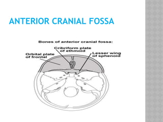
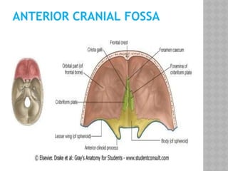
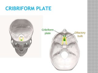
ANTERIOR
CRANIAL
FOSSA
Dr. Maliha Nawar
Resident,Phase-A(year-1)
Community Ophthalmology
Bangabandhu Sheikh Mujib
Medical University
⦿Before we start, lets give a glance on
Anatomy of base of
skull
⦿The base of the skull represents a central
and complex bone structure of the skull that
forms the floor of the cranial cavity on which
the brain lies and separetes the brain from
other facial structures.
⦿Skull base boundaries:
Anterior: Upper incisor teeth
Posterior : Superior nuchal line of occipital
bone.
Lateral:Remaining upper teeth, the zygomatic
arch & its posterior root, the mastoid process
⦿Composed of five bones:
1)Ethmoid 2) Sphenoid 3) Occipital
4) Paired temporal 5) Paired frontal bones
INTERNAL SURFACE OF SKULL:
• Has 3 large depressions:
⦿▫ Anterior cranial fossa
⦿▫ Middle cranial fossa
⦿▫ Posterior cranial fossa
• All lie at different levels within cavity
• Anterior cranial fossa lies at highest level
ANTERIOR CRANIAL FOSSA
⦿consists of three bones: the frontal bone,
ethmoid bone and sphenoid bone.
⦿It is bounded as follows:
⦿Anteriorly and laterally it is bounded by the
inner surface of the frontal bone.
⦿Posteriorly and medially it is bounded by the
limbus of the sphenoid bone. The limbus is a
bony ridge that forms the anterior border of
the prechiasmatic sulcus (a groove running
between the right and left optic canals).
ANTERIOR CRANIAL FOSSA (CONT.)
⦿Posteriorly and laterally it is bounded by
the lesser wings of the sphenoid bone (these
are two triangular projections of bone that
arise from the central sphenoid body).
⦿The floor consists of the frontal bone,
ethmoid bone and the anterior aspects of the
body and lesser wings of the sphenoid bone
ANTERIOR CRANIAL FOSSA
SURGICAL ANATOMY:
⦿Occupied by inferior & anterior parts of
frontal lobes of cerebrum hemispheres..
⦿Most of fossa formed by orbital part of frontal
bone.
⦿Frontal crest is a median body extension of
frontal bone.
⦿At its base, present is Foraman cecum of
frontal bone-important in natal life.
SURGICAL ANATOMY:
⦿The anterior aspect of the sphenoid bone lies
within the anterior cranial fossa.
⦿The rounded ends of the lesser wings are
known as the anterior clinoid processes. They
serve as a place of attachment for the
tentorium cerebelli (a sheet of dura mater
that divides the cerebrum from the
cerebellum).
⦿The lesser wings of the sphenoid bone also
separate the anterior and middle cranial
fossa.
ANTERIOR CRANIAL FOSSA
SURGICAL ANATOMY:
⦿In the midline of the ethmoid bone,
the crista galli (latin for cock’s comb) is
situated. This is an upwards projection of
bone.
⦿On either side of the crista galli is the
cribriform plate which supports the olfactory
bulb and has numerous foramina that
transmit vessels and nerves.
CONTENTS & FORAMINA’S OF ACF:
Anterior Cranial Fossa Contents
1. Foramen Cecum Nasal emissary vein(1%
population)
2. Cribiform foramina Axons of olfactory cells- (CN I)
3. Anterior & Posterior Ethmoidal
Foramina
Anterior & Posterior Ethmoidal
artery , vein and nerve.
4.Frontal crest Midline bony ridge that projects
upwards & provide attachment
to the falx cerebri.
5. Crista galli Provides site for ant. most
attachment of the falx cerebri.
ANTERIOR CRANIAL FOSSA
CLINICAL RELEVANCE:
⦿The cribriform plate of the ethmoid is the
thinnest part of the anterior cranial fossa,
and therefore most likely to fracture. There
are two major consequences of cribriform
plate fracture.
⦿Anosmia – the olfactory nerve fibres run
through the cribriform plate, and can be
‘sheared’, resulting in loss of sense of smell.
CRIBRIFORM PLATE
⦿CSF rhinorrhoea - the fragments of bone can
tear the meningeal coverings of the brain,
causing the leakage of cerebrospinal fluid
into the nasal cavity. This is visible as a
clear fluid.
⦿Fracture involve orbital plate of frontal lead
to subconjunctival hematoma extending to
posterior limit of sclera which may lead to
exopthalmus or frontal sinus may involve.
Sub conjunctival hemorrhage
⦿Traumatic events, or a basal skull fracture,
typically cause Raccoon
eyes(periorbital ecchymosis).
Raccoon
Eyes
⦿A carotid-cavernous fistula results from an
abnormal communication between the
arterial and venous systems within the
cavernous sinus.
⦿Patients usually present with bruit, pain &
sudden or insidious onset of redness in one
eye, associated with progressive proptosis or
bulging.
Carotid-
Cavernous fistula
⦿Meningiomas of the anterior cranial fossa
arise in various location.
⦿They are commonly divided into two
major subgroups: olfactory groove
meningiomas that arise over the
cribriform plate and frontosphenoid
suture and suprasellar meningiomas.
⦿Olfactory groove meningiomas produce
symptoms,as headache, personality
changes, anosmia, or seizures.
⦿ Large olfactory groove meningiomas that
extend posteriorly may encroach upon the
optic apparatus producing visual loss.
Olfactory groove
meningioma
⦿Olfactory neuroblastoma is a rare cancer of
the cribiform plate. It accounts for about 5%
of all cancers of the nasal cavity and
paranasal sinuses.
⦿The most common symptom is blockage of
the nasal passageway Other signs and
symptoms may include:(anosmia),Chronic
sinus infections,Nasal
bleeding&discharge,Pain,Visual changes,Ear
pain.
⦿Glioma is a type of tumor that occurs in the
brain and spinal cord.
⦿Common signs and symptoms of gliomas
include:Headache,Nausea or
vomiting,Confusion,Memory loss,Personality
changes,Difficulty with balance,Urinary
incontinence,Vision problems, such as
blurred vision, double vision or loss of
peripheral vision,Speech difficulties,Seizures
⦿Lesions compromising the superior orbital
fissure give rise to external ophthalmoplegia,
neuropathic pain or dysesthesias of the
forehead, and exophthalmos. When the
orbital apex is involved, visual disturbances,
papilledema or optic atrophy , chemosis ,
and exophthalmos are encountered
This patient has profound
ptosis and ophthalmoplegia in all
directions of gaze
A, Photograph of the patient on the third day of trauma, showing ptosis
and periorbital ecchymosis. B, Preoperative CT demonstrating depressed
fracture of the right sphenoid bone with compression over the right
superior orbital fissure (arrow). D and F, Reconstruction of postoperative
CT showing thorough decompression of the right optical canal and
superior orbital fissure (arrow, circle). C and E, Three, Six months after
surgery, partial recovery of ptosis and extraocular muscle movement
gradually.
SURGICAL APPROACHES:
⦿The location of pathology, its size, and how
it affects one, will help to determine if
surgery is the best treatment choice.
⦿There are two main approaches for anterior
based pathologies:
1. Minimally-invasive surgery involving an
endoscope.
2. Traditional brain surgery, known as an open
craniotomy, where we remove a piece of
bone from the forehead or temple to
access the tumor or other pathology.
⦿Surgical approaches to the anterior cranial
base include methods that are purely
extracranial and those that utilize combined
extracranial and intracranial exposures.
⦿The extracranial techniques - external
ethmoidectomy, frontal sinusotomy, and
intranasalethmoidectomy - are suitable only
for management of discrete, well localized
lesions such as CSF fistulas and some very
limited benign anterior cranial base tumors.
⦿The remaining majority of anterior cranial
base lesions are best managed using the
combined intracranial-extracranial
techniques, of which there are basically two
types:
1. the anterior craniofacial resection and
2. the basal subfrontal approach.
⦿Both of these approaches require bifrontal
craniotomy for obtaining intracranial
exposure.
ANTERIOR SKULL BASE SURGICAL
APPROACHES:
TRANSCRANIAL APPROACHES:
Anterior cranial-fossa

Anterior cranial-fossa

  • 1.
    ANTERIOR CRANIAL FOSSA Dr. Maliha Nawar Resident,Phase-A(year-1) CommunityOphthalmology Bangabandhu Sheikh Mujib Medical University
  • 2.
    ⦿Before we start,lets give a glance on Anatomy of base of skull
  • 3.
    ⦿The base ofthe skull represents a central and complex bone structure of the skull that forms the floor of the cranial cavity on which the brain lies and separetes the brain from other facial structures.
  • 4.
    ⦿Skull base boundaries: Anterior:Upper incisor teeth Posterior : Superior nuchal line of occipital bone. Lateral:Remaining upper teeth, the zygomatic arch & its posterior root, the mastoid process
  • 5.
    ⦿Composed of fivebones: 1)Ethmoid 2) Sphenoid 3) Occipital 4) Paired temporal 5) Paired frontal bones
  • 6.
    INTERNAL SURFACE OFSKULL: • Has 3 large depressions: ⦿▫ Anterior cranial fossa ⦿▫ Middle cranial fossa ⦿▫ Posterior cranial fossa • All lie at different levels within cavity • Anterior cranial fossa lies at highest level
  • 8.
    ANTERIOR CRANIAL FOSSA ⦿consistsof three bones: the frontal bone, ethmoid bone and sphenoid bone. ⦿It is bounded as follows: ⦿Anteriorly and laterally it is bounded by the inner surface of the frontal bone. ⦿Posteriorly and medially it is bounded by the limbus of the sphenoid bone. The limbus is a bony ridge that forms the anterior border of the prechiasmatic sulcus (a groove running between the right and left optic canals).
  • 9.
    ANTERIOR CRANIAL FOSSA(CONT.) ⦿Posteriorly and laterally it is bounded by the lesser wings of the sphenoid bone (these are two triangular projections of bone that arise from the central sphenoid body). ⦿The floor consists of the frontal bone, ethmoid bone and the anterior aspects of the body and lesser wings of the sphenoid bone
  • 10.
  • 11.
    ANTERIOR CRANIAL FOSSA SURGICALANATOMY: ⦿Occupied by inferior & anterior parts of frontal lobes of cerebrum hemispheres.. ⦿Most of fossa formed by orbital part of frontal bone. ⦿Frontal crest is a median body extension of frontal bone. ⦿At its base, present is Foraman cecum of frontal bone-important in natal life.
  • 12.
    SURGICAL ANATOMY: ⦿The anterioraspect of the sphenoid bone lies within the anterior cranial fossa. ⦿The rounded ends of the lesser wings are known as the anterior clinoid processes. They serve as a place of attachment for the tentorium cerebelli (a sheet of dura mater that divides the cerebrum from the cerebellum). ⦿The lesser wings of the sphenoid bone also separate the anterior and middle cranial fossa.
  • 13.
  • 14.
    SURGICAL ANATOMY: ⦿In themidline of the ethmoid bone, the crista galli (latin for cock’s comb) is situated. This is an upwards projection of bone. ⦿On either side of the crista galli is the cribriform plate which supports the olfactory bulb and has numerous foramina that transmit vessels and nerves.
  • 16.
    CONTENTS & FORAMINA’SOF ACF: Anterior Cranial Fossa Contents 1. Foramen Cecum Nasal emissary vein(1% population) 2. Cribiform foramina Axons of olfactory cells- (CN I) 3. Anterior & Posterior Ethmoidal Foramina Anterior & Posterior Ethmoidal artery , vein and nerve. 4.Frontal crest Midline bony ridge that projects upwards & provide attachment to the falx cerebri. 5. Crista galli Provides site for ant. most attachment of the falx cerebri.
  • 17.
  • 19.
    CLINICAL RELEVANCE: ⦿The cribriformplate of the ethmoid is the thinnest part of the anterior cranial fossa, and therefore most likely to fracture. There are two major consequences of cribriform plate fracture. ⦿Anosmia – the olfactory nerve fibres run through the cribriform plate, and can be ‘sheared’, resulting in loss of sense of smell.
  • 20.
  • 21.
    ⦿CSF rhinorrhoea -the fragments of bone can tear the meningeal coverings of the brain, causing the leakage of cerebrospinal fluid into the nasal cavity. This is visible as a clear fluid.
  • 22.
    ⦿Fracture involve orbitalplate of frontal lead to subconjunctival hematoma extending to posterior limit of sclera which may lead to exopthalmus or frontal sinus may involve. Sub conjunctival hemorrhage
  • 23.
    ⦿Traumatic events, ora basal skull fracture, typically cause Raccoon eyes(periorbital ecchymosis). Raccoon Eyes
  • 24.
    ⦿A carotid-cavernous fistularesults from an abnormal communication between the arterial and venous systems within the cavernous sinus. ⦿Patients usually present with bruit, pain & sudden or insidious onset of redness in one eye, associated with progressive proptosis or bulging. Carotid- Cavernous fistula
  • 25.
    ⦿Meningiomas of theanterior cranial fossa arise in various location. ⦿They are commonly divided into two major subgroups: olfactory groove meningiomas that arise over the cribriform plate and frontosphenoid suture and suprasellar meningiomas. ⦿Olfactory groove meningiomas produce symptoms,as headache, personality changes, anosmia, or seizures.
  • 26.
    ⦿ Large olfactorygroove meningiomas that extend posteriorly may encroach upon the optic apparatus producing visual loss. Olfactory groove meningioma
  • 27.
    ⦿Olfactory neuroblastoma isa rare cancer of the cribiform plate. It accounts for about 5% of all cancers of the nasal cavity and paranasal sinuses. ⦿The most common symptom is blockage of the nasal passageway Other signs and symptoms may include:(anosmia),Chronic sinus infections,Nasal bleeding&discharge,Pain,Visual changes,Ear pain.
  • 28.
    ⦿Glioma is atype of tumor that occurs in the brain and spinal cord. ⦿Common signs and symptoms of gliomas include:Headache,Nausea or vomiting,Confusion,Memory loss,Personality changes,Difficulty with balance,Urinary incontinence,Vision problems, such as blurred vision, double vision or loss of peripheral vision,Speech difficulties,Seizures
  • 29.
    ⦿Lesions compromising thesuperior orbital fissure give rise to external ophthalmoplegia, neuropathic pain or dysesthesias of the forehead, and exophthalmos. When the orbital apex is involved, visual disturbances, papilledema or optic atrophy , chemosis , and exophthalmos are encountered This patient has profound ptosis and ophthalmoplegia in all directions of gaze
  • 30.
    A, Photograph ofthe patient on the third day of trauma, showing ptosis and periorbital ecchymosis. B, Preoperative CT demonstrating depressed fracture of the right sphenoid bone with compression over the right superior orbital fissure (arrow). D and F, Reconstruction of postoperative CT showing thorough decompression of the right optical canal and superior orbital fissure (arrow, circle). C and E, Three, Six months after surgery, partial recovery of ptosis and extraocular muscle movement gradually.
  • 31.
    SURGICAL APPROACHES: ⦿The locationof pathology, its size, and how it affects one, will help to determine if surgery is the best treatment choice. ⦿There are two main approaches for anterior based pathologies: 1. Minimally-invasive surgery involving an endoscope. 2. Traditional brain surgery, known as an open craniotomy, where we remove a piece of bone from the forehead or temple to access the tumor or other pathology.
  • 32.
    ⦿Surgical approaches tothe anterior cranial base include methods that are purely extracranial and those that utilize combined extracranial and intracranial exposures. ⦿The extracranial techniques - external ethmoidectomy, frontal sinusotomy, and intranasalethmoidectomy - are suitable only for management of discrete, well localized lesions such as CSF fistulas and some very limited benign anterior cranial base tumors.
  • 33.
    ⦿The remaining majorityof anterior cranial base lesions are best managed using the combined intracranial-extracranial techniques, of which there are basically two types: 1. the anterior craniofacial resection and 2. the basal subfrontal approach. ⦿Both of these approaches require bifrontal craniotomy for obtaining intracranial exposure.
  • 34.
    ANTERIOR SKULL BASESURGICAL APPROACHES:
  • 35.
  • 36.