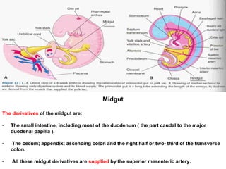
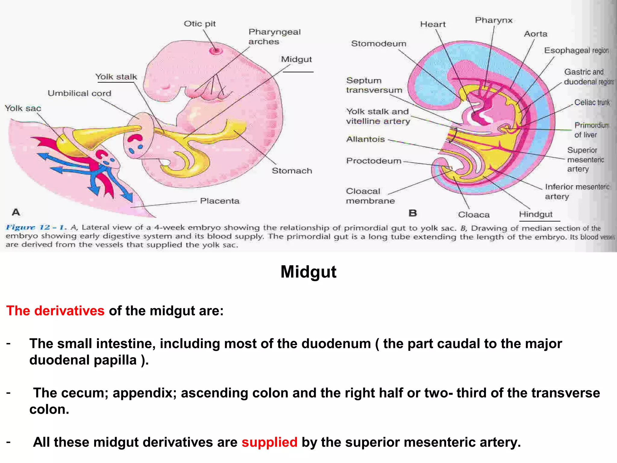
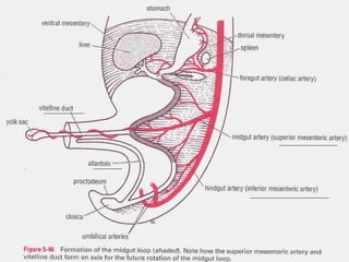
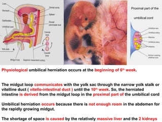
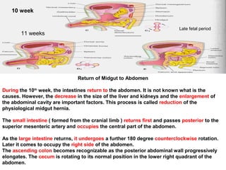
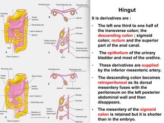
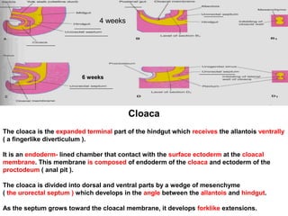
SlideShare a Scribd company logo
1 of 23
Midgut
The derivatives of the midgut are:
- The small intestine, including most of the duodenum ( the part caudal to the major
duodenal papilla ).
- The cecum; appendix; ascending colon and the right half or two- third of the transverse
colon.
- All these midgut derivatives are supplied by the superior mesenteric artery.
5th
week
The midgut loop is suspended from the dorsal abdominal wall by an elongated
mesentery.
As it elongates, the ventral U – shaped loop of gut ( midgut loop ) projects into the
remains of the extraembryonic coelom in the proximal part of the umbilical cord at
the end of the 5th
week.
At this stage, the intraembryonic coelom ( peritoneal cavity ) communicates with
the extraembryonic coelom at the umbilicus.
Physiological umbilical herniation occurs at the beginning of 6th
week.
The midgut loop communicates with the yolk sac through the narrow yolk stalk or
vitelline duct ( vitello-intestinal duct ) until the 10th
week. So, the herniated
intestine is derived from the midgut loop in the proximal part of the umbilical cord
Umbilical herniation occurs because there is not enough room in the abdomen for
the rapidly growing midgut.
The shortage of space is caused by the relatively massive liver and the 2 kidneys
Proximal part of the
umbilical cord
The midgut loop has a cranial limb and a caudal limb. The yolk stalk is attached to the apex
of the midgut loop where the 2 limbs join.
The cranial limb grows rapidly and forms small intestinal loops. The caudal limb undergoes
very little change except for development of the cecal diverticulum ( the primordium of the
cecum and appendix.
Rotation of Midgut Loop
While it is in the umbilical cord, the midgut loop rotates 90 degrees counterclockwise around
the axis of the superior mesenteric artery and yolk stalk. This brings the cranial limb of the
midgut loop to the right and the caudal limb to the left.
During rotation the cranial limb elongates and forms jejunum & ileum ( intestinal loops ).
10 week
Return of Midgut to Abdomen
During the 10th
week, the intestines return to the abdomen. It is not known what is the
causes. However, the decrease in the size of the liver and kidneys and the enlargement of
the abdominal cavity are important factors. This process is called reduction of the
physiological midgut hernia.
The small intestine ( formed from the cranial limb ) returns first and passes posterior to the
superior mesenteric artery and occupies the central part of the abdomen.
As the large intestine returns, it undergoes a further 180 degree counterclockwise rotation.
Later it comes to occupy the right side of the abdomen.
The ascending colon becomes recognizable as the posterior abdominal wall progressively
elongates. The cecum is rotating to its normal position in the lower right quadrant of the
abdomen.
11 weeks
Late fetal period
10 week
Fixation of Intestines
Rotation of the stomach and duodenum causes the duodenum and pancreas to fall to the
right. The enlarged colon presses the duodenum against the posterior abdominal wall. As a
result, most of the duodenal mesentery is absorbed and the duodenum, except for about the
first 2.5 cm ( derived from the foregut ), has no mesentery and lies retroperitoneally.
At first the dorsal mesentery is in the median plane. As the intestines enlarge, lengthen and
assume their final position, their mesenteries are pressed against the posterior abdominal
wall. So, the mesentery of the ascending colon fuses with the parietal peritoneum on this
wall and disappears. The descending colon also becomes retroperitoneal.
Other derivatives of the midgut
loop ( jejunum & ileum ) retain
their mesenteries. The
mesentery is at first attached to
the median plane of the
posterior abdominal wall.
After the mesentery of the
ascending colon disappears,
the fan- shaped mesentery of
the small intestine acquires a
new line of attachment that
passes from the
duodenojejunal junction
inferolaterally to the ileocecal
junction.
Cecum & Appendix
The cecal diverticulum ( primordium of the cecum and vermiform appendix ) appears in the
6th
week as a swelling on the antimesenteric border of the caudal limb of the midgut loop.
The apex of the cecal diverticulum does not grow as rapidly as the rest of it. Thus, the
appendix is initially a small diverticulum of the apex of the cecum.
The appendix increases rapidly in length so that at birth it is a relatively long tube
arising from the distal end of the cecum.
After birth the wall of the cecum grows unequally, with the result that the appendix
comes to enter its medial side. The appendix may pass posterior to the cecum ( retrocecal )
or colon ( retrocolic ). It may descend over the brim of the pelvis ( pelvis appendix ).
In about 64 % of people the appendix is located retrocecally.
Congenital Omphalocele
This anomali is persistence of the herniation
of abdominal contents into the proximal part
of the umbilical cord and failure of the
intestine to return to the abdominal cavity
from the extraembryonic coleom during the
10th
week.
The covering of the hernial sac is the
epithelium of the umbilical cord ( a derivative
of the amnion ).
Herniation of the intestines into the cord
occurs in about 1 of 5000 births and
herniation of the liver and intestines in 1 of
about 10000 births.
The size of the hernia depends on its
contents. When there is small abdominal
cavity, there is omphalocele.
Immediate surgical repair is required
Ileal Diverticulum
An ileal diverticulum is the remnant of the proximal part of the yolk stalk. It
typically appears as a fingerlike pouch about 3 to 6 cm long that arises from the
antimesenteric border of the ileum 40 t0 50 cm from the ileocecal junction.
It is common. Meckel diverticulum occurs in 2 to 4 % of people and is 3 to 5 times
more prevalent in males than females. When it inflames, it causes symptoms that
mimic appendicitis.
The wall of the diverticulum contains all layers of the ileum and may contain small
patches of gastric and pancreatic tissues.
The gastric mucosa often secrets acid producing ulceration and bleeding.
An ileal diverticulum may be connected to the umbilicus by a fibrous cord or an
omphaloenteric fistula which results from persistence of the entire intra-
abdominal portion of the yolk stalk ( vitelline duct ).
On the fibrous remnant of the of the yolk stalk a vitelline cysts is formed.
Umbilical sinus results from the persistence of the yolk stalk near the umbilicus. It
is usually appear with volvulus of the diverticulum.
The yolk stalk has persisted as a fibrous cord connecting the ileum with the
umbilicus and containing a persistent vitelline artery .
Hingut
It is derivatives are :
- The left one third to one half of
the transverse colon; the
descending colon ; sigmoid
colon; rectum and the superior
part of the anal canal.
- The epithelium of the urinary
bladder and most of the urethra.
- These derivatives are supplied
by the inferior mesenteric artery.
- The descending colon becomes
retroperitoneal as its dorsal
mesentery fuses with the
peritoneum on the left posterior
abdominal wall and then
disappears.
- The mesentery of the sigmoid
colon is retained but it is shorter
than in the embryo.
Cloaca
The cloaca is the expanded terminal part of the hindgut which receives the allantois ventrally
( a fingerlike diverticulum ).
It is an endoderm- lined chamber that contact with the surface ectoderm at the cloacal
membrane. This membrane is composed of endoderm of the cloaca and ectoderm of the
proctodeum ( anal pit ).
The cloaca is divided into dorsal and ventral parts by a wedge of mesenchyme
( the urorectal septum ) which develops in the angle between the allantois and hindgut.
As the septum grows toward the cloacal membrane, it develops forklike extensions.
4 weeks
6 weeks
The 2 parts are : A- Rectum and cranial part of the anal canal dorsally.
B- Urogenital sinus ventrally.
The postanal or tailgut degenerates and disappears as the rectum is formed.
By the 7th
week, the urorectal septum has fused with the cloacal membrane, dividing it into a
dorsal anal membrane and a larger ventral urogenital membrane. The area of fusion of the
urorectal septum with the cloacal membrane is represented in the adult by the perineal body
( the tendinous center of the perineum ).
The urorectal septum also, divides the cloacal sphincter into anterior & posterior parts. The
posterior part becomes the external anal sphincter and the anterior part develops into the
superficial transverse perineal ( bulbospongiosus and ischiocavernosus ) muscles. This
developmental fact explains why one nerve ( pudendal nerve ) supplies all these muscles.
7 weeks
Mesenchymal proliferations produce elevations of the surface ectoderm around
the anal membrane. As a result, this membrane is soon located at the bottom of
an ectodermal depression ( the proctodeum ) or anal pit.
The anal membrane usually ruptures at the end of the 8th
week bringing the distal
part of the digestive tract ( anal canal ) into communication with the amniotic
cavity.
7 weeks
Anal Canal
The superior two- thirds ( about 25 cm ) of the adult anal canal are derived from the hindgut
and the inferior one- third ( about 13 cm ) develops from the proctodeum.
The junction of the epithelium derived from the ectoderm of the proctodeum and the
endoderm of the hindgut is roughly indicated by the irregular pectinate line which is located
at the inferior limit of the anal valves. This line indicates the site of the anal membrane.
About 2 cm superior to the anus is an anocutaneous line ( white line ). This is the site where
the composition of the anal epithelium changes from columnar to stratified squamous cells.
At the anus, the epithelium is keratinized and continuous with the skin around the anus. The
other layers of the wall of the anal canal are derived from splanchnic mesenchyme.
Similar to the pyloric sphincter and the ileocecal valve ( sphincter ) the formation of the anal
sphincter appears to be under genetic control.
The lymphatic drainage of the superior part is to the inferior mesenteric lymph nodes. The
lymphatic drainage of the inferior part of the anal canal is to the superficial inguinal lymph
nodes.
Its nerve supply is from the inferior rectal nerve, so, it is sensitive to pain; temperature;
touch and pressure.
The differences in blood; nerve supply and venous & lymphatic drainage of the
anal canal are important clinically for the spread of the metastasis of cancer cells.
The characteristics of the carcinomas in the two parts also differ. Tumors in the
superior part are painless and arise from columnar epithelium. Those in the
inferior part are painful and arise from stratified squamous epithelium.
Imperforate anus ( membranous anal atresia )
It occurs about once in every 5000 newborn infants and is more common in
males.
There is a thin layer of tissue separates the anal canal from the exterior. This anal
membrane is thin enough to bulge on straining and appears blue from the
presence of meconium superior to it. The large intestine is distended with feces
and contrast material.
The anus is in the normal position. This anomaly results from failure of the anal
membrane to perforate at the end of the 8th
week.
Congenital Megacolon
( Hirschsprung Disease )
It is a dominant inherited multigenic disorder. It is
the most common cause of neonatal obstruction of
the colon and occurs for about 33% . Males are
affected more often than females ( 4- 1 ).
A part of the colon is dilated because of the absence
of autonomic ganglia cells in the myenteric plexus
distal to the dilated segment of the colon.
The enlarged colon has the normal number of
ganglion cells. The dilatation results from failure of
peristalsis in the aganglionic segment ( transition
zone ) which prevents movement of the intestinal
contents.
In most cases only the rectum and sigmoid colon are involved. Also, ganglia may be absent
from more proximal parts of the colon.
It results from failure of the neural crest cells to migrate into the wall of the colon during the
5th to 7th weeks. This results in failure of parasympathetic ganglion cells to develop in the
Auerbach and Meissner plexuses.
The cause of failure of some neural crest cells to complete their migration is unknown.

More Related Content

What's hot

Vena cava anatomy and variants
Vena cava anatomy and variantsVena cava anatomy and variants
Vena cava anatomy and variantsMilan Silwal
 
Development of midgut and pancreas
Development of midgut  and pancreas Development of midgut  and pancreas
Development of midgut and pancreas Sahar Hafeez
 
Derivatives of foregut
Derivatives of foregutDerivatives of foregut
Derivatives of foregutLucidante1
 
Embryology liver,pancreas,spleen & respiratory system
Embryology   liver,pancreas,spleen & respiratory systemEmbryology   liver,pancreas,spleen & respiratory system
Embryology liver,pancreas,spleen & respiratory systemMBBS IMS MSU
 
Portal vein & portocaval anastomosis
Portal vein & portocaval anastomosis Portal vein & portocaval anastomosis
Portal vein & portocaval anastomosis Dr Mohammad Amaan
 
Extrahepatic Biliary Apparatus
Extrahepatic Biliary ApparatusExtrahepatic Biliary Apparatus
Extrahepatic Biliary ApparatusDrUroojRehman
 
Development of liver , extrahepatic biliary apparatus , pancreas and spleen.
Development of liver , extrahepatic biliary apparatus , pancreas and spleen.Development of liver , extrahepatic biliary apparatus , pancreas and spleen.
Development of liver , extrahepatic biliary apparatus , pancreas and spleen.SaachiGupta4
 
Gross anatomy and histology of extrahepatic biliary apparatus
Gross anatomy and histology of extrahepatic biliary apparatusGross anatomy and histology of extrahepatic biliary apparatus
Gross anatomy and histology of extrahepatic biliary apparatusDr Laxman Khanal
 
Arterial supply of the Abdomen : Abdominal Aorta
Arterial supply of the Abdomen : Abdominal AortaArterial supply of the Abdomen : Abdominal Aorta
Arterial supply of the Abdomen : Abdominal AortaBitew Mekonnen
 
Development of Midgut & Hindgut
Development of Midgut & HindgutDevelopment of Midgut & Hindgut
Development of Midgut & HindgutNeeta Chhabra
 
Anatomy Of Urinary Bladder
Anatomy Of Urinary BladderAnatomy Of Urinary Bladder
Anatomy Of Urinary BladderUmair Majeed
 
Development of urogenital system I
Development of urogenital system IDevelopment of urogenital system I
Development of urogenital system IPrabhakar Yadav
 
Development of Stomach (Special Embryology)
Development of Stomach (Special Embryology)Development of Stomach (Special Embryology)
Development of Stomach (Special Embryology)Dr. Sherif Fahmy
 
Embryology urogenital system
Embryology    urogenital systemEmbryology    urogenital system
Embryology urogenital systemMBBS IMS MSU
 

What's hot (20)

Vena cava anatomy and variants
Vena cava anatomy and variantsVena cava anatomy and variants
Vena cava anatomy and variants
 
Development of midgut and pancreas
Development of midgut  and pancreas Development of midgut  and pancreas
Development of midgut and pancreas
 
Derivatives of foregut
Derivatives of foregutDerivatives of foregut
Derivatives of foregut
 
Embryology liver,pancreas,spleen & respiratory system
Embryology   liver,pancreas,spleen & respiratory systemEmbryology   liver,pancreas,spleen & respiratory system
Embryology liver,pancreas,spleen & respiratory system
 
Duodenum & pancreas
Duodenum & pancreas   Duodenum & pancreas
Duodenum & pancreas
 
Portal vein & portocaval anastomosis
Portal vein & portocaval anastomosis Portal vein & portocaval anastomosis
Portal vein & portocaval anastomosis
 
Extrahepatic Biliary Apparatus
Extrahepatic Biliary ApparatusExtrahepatic Biliary Apparatus
Extrahepatic Biliary Apparatus
 
Development of liver , extrahepatic biliary apparatus , pancreas and spleen.
Development of liver , extrahepatic biliary apparatus , pancreas and spleen.Development of liver , extrahepatic biliary apparatus , pancreas and spleen.
Development of liver , extrahepatic biliary apparatus , pancreas and spleen.
 
Development of GIT
Development of GITDevelopment of GIT
Development of GIT
 
Gross anatomy and histology of extrahepatic biliary apparatus
Gross anatomy and histology of extrahepatic biliary apparatusGross anatomy and histology of extrahepatic biliary apparatus
Gross anatomy and histology of extrahepatic biliary apparatus
 
Arterial supply of the Abdomen : Abdominal Aorta
Arterial supply of the Abdomen : Abdominal AortaArterial supply of the Abdomen : Abdominal Aorta
Arterial supply of the Abdomen : Abdominal Aorta
 
ANATOMY OF DUODENUM
ANATOMY OF DUODENUMANATOMY OF DUODENUM
ANATOMY OF DUODENUM
 
Development of Midgut & Hindgut
Development of Midgut & HindgutDevelopment of Midgut & Hindgut
Development of Midgut & Hindgut
 
Anatomy Of Urinary Bladder
Anatomy Of Urinary BladderAnatomy Of Urinary Bladder
Anatomy Of Urinary Bladder
 
The duodenum
The duodenumThe duodenum
The duodenum
 
The Peritoneum
The PeritoneumThe Peritoneum
The Peritoneum
 
The jejunum and ileum
The jejunum and ileumThe jejunum and ileum
The jejunum and ileum
 
Development of urogenital system I
Development of urogenital system IDevelopment of urogenital system I
Development of urogenital system I
 
Development of Stomach (Special Embryology)
Development of Stomach (Special Embryology)Development of Stomach (Special Embryology)
Development of Stomach (Special Embryology)
 
Embryology urogenital system
Embryology    urogenital systemEmbryology    urogenital system
Embryology urogenital system
 

Similar to 05. midgut& hindgut embryo

Development Of Mid & Hind Guts
Development Of Mid & Hind GutsDevelopment Of Mid & Hind Guts
Development Of Mid & Hind GutsSara Memon
 
Embryology_and_Anatomy_of_the_Gastrointestinal_Tract_NASPGHAN.pdf
Embryology_and_Anatomy_of_the_Gastrointestinal_Tract_NASPGHAN.pdfEmbryology_and_Anatomy_of_the_Gastrointestinal_Tract_NASPGHAN.pdf
Embryology_and_Anatomy_of_the_Gastrointestinal_Tract_NASPGHAN.pdfelphaswalela
 
developmentofgit-copy-150530164223-lva1-app6892.pdf
developmentofgit-copy-150530164223-lva1-app6892.pdfdevelopmentofgit-copy-150530164223-lva1-app6892.pdf
developmentofgit-copy-150530164223-lva1-app6892.pdfkarthikeyan104187
 
Organogenesis of Alimentry Canal
Organogenesis of Alimentry CanalOrganogenesis of Alimentry Canal
Organogenesis of Alimentry CanalPoojaVishnoi7
 
GI presentation embryo
GI presentation embryo  GI presentation embryo
GI presentation embryo naba al wazen
 
Embryology Course VIII - Digestive System
Embryology Course VIII - Digestive SystemEmbryology Course VIII - Digestive System
Embryology Course VIII - Digestive SystemRawa Muhsin
 
Embryology alimentary system 2
Embryology   alimentary system 2Embryology   alimentary system 2
Embryology alimentary system 2MBBS IMS MSU
 
DEVELOPMENT OF DIGESTIVE SYSTEM.pptx
DEVELOPMENT OF DIGESTIVE SYSTEM.pptxDEVELOPMENT OF DIGESTIVE SYSTEM.pptx
DEVELOPMENT OF DIGESTIVE SYSTEM.pptxwmdpr4x64f
 
GIT embryology By Dr Parashuram Waddar Pediatrician
GIT embryology By Dr Parashuram Waddar Pediatrician GIT embryology By Dr Parashuram Waddar Pediatrician
GIT embryology By Dr Parashuram Waddar Pediatrician ParasuramWaddar2
 
The jejunum, ileum & mesentery
The jejunum, ileum & mesenteryThe jejunum, ileum & mesentery
The jejunum, ileum & mesenteryDr Mohammad Amaan
 
Appendix ,Colon, Rectums ana Anal Canal PRESENTATION POWERPOINT.pptx
Appendix ,Colon, Rectums ana Anal Canal PRESENTATION POWERPOINT.pptxAppendix ,Colon, Rectums ana Anal Canal PRESENTATION POWERPOINT.pptx
Appendix ,Colon, Rectums ana Anal Canal PRESENTATION POWERPOINT.pptxAnimasahunOlawale
 
Approach to a vomiting Infant
Approach to a vomiting InfantApproach to a vomiting Infant
Approach to a vomiting InfantSameer Peer
 
Development of FOREGUT MIDGUT AND HINDGUT.ppt
Development of FOREGUT MIDGUT AND HINDGUT.pptDevelopment of FOREGUT MIDGUT AND HINDGUT.ppt
Development of FOREGUT MIDGUT AND HINDGUT.pptJairajSinghChauhan1
 

Similar to 05. midgut& hindgut embryo (20)

Development Of Mid & Hind Guts
Development Of Mid & Hind GutsDevelopment Of Mid & Hind Guts
Development Of Mid & Hind Guts
 
Embryology part 5
Embryology part 5Embryology part 5
Embryology part 5
 
Embryology part 6
Embryology part 6Embryology part 6
Embryology part 6
 
Embryology_and_Anatomy_of_the_Gastrointestinal_Tract_NASPGHAN.pdf
Embryology_and_Anatomy_of_the_Gastrointestinal_Tract_NASPGHAN.pdfEmbryology_and_Anatomy_of_the_Gastrointestinal_Tract_NASPGHAN.pdf
Embryology_and_Anatomy_of_the_Gastrointestinal_Tract_NASPGHAN.pdf
 
developmentofgit-copy-150530164223-lva1-app6892.pdf
developmentofgit-copy-150530164223-lva1-app6892.pdfdevelopmentofgit-copy-150530164223-lva1-app6892.pdf
developmentofgit-copy-150530164223-lva1-app6892.pdf
 
Organogenesis of Alimentry Canal
Organogenesis of Alimentry CanalOrganogenesis of Alimentry Canal
Organogenesis of Alimentry Canal
 
GI presentation embryo
GI presentation embryo  GI presentation embryo
GI presentation embryo
 
Abdomen.ppt
Abdomen.pptAbdomen.ppt
Abdomen.ppt
 
Embryology Course VIII - Digestive System
Embryology Course VIII - Digestive SystemEmbryology Course VIII - Digestive System
Embryology Course VIII - Digestive System
 
12. git[1]
12. git[1]12. git[1]
12. git[1]
 
Embryology alimentary system 2
Embryology   alimentary system 2Embryology   alimentary system 2
Embryology alimentary system 2
 
DEVELOPMENT OF DIGESTIVE SYSTEM.pptx
DEVELOPMENT OF DIGESTIVE SYSTEM.pptxDEVELOPMENT OF DIGESTIVE SYSTEM.pptx
DEVELOPMENT OF DIGESTIVE SYSTEM.pptx
 
GIT embryology By Dr Parashuram Waddar Pediatrician
GIT embryology By Dr Parashuram Waddar Pediatrician GIT embryology By Dr Parashuram Waddar Pediatrician
GIT embryology By Dr Parashuram Waddar Pediatrician
 
The jejunum, ileum & mesentery
The jejunum, ileum & mesenteryThe jejunum, ileum & mesentery
The jejunum, ileum & mesentery
 
Midgut
MidgutMidgut
Midgut
 
Midgut
MidgutMidgut
Midgut
 
Anatomy of digestive system-III.pptx
Anatomy of digestive system-III.pptxAnatomy of digestive system-III.pptx
Anatomy of digestive system-III.pptx
 
Appendix ,Colon, Rectums ana Anal Canal PRESENTATION POWERPOINT.pptx
Appendix ,Colon, Rectums ana Anal Canal PRESENTATION POWERPOINT.pptxAppendix ,Colon, Rectums ana Anal Canal PRESENTATION POWERPOINT.pptx
Appendix ,Colon, Rectums ana Anal Canal PRESENTATION POWERPOINT.pptx
 
Approach to a vomiting Infant
Approach to a vomiting InfantApproach to a vomiting Infant
Approach to a vomiting Infant
 
Development of FOREGUT MIDGUT AND HINDGUT.ppt
Development of FOREGUT MIDGUT AND HINDGUT.pptDevelopment of FOREGUT MIDGUT AND HINDGUT.ppt
Development of FOREGUT MIDGUT AND HINDGUT.ppt
 

05. midgut& hindgut embryo

  • 1. Midgut The derivatives of the midgut are: - The small intestine, including most of the duodenum ( the part caudal to the major duodenal papilla ). - The cecum; appendix; ascending colon and the right half or two- third of the transverse colon. - All these midgut derivatives are supplied by the superior mesenteric artery.
  • 2.
  • 3. 5th week The midgut loop is suspended from the dorsal abdominal wall by an elongated mesentery. As it elongates, the ventral U – shaped loop of gut ( midgut loop ) projects into the remains of the extraembryonic coelom in the proximal part of the umbilical cord at the end of the 5th week. At this stage, the intraembryonic coelom ( peritoneal cavity ) communicates with the extraembryonic coelom at the umbilicus.
  • 4. Physiological umbilical herniation occurs at the beginning of 6th week. The midgut loop communicates with the yolk sac through the narrow yolk stalk or vitelline duct ( vitello-intestinal duct ) until the 10th week. So, the herniated intestine is derived from the midgut loop in the proximal part of the umbilical cord Umbilical herniation occurs because there is not enough room in the abdomen for the rapidly growing midgut. The shortage of space is caused by the relatively massive liver and the 2 kidneys Proximal part of the umbilical cord
  • 5.
  • 6. The midgut loop has a cranial limb and a caudal limb. The yolk stalk is attached to the apex of the midgut loop where the 2 limbs join. The cranial limb grows rapidly and forms small intestinal loops. The caudal limb undergoes very little change except for development of the cecal diverticulum ( the primordium of the cecum and appendix. Rotation of Midgut Loop While it is in the umbilical cord, the midgut loop rotates 90 degrees counterclockwise around the axis of the superior mesenteric artery and yolk stalk. This brings the cranial limb of the midgut loop to the right and the caudal limb to the left. During rotation the cranial limb elongates and forms jejunum & ileum ( intestinal loops ). 10 week
  • 7. Return of Midgut to Abdomen During the 10th week, the intestines return to the abdomen. It is not known what is the causes. However, the decrease in the size of the liver and kidneys and the enlargement of the abdominal cavity are important factors. This process is called reduction of the physiological midgut hernia. The small intestine ( formed from the cranial limb ) returns first and passes posterior to the superior mesenteric artery and occupies the central part of the abdomen. As the large intestine returns, it undergoes a further 180 degree counterclockwise rotation. Later it comes to occupy the right side of the abdomen. The ascending colon becomes recognizable as the posterior abdominal wall progressively elongates. The cecum is rotating to its normal position in the lower right quadrant of the abdomen. 11 weeks Late fetal period 10 week
  • 8. Fixation of Intestines Rotation of the stomach and duodenum causes the duodenum and pancreas to fall to the right. The enlarged colon presses the duodenum against the posterior abdominal wall. As a result, most of the duodenal mesentery is absorbed and the duodenum, except for about the first 2.5 cm ( derived from the foregut ), has no mesentery and lies retroperitoneally. At first the dorsal mesentery is in the median plane. As the intestines enlarge, lengthen and assume their final position, their mesenteries are pressed against the posterior abdominal wall. So, the mesentery of the ascending colon fuses with the parietal peritoneum on this wall and disappears. The descending colon also becomes retroperitoneal.
  • 9. Other derivatives of the midgut loop ( jejunum & ileum ) retain their mesenteries. The mesentery is at first attached to the median plane of the posterior abdominal wall. After the mesentery of the ascending colon disappears, the fan- shaped mesentery of the small intestine acquires a new line of attachment that passes from the duodenojejunal junction inferolaterally to the ileocecal junction.
  • 10. Cecum & Appendix The cecal diverticulum ( primordium of the cecum and vermiform appendix ) appears in the 6th week as a swelling on the antimesenteric border of the caudal limb of the midgut loop. The apex of the cecal diverticulum does not grow as rapidly as the rest of it. Thus, the appendix is initially a small diverticulum of the apex of the cecum. The appendix increases rapidly in length so that at birth it is a relatively long tube arising from the distal end of the cecum. After birth the wall of the cecum grows unequally, with the result that the appendix comes to enter its medial side. The appendix may pass posterior to the cecum ( retrocecal ) or colon ( retrocolic ). It may descend over the brim of the pelvis ( pelvis appendix ). In about 64 % of people the appendix is located retrocecally.
  • 11. Congenital Omphalocele This anomali is persistence of the herniation of abdominal contents into the proximal part of the umbilical cord and failure of the intestine to return to the abdominal cavity from the extraembryonic coleom during the 10th week. The covering of the hernial sac is the epithelium of the umbilical cord ( a derivative of the amnion ). Herniation of the intestines into the cord occurs in about 1 of 5000 births and herniation of the liver and intestines in 1 of about 10000 births. The size of the hernia depends on its contents. When there is small abdominal cavity, there is omphalocele. Immediate surgical repair is required
  • 12.
  • 13. Ileal Diverticulum An ileal diverticulum is the remnant of the proximal part of the yolk stalk. It typically appears as a fingerlike pouch about 3 to 6 cm long that arises from the antimesenteric border of the ileum 40 t0 50 cm from the ileocecal junction. It is common. Meckel diverticulum occurs in 2 to 4 % of people and is 3 to 5 times more prevalent in males than females. When it inflames, it causes symptoms that mimic appendicitis. The wall of the diverticulum contains all layers of the ileum and may contain small patches of gastric and pancreatic tissues. The gastric mucosa often secrets acid producing ulceration and bleeding.
  • 14. An ileal diverticulum may be connected to the umbilicus by a fibrous cord or an omphaloenteric fistula which results from persistence of the entire intra- abdominal portion of the yolk stalk ( vitelline duct ). On the fibrous remnant of the of the yolk stalk a vitelline cysts is formed. Umbilical sinus results from the persistence of the yolk stalk near the umbilicus. It is usually appear with volvulus of the diverticulum. The yolk stalk has persisted as a fibrous cord connecting the ileum with the umbilicus and containing a persistent vitelline artery .
  • 15. Hingut It is derivatives are : - The left one third to one half of the transverse colon; the descending colon ; sigmoid colon; rectum and the superior part of the anal canal. - The epithelium of the urinary bladder and most of the urethra. - These derivatives are supplied by the inferior mesenteric artery. - The descending colon becomes retroperitoneal as its dorsal mesentery fuses with the peritoneum on the left posterior abdominal wall and then disappears. - The mesentery of the sigmoid colon is retained but it is shorter than in the embryo.
  • 16. Cloaca The cloaca is the expanded terminal part of the hindgut which receives the allantois ventrally ( a fingerlike diverticulum ). It is an endoderm- lined chamber that contact with the surface ectoderm at the cloacal membrane. This membrane is composed of endoderm of the cloaca and ectoderm of the proctodeum ( anal pit ). The cloaca is divided into dorsal and ventral parts by a wedge of mesenchyme ( the urorectal septum ) which develops in the angle between the allantois and hindgut. As the septum grows toward the cloacal membrane, it develops forklike extensions. 4 weeks 6 weeks
  • 17. The 2 parts are : A- Rectum and cranial part of the anal canal dorsally. B- Urogenital sinus ventrally. The postanal or tailgut degenerates and disappears as the rectum is formed. By the 7th week, the urorectal septum has fused with the cloacal membrane, dividing it into a dorsal anal membrane and a larger ventral urogenital membrane. The area of fusion of the urorectal septum with the cloacal membrane is represented in the adult by the perineal body ( the tendinous center of the perineum ). The urorectal septum also, divides the cloacal sphincter into anterior & posterior parts. The posterior part becomes the external anal sphincter and the anterior part develops into the superficial transverse perineal ( bulbospongiosus and ischiocavernosus ) muscles. This developmental fact explains why one nerve ( pudendal nerve ) supplies all these muscles. 7 weeks
  • 18. Mesenchymal proliferations produce elevations of the surface ectoderm around the anal membrane. As a result, this membrane is soon located at the bottom of an ectodermal depression ( the proctodeum ) or anal pit. The anal membrane usually ruptures at the end of the 8th week bringing the distal part of the digestive tract ( anal canal ) into communication with the amniotic cavity. 7 weeks
  • 19. Anal Canal The superior two- thirds ( about 25 cm ) of the adult anal canal are derived from the hindgut and the inferior one- third ( about 13 cm ) develops from the proctodeum. The junction of the epithelium derived from the ectoderm of the proctodeum and the endoderm of the hindgut is roughly indicated by the irregular pectinate line which is located at the inferior limit of the anal valves. This line indicates the site of the anal membrane. About 2 cm superior to the anus is an anocutaneous line ( white line ). This is the site where the composition of the anal epithelium changes from columnar to stratified squamous cells.
  • 20. At the anus, the epithelium is keratinized and continuous with the skin around the anus. The other layers of the wall of the anal canal are derived from splanchnic mesenchyme. Similar to the pyloric sphincter and the ileocecal valve ( sphincter ) the formation of the anal sphincter appears to be under genetic control. The lymphatic drainage of the superior part is to the inferior mesenteric lymph nodes. The lymphatic drainage of the inferior part of the anal canal is to the superficial inguinal lymph nodes. Its nerve supply is from the inferior rectal nerve, so, it is sensitive to pain; temperature; touch and pressure.
  • 21. The differences in blood; nerve supply and venous & lymphatic drainage of the anal canal are important clinically for the spread of the metastasis of cancer cells. The characteristics of the carcinomas in the two parts also differ. Tumors in the superior part are painless and arise from columnar epithelium. Those in the inferior part are painful and arise from stratified squamous epithelium.
  • 22. Imperforate anus ( membranous anal atresia ) It occurs about once in every 5000 newborn infants and is more common in males. There is a thin layer of tissue separates the anal canal from the exterior. This anal membrane is thin enough to bulge on straining and appears blue from the presence of meconium superior to it. The large intestine is distended with feces and contrast material. The anus is in the normal position. This anomaly results from failure of the anal membrane to perforate at the end of the 8th week.
  • 23. Congenital Megacolon ( Hirschsprung Disease ) It is a dominant inherited multigenic disorder. It is the most common cause of neonatal obstruction of the colon and occurs for about 33% . Males are affected more often than females ( 4- 1 ). A part of the colon is dilated because of the absence of autonomic ganglia cells in the myenteric plexus distal to the dilated segment of the colon. The enlarged colon has the normal number of ganglion cells. The dilatation results from failure of peristalsis in the aganglionic segment ( transition zone ) which prevents movement of the intestinal contents. In most cases only the rectum and sigmoid colon are involved. Also, ganglia may be absent from more proximal parts of the colon. It results from failure of the neural crest cells to migrate into the wall of the colon during the 5th to 7th weeks. This results in failure of parasympathetic ganglion cells to develop in the Auerbach and Meissner plexuses. The cause of failure of some neural crest cells to complete their migration is unknown.