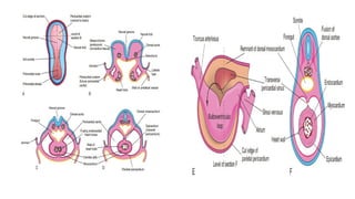
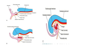
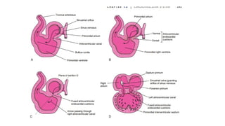
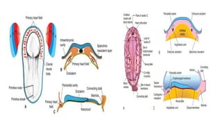
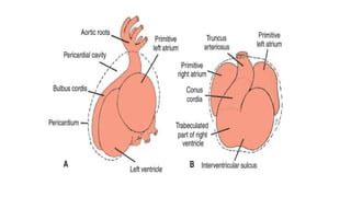
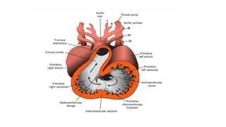
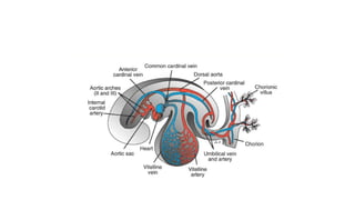
HEART DEVELOPMENT
• By day 18, the lateral
mesoderm has
somatopleure and
splanchnopleure
components
• The splanchnopleure gives
rise to almost all of the
heart components.
• These early endocardial
cells separate from the
mesoderm to create paired
heart tubes.
At the end of the second week, embryonic nutrition is
obtained from the maternal blood by diffusion through
the extraembryonic coelom and umbilical vesicle.
At the beginning of the third week, blood vessel
formation begins in the extraembryonic mesoderm of
the umbilical vesicle, connecting stalk, and chorion.
Embryonic blood vessels begin to develop approximately 2
days later.
•The early formation of the cardiovascular
system is correlated with the urgent need for
blood vessels to bring oxygen and
•nourishment to the embryo from the maternal
circulation through the placenta.
•During the third week, a primordial(the first formed)
uteroplacental circulation develops
Primordial Cardiovascular System
• The heart and great vessels form from mesenchymal
cells in the cardiogenic area
• Paired, endocardial heart tubes, develop during the third
week and fuse to form a primordial heart tube.
• The tubular heart joins with blood vessels in the embryo,
connecting the stalk, chorion, and umbilical vesicle to form a
primordial cardiovascular system.
• By the end of the third week, the blood is circulating, and the
heart begins to beat on the 21st or 22nd day.
Mesodermal cells from the primitive streak
migrate to form bilateral paired strands of the
primary heart field.
Cardiac progenitor cells from the pharyngeal
mesoderm are constituted as the second heart field,
which is located medial to the first heart field.
• The primordial myocardium,
is formed from splanchnic
mesoderm surrounding the
pericardial cavity .
• At this stage, the developing
heart is composed of a thin
endothelial tube, separated
from a thick myocardium by a
gelatinous matrix of
connective tissue, cardiac
jelly.
The endothelial tube become endocardium
The visceral pericardium from sinus venosus
and spread over the myocardium
The remainder of the right ventricle and outflow
tract are derived from the secondary heart field
(SHF), which also contributes cells to formation of
the atria at the caudal end of the heart.
FORMATION OF THE HEART TUBE
• The heart is the first functional organ to develop.
• It develops from splanchnic mesoderm (cardiogenic
area), cranial to the developing mouth and nervous
system.
• It lies ventral to the developing pericardial sac.
• The heart primordium is first evident at 18 days (as an
angioplasty cords which soon canalize to form the 2
heart tubes).
• After completion of the head fold, the developing
heart tubes lie in the ventral aspect of the embryo
and dorsal to the developing pericardial sac.
• After lateral folding of the embryo:
• The 2 heart tubes fuse together to
form a single endocardial heart tube.
• It begins to beat at 22 to 23 days.
Blood flow begins during the beginning of 4th
week (22 or 23 day) and can be visualized by
Ultrasound Doppler.
Development of the
Heart tube
• After lateral folding of the
embryo, the 2 heart tubes
approach each other and fuse
in a craniocaudal direction to
form a single endocardial
heart tube within the
pericardial sac.
What is the fate of
the Heart Tube?
• The heart tube grows faster than
the pericardial sac, so it shows 5
dilations separated by
constrictions.
• These are:
1. Sinus Venosus.
2. Truncus Arteriosus.
3. Bulbus Cordis.
4. Common Ventricle.
5. Common Atrium.
The endocardial heart tube has 2
ends:
1. Venous end; Sinus Venosus.
2. Arterial end; Truncus arteriosus.
U-SHAPED HEART TUBE
• Bulbus cordis and ventricle
grow faster than the other
chambers.
• So the heart bends upon
itself, forming what is called:
• The U-shaped heart tube, or
(Bulboventricular loop).
Bulboventricular
loop
Loop formation Or S-Shaped Heart Tube
• With further development the heart tube bends, upon itself:
SO, the atrium and sinus venosus become dorsal to the truncus
arteriosus, bulbus cordis, and ventricle.
• By this stage the sinus venosus has developed into a body and 2
lateral expansions, called the 2 horns ( right and left horns).
Veins Associated With Heart Development
Each horn
of the sinus
venosus
receives
3 veins:
1.Common
cardinal.
2.Vitelline.
3.Umbilical.
Cardinal vein
from the fetal
body.
Vitelline from
the yolk sac.
Umbilical from
the placenta.
Fate of Sinus
Venosus
• The right horn of the sinus
venosus forms the smooth
posterior wall of the right
atrium.
• The left horn and the body of
the sinus venosus atrophy
and form the coronary sinus.
• The left common cardinal vein
forms the oblique vein of the
left atrium.
Right Atrium
• The right horn of the
sinus venosus forms the
smooth posterior part of
the right atrium.
• Rough Trabeculated
anterior part of the right
atrium is derived from
the primitive or
primordial common
atrium.
• These two parts are
demarcated by the crista
terminalis internally and
sulcus terminalis
externally.
• Rough Trabeculated
part: derived from the
primitive or common
primordial atrium.
• The smooth part:
derived from the
absorbed part of the
Pulmonary Veins.
Left Atrium
Partitioning of Primordial Heart
Partitioning of:
1- Atrioventricular
canal.
2- Common atrium.
3- Common
ventricle.
4- Bulbus cordis.
5- Truncus
Arteriosus.
It begins by the
middle of 4th
week.
It is completed by
the end of 5th
week.
Partitioning of the atrioventricular canal
• Two anterior and
posterior (ventral &
dorsal) subendocardial
cushions are formed on
walls of the AV canal.
• The AV subendocardial
cushions approach each
other and fuse together
to form the septum
intermedium.
• Dividing the AV canal into
right & left canals.
• These canals partially
connect the primordial
atrium and primordial
ventricle.
Partition of the common atrium
Septum Primum
• A sickle- shaped septum
grows from the roof of the
common atrium towards
the septum intermedium.
• So the common atrium is
divides into right & left
halves.
Ostium Primum
• At first the two ends of the
septum primum reach to the
growing subendocardial
cushions before its central part.
• So the septum primum bounds a
foramen called ostium primum.
• It serves as a shunt, enabling the
oxygenated blood to pass from
right atrium to left atrium.
• The ostium primum become
smaller and disappears as the
septum primum completely
fused with subendocardial
cushions (septum intermedium)
to form the interatrial septum.
Septum Secundum
• The upper part of septum
primum that is attached to
the roof of the common
atrium shows gradual
resorption forming an
opening called ostium
secundum.
• Another septum descends
on the right side of the
septum primum called
septum secundum.
• It forms an incomplete
partition between the two
atria.
• Consequently a valvular
foramen forms, (foramen
ovale).
• At birth when the lungs
inflated and pulmonary
circulation begins the
pressure in the left atrium
increases and exceeds that
of the right atrium.
• So the two septae oppose
each other.
• Its site is represented by the
Fossa Ovalis.
• The septum primum forms
the floor of the fossa
ovalis.
• The septum secondum
forms the margin of the
fossa ovalis which is called
the limbus ovalis or
(annulus) ovalis.
Fate of foramen Ovale
Partitioning of Primordial Ventricle
Muscular part of the
interventricular septum
• Division of the primordial
ventricle is first indicated
by a median muscular
ridge, the primordial
interventricular septum.
• It is a thick crescentic fold
which has a concave
upper free edge.
• This septum bounds a
temporary connection
between the 2 ventricles
called interventricular
foramen, (IVF).
Interventricular Septum
Membranous
part of IV septum:
It is derived from:
1- A tissue extension
from the right side
of the endocardial
cushion.
2- Aorticopulmonary
septum.
3- Thick muscular
part of IV septum.
Spiral Aorticopulmonary Septum
• A spiral septum develops
in the Truncus arteriosus
dividing it into aorta and
pulmonary trunk.
• So, now the pulmonary
artery joins the right
ventricle while the aorta
joins the left ventricle.
BULBUS CORDIS
• The bulbus cordis forms the smooth
upper part of the two ventricles.
• Right Ventricle:
• Conus Arteriosus or (Infundibulum)
which leads to the pulmonary trunk.
• Left ventricle:
• Aortic Vestibule leading to
ascending aorta.
• Blood cells develop from specialized endothelial cells
(hemangiogenic epithelium) of vessels as they grow on the
umbilical vesicle and allantois
• At the end of the third week and later in specialized sites
along the dorsal aorta.
• Blood formation (hematogenesis) does not begin in the
embryo until the fifth week.
• It occurs first along the aorta and then in various parts of
the embryonic mesenchyme,
• Mainly the liver and later in the spleen, bone marrow, and
lymph nodes.
• Fetal and adult erythrocytes are derived from hematopoietic
progenitor cell
B, Dorsal view of the embryo
exposed by removing the
amnion (approximately 20 days)
Diagram of the primordial cardiovascular system in an embryo of
approximately 21 days, viewed from the left side.
Observe the transitory stage of the paired symmetric vessels.
Each heart tube continues dorsally into a dorsal aorta that passes
The heart tube remains attached to the dorsal side of
the pericardial cavity by the dorsal mesocardium that
is derived from the SHF
No ventral mesocardium is ever formed.
the dorsal mesocardium disappears, creating the
transverse pericardial sinus, which connects both
sides of the pericardial cavity.
The heart is now suspended in the cavity by blood
vessels at its cranial and caudal poles
 FORMATION OF THE CARDIAC LOOP
 The heart tube continuous to elongate as cells are
added from the SHF to its cranial end.
 This lengthening process is essential for normal
formation of part of the right ventricle and the
outflow tract and for the looping process.
The cephalic portion of the tube bends ventrally, caudally,
and to the right and the atrial (caudal) portion shifts
dorsocranially and to the left.
This bending, creates the cardiac loop.
While the cardiac loop is forming, local expansions become
visible throughout the length of the tube.
The atrial portion, initially a paired structure outside the
pericardial cavity, forms a common atrium and is
incorporated into the pericardial cavity
The atrioventricular junction remains narrow and forms
the atrioventricular canal, which connects the common
atrium and the early embryonic ventricle.
The midportion, the conus cordis, will form the outflow
tracts of both ventricles.
The distal part of the bulbus, the truncus arteriosus,
will form the roots and proximal portion o f the aorta and
pulmonary artery
The junction between the ventricle and the bulbus cordis,
externally indicated by the bulboventricular sulcus
remains narrow.
Thus, the cardiac tube is organized by regions along its
craniocaudal axis from the conotruncus to the right
ventricle to the left ventricle
•With obliteration of the right umbilical vein and
the left vitelline vein during the fifth week, the
left sinus horn rapidly loses its importance
•When the left common cardinal vein is
obliterated at 10 weeks, all that remains of the left
sinus horn is the oblique vein of the left
atrium and the coronary sinus
VASCULAR DEVELOPMENT
Blood vessel development occurs by two mechanisms:
(1) Vasculogenesis in which vessels arise by coalescence(union)
o f angioblasts and
(2) Angiogenesis whereby vessels sprout from existing vessels.
The major vessels, including the dorsal aorta and cardinal veins,
are formed by vasculogenesis.
The remainder of the vascular system then forms by
angiogenesis.
The entire system is patterned by guidance cues involving
vascular endothelial growth factor (VEGF) and other growth
factors
Arterial System
Aortic Arches
When pharyngeal arches form during the fourth and fifth weeks of
development, each arch receives its own cranial nerve and its own
artery.
These arteries, the aortic arches, arise from the aortic sac, the
most distal part of the truncus arteriosus.
The aortic arches are embedded in mesenchyme of the pharyngeal
arches and terminate in the right and left dorsal aortae.
(In the region of the arches, the dorsal aortae remain paired, but
caudal to this region, they fuse to form a single vessel.)
•The pharyngeal arches and their vessels appear in a
cranial-to-caudal sequence, so that they are not all
present simultaneously.
•The aortic sac contributes a branch to each new arch as
it forms, giving rise to a total of five pairs of arteries.
(The fifth arch either never forms or forms incompletely
and then regresses
Division of the truncus arteriosus by the aorticopulmonary
septum divides the outflow channel of the heart into the
ventral aorta and the pulmonary trunk.
The aortic sac then forms right and left horns, which
subsequently give rise to the brachiocephalic artery and the
proximal segment of the aortic arch, respectively.
The first aortic arch has disappeared, although a small
portion persists to form the maxillary artery.
Similarly, the second aortic arch soon disappears,
remaining portions of this arch are the hyoid and stapedial
arteries.
The third arch is large; the fourth and sixth arches are in
the process of formation.
Even though the sixth arch is not completed, the primitive
pulmonary artery is already present as a major branch
The third aortic arch forms the common carotid
artery and the first part of the internal carotid artery.
The remainder of the internal carotid is formed by the
cranial portion of the dorsal aorta.
The external carotid artery is a sprout of the third
aortic arch.
The fourth aortic arch persists on
both sides, but its ultimate fate is
different on the right and left sides.
On the left, it forms part of the arch of
the aorta, between the left common
carotid and the left subclavian arteries.
On the right, it forms the most
proximal segment of the right
subclavian artery, the distal part of
which is formed by a portion of the
right dorsal aorta and the seventh
intersegmental artery.
• The fifth aortic arch either never forms
or forms incompletely and then regresses.
• The sixth aortic arch, also known as the
pulmonary arch, gives off' an important
branch that grows toward the developing
lung bud.
• On the right side, the proximal part
becomes the proximal segment of the right
pulmonary artery.
• The distal portion of this arch loses its
connection with the dorsal aorta and
disappears.
• On the left, the distal part persists during
intrauterine life as the ductus arteriosas.
• A number of other changes occur along with alterations
in the aortic arch system:
• (1) the dorsal aorta between the entrance of the third and
fourth arches, known as the carotid duct, is obliterated
• (2) the right dorsal aorta disappears between the origin of
the seventh intersegmental artery and the junction with the
left dorsal aorta
• (3) cephalic folding, growth of the forebrain, and
elongation of the neck push the heart into the thoracic
cavity.
Hence, the carotid and brachiocephalic arteries elongate
considerably
• As a further result of this caudal shift,
• the left subclavian artery, distally fixed in the arm
bud,
• shifts its point of origin from the aorta at the level
of the seventh intersegmental artery to an
increasingly higher point until it comes close to the
origin of the left common carotid artery ; and
• as a result of the caudal shift of the heart and the
disappearance o f various portions of the aortic
arches, the course of the recurrent laryngeal
nerves becomes different on the right and left
sides.
• When the heart descends, the LRLNs hook around the sixth
aortic arches and ascend again to the larynx, which accounts
for their recurrent course.
• On the right, when the distal part of the sixth aortic arch and
the fifth aortic arch disappear, the recurrent laryngeal nerve
moves up and hooks around the right subclavian artery.
• On the left, the nerve does not move up because the distal
part of the sixth aortic arch persists as the ductus
arteriosus, which later forms the ligamentum
arteriosum
1
• 1
• Vitelline and Umbilical Arteries
• The vitelline arteries, initially a number of paired
vessels supplying the yolk sac, gradually fuse and form
the arteries in the dorsal mesentery of the gut.
• In the adult, they are represented by the celiac and
superior mesenteric arteries.
• The inferior mesenteric arteries are derived from
the umbilical arteries.
• These three vessels supply derivatives of the foregut,
midgut, and hindgut, respectively
• The umbilical arteries, initially paired ventral branches
of the dorsal aorta, course to the placenta in close
association with the allantois.
• During the fourth week, however, each artery acquires a
secondary connection with the dorsal branch of the aorta,
the common iliac artery, and loses its earliest origin.
• After birth, the proximal portions o f the umbilical
arteries persist as the internal iliac and superior
vesical arteries, and the distal parts are obliterated to
form the medial umbilical ligaments.
Venous System
In the fifth week, three pairs o f major veins can be
distinguished:
• (1) the vitelline veins, or omphalomesenteric veins,
carrying blood from the yolk sac to the sinus venosus;
• (2) the umbilical veins, originating in the chorionic villi and
carrying oxygenated blood to the embryo; and
• (3) the cardinal veins, draining the body of the embryo
proper
DEVELOPMENT OF THE SINUS VENOSUS
In the middle of the fourth week, the sinus venosus receives venous
blood from the right and left sinus horns.
• Each horn receives blood from three important veins:
• (1) the vitelline or the omphalomesenteric vein,
• (2) the umbilical vein, and
• (3) the common cardinal vein.
• At first, communication between the sinus and the atrium is wide.
• Soon, however, the entrance of the sinus shifts to the right
• This shift is caused primarily by left-to-right shunts of blood, which
occur in the venous system during the fourth and fifth weeks of
development.
Vitelline Veins
Before entering the sinus venosus they
form a plexus around the duodenum
and pass through the septum
transversum.
The liver cords growing into the septum
interrupt the course of the veins, and an
extensive vascular network, the hepatic
sinusoids, forms .
The left vitelline vein regresses, and
the right vitelline vein forms most of
the hepatic portal system as well as
a portion of the inferior vena cava
(IVC).
• With reduction of the left
sinus horn, blood from the
left side of the liver is
rechanneled toward the
right, resulting in an
enlargement of the right
vitelline vein (right
hepatocardiac channel).
• Ultimately, the right
hepatocardiac channel
forms the hepatocardiac
portion of the inferior
vena cava.
• The proximal part of the
left vitelline vein
disappears
The anastomotic network around the duodenum
develops into a single vessel, the portal vein.
The superior mesenteric vein, which drains the
primary intestinal loop, derives from the right
vitelline vein.
The distal portion o f the left vitelline vein also
disappears.
Umbilical Veins
• Initially, the umbilical veins pass on each side of the
liver, but some connect to the hepatic sinusoids.
• The proximal part of both umbilical veins and the
remainder of the right umbilical vein then disappear, so
that the left vein is the only one to carry blood from the
placenta to the liver .
• With the increase of the placental circulation, a direct
communication forms between the left umbilical vein
and the right hepatocardiac channel, the ductus
venosus
•This vessel bypasses the sinusoidal plexus o f the
liver.
•After birth, the left umbilical vein and ductus
venosus are obliterated and form the
ligamentum teres hepatis and ligamentum
venosum, respectively.
Cardinal Veins
Initially, the cardinal veins form the main venous drainage
system of the embryo.
This system consists of the anterior cardinal veins, which
drain the cephalic part of the embryo, and the posterior
cardinal veins, which drain the rest of the embryo.
The anterior and posterior veins join before entering the
sinus horn and form the short common cardinal veins.
During the fourth week, the cardinal veins form a
symmetrical system
• During the fifth to the seventh weeks, a number of additional veins
are formed:
• (1) the subcardinal veins, which mainly drain the kidneys
• (2) the sacrocardinal veins, which drain the lower extremities;
and
• (3) the supracardinal veins, which drain the body wall by way of
the intercostal veins, taking over the functions of the posterior
cardinal veins
•Formation of the vena cava system is
characterized by the appearance of
anastomoses between left and right in such a
manner that the blood from the left is
channeled to the right side.
• Development of Inferior Vena Cava
occur when blood, returning from the caudal part of the
embryo, is shifted from the left to the right side of the body.
• The IVC is composed of four main segments:
● A hepatic segment derived from the hepatic vein (proximal
part of the right vitelline vein) and hepatic sinusoids
● A prerenal segment derived from the right subcardinal
vein
● A renal segment derived from the subcardinal–
supracardinal anastomosis
● A postrenal segment derived from the right supracardinal
vein
• The anastomosis between the
anterior cardinal veins develops
into the left brachiocephalic vein
• Most of the blood from the left side of
the head and the left upper extremity
is then channeled to the right.
• The terminal portion of the left
posterior cardinal vein entering into
the left brachiocephalic vein is
retained as a small vessel, the left
superior intercostal vein
• This vessel receives blood from
the second and third intercostal
spaces.
• The superior vena cava is formed by the right common
cardinal vein and the proximal portion of the right anterior
cardinal vein.
• The anterior cardinal veins provide the primary venous
drainage of the head during the fourth week of development
and ultimately form the internal jugular veins
• The anastomosis
between the
subcardinal veins
forms the left renal
vein.
• When this
communication has
been established, the
left subcardinal vein
disappears, and only
its distal portion
remains as the left
gonadal vein.
•Hence, the right
subcardinal vein
becomes the main
drainage channel
and develops into
the renal
segment of the
inferior vena
cava.
• The anastomosis between the sacrocardinal veins
forms the left common iliac vein
• The right sacrocardinal vein becomes the sacrocardinal
segment of the inferior vena cava.
• When the renal segment of the inferior vena cava
connects with the hepatic segment, which is derived
from the right vitelline vein, the inferior vena cava,
consisting of hepatic, renal, and sacrocardinal segments,
is complete
• With obliteration of the major portion of the posterior
cardinal veins, the supracardinal veins assume a greater
role in draining the body wall.
• The 4th to 11th right intercostal veins empty into the
right supracardinal vein, which together with a portion
of the posterior cardinal vein forms the azygos vein
On the left, the 4th to 7th intercostal veins enter into the
left supracardinal vein, and the left supracardinal vein,
then known as the hemiazygos vein, empties into the
azygos vein
Development of Cardiac Valves
• When partitioning of the truncus arteriosus is nearly
completed, the semilunar valves begin to develop from
three swellings of subendocardial tissue around the orifices
of the aorta and pulmonary trunk.
• Cardiac precursor neural crest cells also contribute to this
tissue.
• These swellings are hollowed out and reshaped to form
three thin-walled cusps
• The AV valves (tricuspid and mitral valves) develop similarly
from localized proliferations of tissue around the AV canals
The atrioventricular valves are formed from a complex arrangement of an annulus and leaflets,
supported by a subvalvar apparatus that is composed of tendinous cords and papillary muscles.
• Although much has been said and written about their development, the exact nature of the
process has yet to be fully clarified
• Development of Spleen and Tonsils
The spleen develops from an aggregation of mesenchymal cells
in the dorsal mesogastrium .
• The palatine tonsils develop from the endoderm of the second
pair of pharyngeal pouches and nearby mesenchyme.
• The tubal tonsils develop from aggregations of lymph nodules
around the pharyngeal openings of the pharyngotympanic tubes.
• The pharyngeal tonsils (adenoids) develop from an aggregation
of lymph nodules in the wall of the
nasopharynx.
• The lingual tonsil lymph develops from an aggregation of lymph
nodules in the root of the tongue.
• Lymph nodules also develop in the mucosa of the respiratory and alimentary systems.
Atrial Septal
Defects (ASD)
• Absence of septum
primum and septum
secundum, leads to
common atrium.
• Absence of Septum
Secundum
Excessive resorption
of septum primum
(ASD)
Patent
foramen
ovale
(ASD)
VENTRICULAR SEPTAL DEFECT (VSD)
• Roger’s disease
• Absence of the
membranous part of
the interventricular
septum.
• Usually accompanied
by other cardiac
defects.
TETRALOGY OF FALLOT
• Fallot’s Tetralogy:
• 1-VSD.
• 2- Pulmonary stenosis.
• 3-Overriding of the
aorta
• 4- Right ventricular
hypertrophy.
TETRALOGY
OF
FALLOT
Blue Baby
(TGA) TRANSPOSITION OF GREAT ARTERIES
• TGA is due to abnormal rotation or
malformation of the
aorticopulmonary septum.
• So the right ventricle joins the
aorta, while the left ventricle joins
the pulmonary artery.
• One of the most common cause of
cyanotic heart disease in the
newborn.
• Often associated with ASD or VSD
or PDA. Blue
Baby
Persistent Truncus Arteriosus
 It is due to
failure of the
development of the
aorticopulmonary
(spiral) septum.
 It is usually
accompanied with
VSD.
99
THANK
YOU
AND GOOD
LUCK

cardiovascular system embryology ppt pptx

  • 1.
  • 3.
    • By day18, the lateral mesoderm has somatopleure and splanchnopleure components • The splanchnopleure gives rise to almost all of the heart components. • These early endocardial cells separate from the mesoderm to create paired heart tubes.
  • 4.
    At the endof the second week, embryonic nutrition is obtained from the maternal blood by diffusion through the extraembryonic coelom and umbilical vesicle. At the beginning of the third week, blood vessel formation begins in the extraembryonic mesoderm of the umbilical vesicle, connecting stalk, and chorion. Embryonic blood vessels begin to develop approximately 2 days later.
  • 5.
    •The early formationof the cardiovascular system is correlated with the urgent need for blood vessels to bring oxygen and •nourishment to the embryo from the maternal circulation through the placenta. •During the third week, a primordial(the first formed) uteroplacental circulation develops
  • 6.
    Primordial Cardiovascular System •The heart and great vessels form from mesenchymal cells in the cardiogenic area • Paired, endocardial heart tubes, develop during the third week and fuse to form a primordial heart tube. • The tubular heart joins with blood vessels in the embryo, connecting the stalk, chorion, and umbilical vesicle to form a primordial cardiovascular system. • By the end of the third week, the blood is circulating, and the heart begins to beat on the 21st or 22nd day.
  • 7.
    Mesodermal cells fromthe primitive streak migrate to form bilateral paired strands of the primary heart field. Cardiac progenitor cells from the pharyngeal mesoderm are constituted as the second heart field, which is located medial to the first heart field.
  • 8.
    • The primordialmyocardium, is formed from splanchnic mesoderm surrounding the pericardial cavity . • At this stage, the developing heart is composed of a thin endothelial tube, separated from a thick myocardium by a gelatinous matrix of connective tissue, cardiac jelly.
  • 9.
    The endothelial tubebecome endocardium The visceral pericardium from sinus venosus and spread over the myocardium The remainder of the right ventricle and outflow tract are derived from the secondary heart field (SHF), which also contributes cells to formation of the atria at the caudal end of the heart.
  • 10.
    FORMATION OF THEHEART TUBE • The heart is the first functional organ to develop. • It develops from splanchnic mesoderm (cardiogenic area), cranial to the developing mouth and nervous system. • It lies ventral to the developing pericardial sac. • The heart primordium is first evident at 18 days (as an angioplasty cords which soon canalize to form the 2 heart tubes). • After completion of the head fold, the developing heart tubes lie in the ventral aspect of the embryo and dorsal to the developing pericardial sac. • After lateral folding of the embryo: • The 2 heart tubes fuse together to form a single endocardial heart tube. • It begins to beat at 22 to 23 days.
  • 11.
    Blood flow beginsduring the beginning of 4th week (22 or 23 day) and can be visualized by Ultrasound Doppler.
  • 12.
    Development of the Hearttube • After lateral folding of the embryo, the 2 heart tubes approach each other and fuse in a craniocaudal direction to form a single endocardial heart tube within the pericardial sac.
  • 13.
    What is thefate of the Heart Tube? • The heart tube grows faster than the pericardial sac, so it shows 5 dilations separated by constrictions. • These are: 1. Sinus Venosus. 2. Truncus Arteriosus. 3. Bulbus Cordis. 4. Common Ventricle. 5. Common Atrium. The endocardial heart tube has 2 ends: 1. Venous end; Sinus Venosus. 2. Arterial end; Truncus arteriosus.
  • 14.
    U-SHAPED HEART TUBE •Bulbus cordis and ventricle grow faster than the other chambers. • So the heart bends upon itself, forming what is called: • The U-shaped heart tube, or (Bulboventricular loop). Bulboventricular loop
  • 15.
    Loop formation OrS-Shaped Heart Tube • With further development the heart tube bends, upon itself: SO, the atrium and sinus venosus become dorsal to the truncus arteriosus, bulbus cordis, and ventricle. • By this stage the sinus venosus has developed into a body and 2 lateral expansions, called the 2 horns ( right and left horns).
  • 16.
    Veins Associated WithHeart Development Each horn of the sinus venosus receives 3 veins: 1.Common cardinal. 2.Vitelline. 3.Umbilical. Cardinal vein from the fetal body. Vitelline from the yolk sac. Umbilical from the placenta.
  • 17.
    Fate of Sinus Venosus •The right horn of the sinus venosus forms the smooth posterior wall of the right atrium. • The left horn and the body of the sinus venosus atrophy and form the coronary sinus. • The left common cardinal vein forms the oblique vein of the left atrium.
  • 18.
    Right Atrium • Theright horn of the sinus venosus forms the smooth posterior part of the right atrium. • Rough Trabeculated anterior part of the right atrium is derived from the primitive or primordial common atrium. • These two parts are demarcated by the crista terminalis internally and sulcus terminalis externally.
  • 19.
    • Rough Trabeculated part:derived from the primitive or common primordial atrium. • The smooth part: derived from the absorbed part of the Pulmonary Veins. Left Atrium
  • 20.
    Partitioning of PrimordialHeart Partitioning of: 1- Atrioventricular canal. 2- Common atrium. 3- Common ventricle. 4- Bulbus cordis. 5- Truncus Arteriosus. It begins by the middle of 4th week. It is completed by the end of 5th week.
  • 21.
    Partitioning of theatrioventricular canal • Two anterior and posterior (ventral & dorsal) subendocardial cushions are formed on walls of the AV canal. • The AV subendocardial cushions approach each other and fuse together to form the septum intermedium. • Dividing the AV canal into right & left canals. • These canals partially connect the primordial atrium and primordial ventricle.
  • 22.
    Partition of thecommon atrium Septum Primum • A sickle- shaped septum grows from the roof of the common atrium towards the septum intermedium. • So the common atrium is divides into right & left halves.
  • 23.
    Ostium Primum • Atfirst the two ends of the septum primum reach to the growing subendocardial cushions before its central part. • So the septum primum bounds a foramen called ostium primum. • It serves as a shunt, enabling the oxygenated blood to pass from right atrium to left atrium. • The ostium primum become smaller and disappears as the septum primum completely fused with subendocardial cushions (septum intermedium) to form the interatrial septum.
  • 24.
    Septum Secundum • Theupper part of septum primum that is attached to the roof of the common atrium shows gradual resorption forming an opening called ostium secundum. • Another septum descends on the right side of the septum primum called septum secundum. • It forms an incomplete partition between the two atria. • Consequently a valvular foramen forms, (foramen ovale).
  • 26.
    • At birthwhen the lungs inflated and pulmonary circulation begins the pressure in the left atrium increases and exceeds that of the right atrium. • So the two septae oppose each other. • Its site is represented by the Fossa Ovalis. • The septum primum forms the floor of the fossa ovalis. • The septum secondum forms the margin of the fossa ovalis which is called the limbus ovalis or (annulus) ovalis. Fate of foramen Ovale
  • 27.
    Partitioning of PrimordialVentricle Muscular part of the interventricular septum • Division of the primordial ventricle is first indicated by a median muscular ridge, the primordial interventricular septum. • It is a thick crescentic fold which has a concave upper free edge. • This septum bounds a temporary connection between the 2 ventricles called interventricular foramen, (IVF).
  • 28.
    Interventricular Septum Membranous part ofIV septum: It is derived from: 1- A tissue extension from the right side of the endocardial cushion. 2- Aorticopulmonary septum. 3- Thick muscular part of IV septum.
  • 29.
    Spiral Aorticopulmonary Septum •A spiral septum develops in the Truncus arteriosus dividing it into aorta and pulmonary trunk. • So, now the pulmonary artery joins the right ventricle while the aorta joins the left ventricle.
  • 30.
    BULBUS CORDIS • Thebulbus cordis forms the smooth upper part of the two ventricles. • Right Ventricle: • Conus Arteriosus or (Infundibulum) which leads to the pulmonary trunk. • Left ventricle: • Aortic Vestibule leading to ascending aorta.
  • 31.
    • Blood cellsdevelop from specialized endothelial cells (hemangiogenic epithelium) of vessels as they grow on the umbilical vesicle and allantois • At the end of the third week and later in specialized sites along the dorsal aorta. • Blood formation (hematogenesis) does not begin in the embryo until the fifth week. • It occurs first along the aorta and then in various parts of the embryonic mesenchyme, • Mainly the liver and later in the spleen, bone marrow, and lymph nodes. • Fetal and adult erythrocytes are derived from hematopoietic progenitor cell
  • 32.
    B, Dorsal viewof the embryo exposed by removing the amnion (approximately 20 days)
  • 33.
    Diagram of theprimordial cardiovascular system in an embryo of approximately 21 days, viewed from the left side. Observe the transitory stage of the paired symmetric vessels. Each heart tube continues dorsally into a dorsal aorta that passes
  • 39.
    The heart tuberemains attached to the dorsal side of the pericardial cavity by the dorsal mesocardium that is derived from the SHF No ventral mesocardium is ever formed. the dorsal mesocardium disappears, creating the transverse pericardial sinus, which connects both sides of the pericardial cavity. The heart is now suspended in the cavity by blood vessels at its cranial and caudal poles
  • 40.
     FORMATION OFTHE CARDIAC LOOP  The heart tube continuous to elongate as cells are added from the SHF to its cranial end.  This lengthening process is essential for normal formation of part of the right ventricle and the outflow tract and for the looping process.
  • 41.
    The cephalic portionof the tube bends ventrally, caudally, and to the right and the atrial (caudal) portion shifts dorsocranially and to the left. This bending, creates the cardiac loop. While the cardiac loop is forming, local expansions become visible throughout the length of the tube. The atrial portion, initially a paired structure outside the pericardial cavity, forms a common atrium and is incorporated into the pericardial cavity The atrioventricular junction remains narrow and forms the atrioventricular canal, which connects the common atrium and the early embryonic ventricle.
  • 42.
    The midportion, theconus cordis, will form the outflow tracts of both ventricles. The distal part of the bulbus, the truncus arteriosus, will form the roots and proximal portion o f the aorta and pulmonary artery The junction between the ventricle and the bulbus cordis, externally indicated by the bulboventricular sulcus remains narrow. Thus, the cardiac tube is organized by regions along its craniocaudal axis from the conotruncus to the right ventricle to the left ventricle
  • 46.
    •With obliteration ofthe right umbilical vein and the left vitelline vein during the fifth week, the left sinus horn rapidly loses its importance •When the left common cardinal vein is obliterated at 10 weeks, all that remains of the left sinus horn is the oblique vein of the left atrium and the coronary sinus
  • 47.
    VASCULAR DEVELOPMENT Blood vesseldevelopment occurs by two mechanisms: (1) Vasculogenesis in which vessels arise by coalescence(union) o f angioblasts and (2) Angiogenesis whereby vessels sprout from existing vessels. The major vessels, including the dorsal aorta and cardinal veins, are formed by vasculogenesis. The remainder of the vascular system then forms by angiogenesis. The entire system is patterned by guidance cues involving vascular endothelial growth factor (VEGF) and other growth factors
  • 48.
    Arterial System Aortic Arches Whenpharyngeal arches form during the fourth and fifth weeks of development, each arch receives its own cranial nerve and its own artery. These arteries, the aortic arches, arise from the aortic sac, the most distal part of the truncus arteriosus. The aortic arches are embedded in mesenchyme of the pharyngeal arches and terminate in the right and left dorsal aortae. (In the region of the arches, the dorsal aortae remain paired, but caudal to this region, they fuse to form a single vessel.)
  • 49.
    •The pharyngeal archesand their vessels appear in a cranial-to-caudal sequence, so that they are not all present simultaneously. •The aortic sac contributes a branch to each new arch as it forms, giving rise to a total of five pairs of arteries. (The fifth arch either never forms or forms incompletely and then regresses
  • 52.
    Division of thetruncus arteriosus by the aorticopulmonary septum divides the outflow channel of the heart into the ventral aorta and the pulmonary trunk. The aortic sac then forms right and left horns, which subsequently give rise to the brachiocephalic artery and the proximal segment of the aortic arch, respectively.
  • 53.
    The first aorticarch has disappeared, although a small portion persists to form the maxillary artery. Similarly, the second aortic arch soon disappears, remaining portions of this arch are the hyoid and stapedial arteries. The third arch is large; the fourth and sixth arches are in the process of formation. Even though the sixth arch is not completed, the primitive pulmonary artery is already present as a major branch
  • 54.
    The third aorticarch forms the common carotid artery and the first part of the internal carotid artery. The remainder of the internal carotid is formed by the cranial portion of the dorsal aorta. The external carotid artery is a sprout of the third aortic arch.
  • 55.
    The fourth aorticarch persists on both sides, but its ultimate fate is different on the right and left sides. On the left, it forms part of the arch of the aorta, between the left common carotid and the left subclavian arteries. On the right, it forms the most proximal segment of the right subclavian artery, the distal part of which is formed by a portion of the right dorsal aorta and the seventh intersegmental artery.
  • 56.
    • The fifthaortic arch either never forms or forms incompletely and then regresses. • The sixth aortic arch, also known as the pulmonary arch, gives off' an important branch that grows toward the developing lung bud. • On the right side, the proximal part becomes the proximal segment of the right pulmonary artery. • The distal portion of this arch loses its connection with the dorsal aorta and disappears. • On the left, the distal part persists during intrauterine life as the ductus arteriosas.
  • 57.
    • A numberof other changes occur along with alterations in the aortic arch system: • (1) the dorsal aorta between the entrance of the third and fourth arches, known as the carotid duct, is obliterated • (2) the right dorsal aorta disappears between the origin of the seventh intersegmental artery and the junction with the left dorsal aorta • (3) cephalic folding, growth of the forebrain, and elongation of the neck push the heart into the thoracic cavity. Hence, the carotid and brachiocephalic arteries elongate considerably
  • 58.
    • As afurther result of this caudal shift, • the left subclavian artery, distally fixed in the arm bud, • shifts its point of origin from the aorta at the level of the seventh intersegmental artery to an increasingly higher point until it comes close to the origin of the left common carotid artery ; and • as a result of the caudal shift of the heart and the disappearance o f various portions of the aortic arches, the course of the recurrent laryngeal nerves becomes different on the right and left sides.
  • 60.
    • When theheart descends, the LRLNs hook around the sixth aortic arches and ascend again to the larynx, which accounts for their recurrent course. • On the right, when the distal part of the sixth aortic arch and the fifth aortic arch disappear, the recurrent laryngeal nerve moves up and hooks around the right subclavian artery. • On the left, the nerve does not move up because the distal part of the sixth aortic arch persists as the ductus arteriosus, which later forms the ligamentum arteriosum
  • 61.
  • 63.
    • Vitelline andUmbilical Arteries • The vitelline arteries, initially a number of paired vessels supplying the yolk sac, gradually fuse and form the arteries in the dorsal mesentery of the gut. • In the adult, they are represented by the celiac and superior mesenteric arteries. • The inferior mesenteric arteries are derived from the umbilical arteries. • These three vessels supply derivatives of the foregut, midgut, and hindgut, respectively
  • 65.
    • The umbilicalarteries, initially paired ventral branches of the dorsal aorta, course to the placenta in close association with the allantois. • During the fourth week, however, each artery acquires a secondary connection with the dorsal branch of the aorta, the common iliac artery, and loses its earliest origin. • After birth, the proximal portions o f the umbilical arteries persist as the internal iliac and superior vesical arteries, and the distal parts are obliterated to form the medial umbilical ligaments.
  • 66.
    Venous System In thefifth week, three pairs o f major veins can be distinguished: • (1) the vitelline veins, or omphalomesenteric veins, carrying blood from the yolk sac to the sinus venosus; • (2) the umbilical veins, originating in the chorionic villi and carrying oxygenated blood to the embryo; and • (3) the cardinal veins, draining the body of the embryo proper
  • 67.
    DEVELOPMENT OF THESINUS VENOSUS In the middle of the fourth week, the sinus venosus receives venous blood from the right and left sinus horns. • Each horn receives blood from three important veins: • (1) the vitelline or the omphalomesenteric vein, • (2) the umbilical vein, and • (3) the common cardinal vein. • At first, communication between the sinus and the atrium is wide. • Soon, however, the entrance of the sinus shifts to the right • This shift is caused primarily by left-to-right shunts of blood, which occur in the venous system during the fourth and fifth weeks of development.
  • 69.
    Vitelline Veins Before enteringthe sinus venosus they form a plexus around the duodenum and pass through the septum transversum. The liver cords growing into the septum interrupt the course of the veins, and an extensive vascular network, the hepatic sinusoids, forms . The left vitelline vein regresses, and the right vitelline vein forms most of the hepatic portal system as well as a portion of the inferior vena cava (IVC).
  • 70.
    • With reductionof the left sinus horn, blood from the left side of the liver is rechanneled toward the right, resulting in an enlargement of the right vitelline vein (right hepatocardiac channel). • Ultimately, the right hepatocardiac channel forms the hepatocardiac portion of the inferior vena cava. • The proximal part of the left vitelline vein disappears
  • 74.
    The anastomotic networkaround the duodenum develops into a single vessel, the portal vein. The superior mesenteric vein, which drains the primary intestinal loop, derives from the right vitelline vein. The distal portion o f the left vitelline vein also disappears.
  • 75.
    Umbilical Veins • Initially,the umbilical veins pass on each side of the liver, but some connect to the hepatic sinusoids. • The proximal part of both umbilical veins and the remainder of the right umbilical vein then disappear, so that the left vein is the only one to carry blood from the placenta to the liver . • With the increase of the placental circulation, a direct communication forms between the left umbilical vein and the right hepatocardiac channel, the ductus venosus
  • 78.
    •This vessel bypassesthe sinusoidal plexus o f the liver. •After birth, the left umbilical vein and ductus venosus are obliterated and form the ligamentum teres hepatis and ligamentum venosum, respectively.
  • 79.
    Cardinal Veins Initially, thecardinal veins form the main venous drainage system of the embryo. This system consists of the anterior cardinal veins, which drain the cephalic part of the embryo, and the posterior cardinal veins, which drain the rest of the embryo. The anterior and posterior veins join before entering the sinus horn and form the short common cardinal veins. During the fourth week, the cardinal veins form a symmetrical system
  • 80.
    • During thefifth to the seventh weeks, a number of additional veins are formed: • (1) the subcardinal veins, which mainly drain the kidneys • (2) the sacrocardinal veins, which drain the lower extremities; and • (3) the supracardinal veins, which drain the body wall by way of the intercostal veins, taking over the functions of the posterior cardinal veins
  • 82.
    •Formation of thevena cava system is characterized by the appearance of anastomoses between left and right in such a manner that the blood from the left is channeled to the right side.
  • 83.
    • Development ofInferior Vena Cava occur when blood, returning from the caudal part of the embryo, is shifted from the left to the right side of the body. • The IVC is composed of four main segments: ● A hepatic segment derived from the hepatic vein (proximal part of the right vitelline vein) and hepatic sinusoids ● A prerenal segment derived from the right subcardinal vein ● A renal segment derived from the subcardinal– supracardinal anastomosis ● A postrenal segment derived from the right supracardinal vein
  • 84.
    • The anastomosisbetween the anterior cardinal veins develops into the left brachiocephalic vein • Most of the blood from the left side of the head and the left upper extremity is then channeled to the right. • The terminal portion of the left posterior cardinal vein entering into the left brachiocephalic vein is retained as a small vessel, the left superior intercostal vein • This vessel receives blood from the second and third intercostal spaces.
  • 85.
    • The superiorvena cava is formed by the right common cardinal vein and the proximal portion of the right anterior cardinal vein. • The anterior cardinal veins provide the primary venous drainage of the head during the fourth week of development and ultimately form the internal jugular veins
  • 86.
    • The anastomosis betweenthe subcardinal veins forms the left renal vein. • When this communication has been established, the left subcardinal vein disappears, and only its distal portion remains as the left gonadal vein.
  • 87.
    •Hence, the right subcardinalvein becomes the main drainage channel and develops into the renal segment of the inferior vena cava.
  • 88.
    • The anastomosisbetween the sacrocardinal veins forms the left common iliac vein • The right sacrocardinal vein becomes the sacrocardinal segment of the inferior vena cava. • When the renal segment of the inferior vena cava connects with the hepatic segment, which is derived from the right vitelline vein, the inferior vena cava, consisting of hepatic, renal, and sacrocardinal segments, is complete
  • 89.
    • With obliterationof the major portion of the posterior cardinal veins, the supracardinal veins assume a greater role in draining the body wall. • The 4th to 11th right intercostal veins empty into the right supracardinal vein, which together with a portion of the posterior cardinal vein forms the azygos vein On the left, the 4th to 7th intercostal veins enter into the left supracardinal vein, and the left supracardinal vein, then known as the hemiazygos vein, empties into the azygos vein
  • 90.
    Development of CardiacValves • When partitioning of the truncus arteriosus is nearly completed, the semilunar valves begin to develop from three swellings of subendocardial tissue around the orifices of the aorta and pulmonary trunk. • Cardiac precursor neural crest cells also contribute to this tissue. • These swellings are hollowed out and reshaped to form three thin-walled cusps • The AV valves (tricuspid and mitral valves) develop similarly from localized proliferations of tissue around the AV canals The atrioventricular valves are formed from a complex arrangement of an annulus and leaflets, supported by a subvalvar apparatus that is composed of tendinous cords and papillary muscles. • Although much has been said and written about their development, the exact nature of the process has yet to be fully clarified
  • 91.
    • Development ofSpleen and Tonsils The spleen develops from an aggregation of mesenchymal cells in the dorsal mesogastrium . • The palatine tonsils develop from the endoderm of the second pair of pharyngeal pouches and nearby mesenchyme. • The tubal tonsils develop from aggregations of lymph nodules around the pharyngeal openings of the pharyngotympanic tubes. • The pharyngeal tonsils (adenoids) develop from an aggregation of lymph nodules in the wall of the nasopharynx. • The lingual tonsil lymph develops from an aggregation of lymph nodules in the root of the tongue. • Lymph nodules also develop in the mucosa of the respiratory and alimentary systems.
  • 92.
    Atrial Septal Defects (ASD) •Absence of septum primum and septum secundum, leads to common atrium. • Absence of Septum Secundum
  • 93.
    Excessive resorption of septumprimum (ASD) Patent foramen ovale (ASD)
  • 94.
    VENTRICULAR SEPTAL DEFECT(VSD) • Roger’s disease • Absence of the membranous part of the interventricular septum. • Usually accompanied by other cardiac defects.
  • 95.
    TETRALOGY OF FALLOT •Fallot’s Tetralogy: • 1-VSD. • 2- Pulmonary stenosis. • 3-Overriding of the aorta • 4- Right ventricular hypertrophy.
  • 96.
  • 97.
    (TGA) TRANSPOSITION OFGREAT ARTERIES • TGA is due to abnormal rotation or malformation of the aorticopulmonary septum. • So the right ventricle joins the aorta, while the left ventricle joins the pulmonary artery. • One of the most common cause of cyanotic heart disease in the newborn. • Often associated with ASD or VSD or PDA. Blue Baby
  • 98.
    Persistent Truncus Arteriosus It is due to failure of the development of the aorticopulmonary (spiral) septum.  It is usually accompanied with VSD.
  • 99.

Editor's Notes

  • #4 You've got the right idea! Here's a breakdown of the development process you described: The Splitting of the Lateral Mesoderm: By day 18 of embryonic development, the lateral mesoderm, a layer of cells that will form many important structures, undergoes a division. This division creates two layers: Somatopleure: This layer lies closer to the ectoderm (outer germ layer) and contributes to the formation of the body wall, including the dermis and skeletal muscles. Splanchnopleure: This layer is closer to the endoderm (inner germ layer) and plays a crucial role in developing internal organs. The Splanchnopleure and the Heart: The splanchnopleure, as you mentioned, gives rise to most of the components of the heart. Early in development, specific cells within the splanchnopleure, called precardiac splanchnopleure or cardiogenic mesoderm, become specialized to form the heart. These precardiac cells separate from the surrounding mesoderm and come together to form paired structures called the cardiac tubes. The cardiac tubes will eventually fuse and undergo further development to create the mature heart. Endocardial Cells: It's important to note that while the splanchnopleure gives rise to most of the heart structures, another cell layer plays a vital role: the endoderm. The endoderm contributes to the formation of the inner lining of the heart, called the endocardium. Endocardial cells migrate from the endoderm and line the inner surface of the developing heart chambers, contributing to a smooth and functional surface for blood flow. In summary, the lateral mesoderm's splanchnopleure plays a key role in heart development by forming the initial building blocks, the cardiac tubes, while the endoderm contributes the endocardial lining.
  • #5 That's exactly right! You've described the critical transition in embryonic development where the embryo shifts its source of nutrition from diffusion to a primitive circulatory system. Here's a breakdown of the events: Week 2: Diffusion-Based Nutrition At the end of the second week of development, the embryo is still very small and relies on a process called diffusion to obtain nutrients and oxygen from the maternal blood. Diffusion is a passive process where molecules move from an area of high concentration to an area of low concentration. The maternal blood bathes the developing embryo in the extraembryonic coelom, a fluid-filled cavity surrounding the embryo. Nutrients and oxygen from the maternal blood can then passively diffuse across the thin wall of the umbilical vesicle, a yolk sac attached to the embryo, and reach the developing tissues. Waste products from the embryo diffuse back into the extraembryonic coelom and eventually reach the maternal circulation. Week 3: The Rise of Blood Vessels By the beginning of the third week, this diffusion-based system becomes insufficient to meet the growing needs of the embryo. To address this, a critical development occurs: the formation of blood vessels. Blood vessel formation, also known as vasculogenesis, begins in the extraembryonic mesoderm. The extraembryonic mesoderm is a layer of cells surrounding the embryo that contributes to the development of structures like the umbilical cord and yolk sac. These newly formed blood vessels first appear in the: Umbilical vesicle: This allows for a more efficient exchange of nutrients and waste products. Connecting stalk: This stalk will eventually become the umbilical cord, providing a direct connection to the placenta for nutrient and oxygen exchange in later development. Chorion: This structure will form part of the placenta, facilitating exchange with maternal blood. Embryonic Blood Vessels Take Over Approximately two days after the establishment of these initial blood vessels in the extraembryonic tissues, blood vessel formation begins within the embryo itself. This process, also known as vasculogenesis, creates a network of blood vessels that will deliver nutrients and oxygen directly to the developing organs and tissues. The formation of a functional circulatory system within the embryo is a crucial step, marking the transition from a reliance on diffusion to a more efficient and sustainable way to support growth and development.
  • #6 Connecting Stalk: The connecting stalk, which will eventually become the umbilical cord, is one of the first sites where blood vessel formation (vasculogenesis) occurs within the embryo. These early blood vessels connect the embryo to the developing placenta, allowing for a more efficient exchange of nutrients and waste products. Chorion: The chorion is another crucial player in the developing placenta. Blood vessels formed within the chorionic mesoderm contribute to the formation of chorionic villi, finger-like projections that increase the surface area for exchange with maternal blood. Umbilical Vesicle: The umbilical vesicle, also known as the yolk sac, plays a dual role in early development. Initially, it serves as a source of nutrients through diffusion in the second week. In the third week, blood vessels develop within the umbilical vesicle mesoderm, allowing for a more direct exchange of nutrients and waste products between the embryo and the maternal blood through the developing circulation. Cardiogenic Area: This is a horseshoe-shaped region located cranial (towards the head) to the developing neural tube. It's within this area that the specialized mesenchymal cells undergo differentiation to form the building blocks of the heart and major blood vessels.
  • #25 I apologize for the confusion. You are right, the image you sent previously is indeed a depiction of the circulatory system of a fetus, not directly related to the dorsal aorta or recurrent laryngeal nerve. The circulatory system in a fetus is different from an adult’s system. The fetus receives oxygenated blood from the mother through the umbilical cord. The blood vessels labeled in the image you sent likely represent parts of the fetal circulatory system: Umbilical vein: This large vessel carries oxygen-rich blood from the placenta to the fetus. Ductus venosus: This vessel bypasses the liver and carries blood from the umbilical vein to the inferior vena cava. Inferior vena cava: This major vein carries blood from the lower body back to the heart. Portal vein: This vein carries blood from the intestines and spleen to the liver. Liver: The liver plays a critical role in processing nutrients and filtering blood in the fetus. Umbilical arteries: These arteries carry deoxygenated blood from the fetus back to the placenta. Aorta: The aorta is the largest artery in the body and carries oxygenated blood away from the heart. In the fetus, the aorta receives blood from the ductus arteriosus. Ductus arteriosus: This vessel connects the pulmonary artery to the aorta, bypassing the lungs of the fetus. Pulmonary artery: This artery carries blood from the right ventricle of the heart to the lungs. Let me know if you have any other questions about the fetal circulatory system or the adult dorsal aorta and recurrent laryngeal nerve.