Organogenesis / SystemicEmbryology
Organogenesis / Systemic Embryology
2nd semester
2nd semester
Embryology
Embryology of the
of the Cardiovascular
Cardiovascular
System {CVS}
System {CVS}
ADDAIYY
ADDAIYY
Department of Human Anatomy
Department of Human Anatomy
Federal University of Health Sciences
Federal University of Health Sciences
Azare,
Azare, FUHSA
FUHSA
2
2
Development of theheart and blood
Development of the heart and blood
vessels
vessels
Blood isl
Blood islands
ands and constitution of the primitive
and constitution of the primitive blood
blood
circulation in the embryo
circulation in the embryo
Development of the heart and large arteries,
Development of the heart and large arteries, especially aortic
especially aortic
arches
arches
Fetal blood circulation
Fetal blood circulation
Congenital malformations of the heart and
Congenital malformations of the heart and major blood
major blood
vessels
vessels
5
5
6.
CVS is thefirst system to function in embryos
blood begins to circulate by the end of the 3rd week
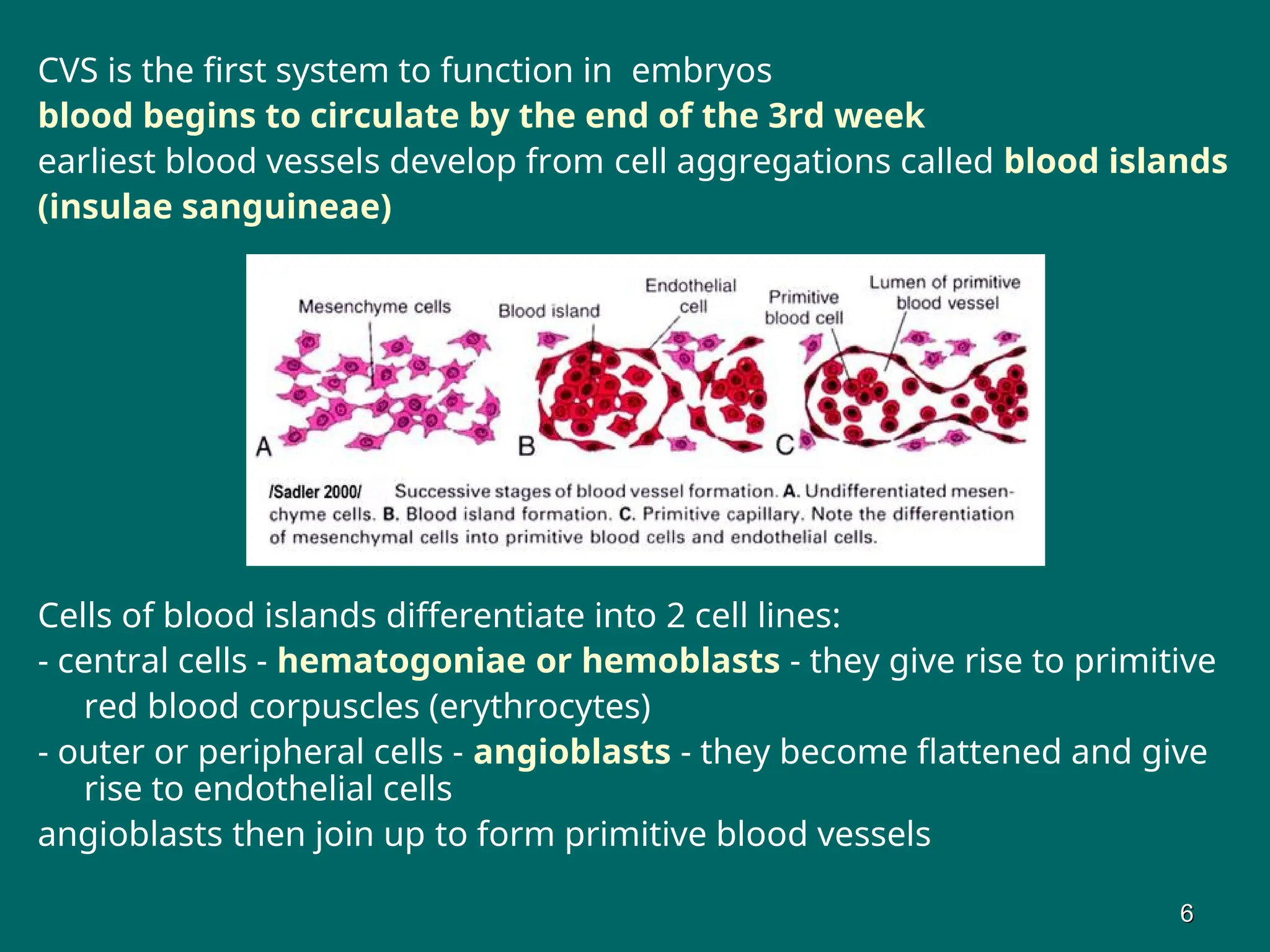
earliest blood vessels develop from cell aggregations called blood islands
(insulae sanguineae)
Cells of blood islands differentiate into 2 cell lines:
- central cells - hematogoniae or hemoblasts - they give rise to primitive
red blood corpuscles (erythrocytes)
- outer or peripheral cells - angioblasts - they become flattened and give
rise to endothelial cells
angioblasts then join up to form primitive blood vessels
6
6
7.
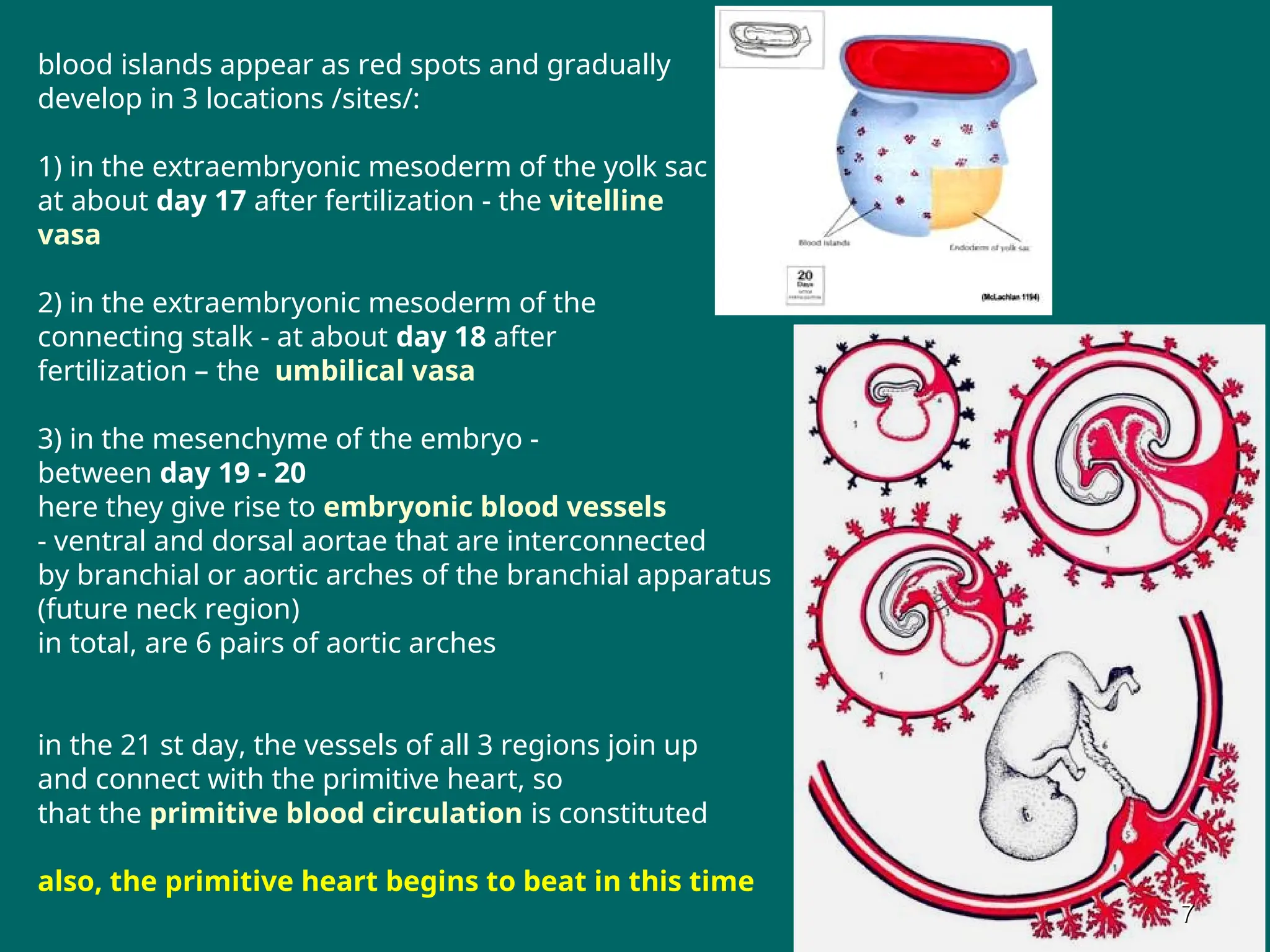
blood islands appearas red spots and gradually
develop in 3 locations /sites/:
1) in the extraembryonic mesoderm of the yolk sac -
at about day 17 after fertilization - the vitelline
vasa
2) in the extraembryonic mesoderm of the
connecting stalk - at about day 18 after
fertilization – the umbilical vasa
3) in the mesenchyme of the embryo -
between day 19 - 20
here they give rise to embryonic blood vessels
- ventral and dorsal aortae that are interconnected
by branchial or aortic arches of the branchial apparatus
(future neck region)
in total, are 6 pairs of aortic arches
in the 21 st day, the vessels of all 3 regions join up
and connect with the primitive heart, so
that the primitive blood circulation is constituted
also, the primitive heart begins to beat in this time
7
7
8.
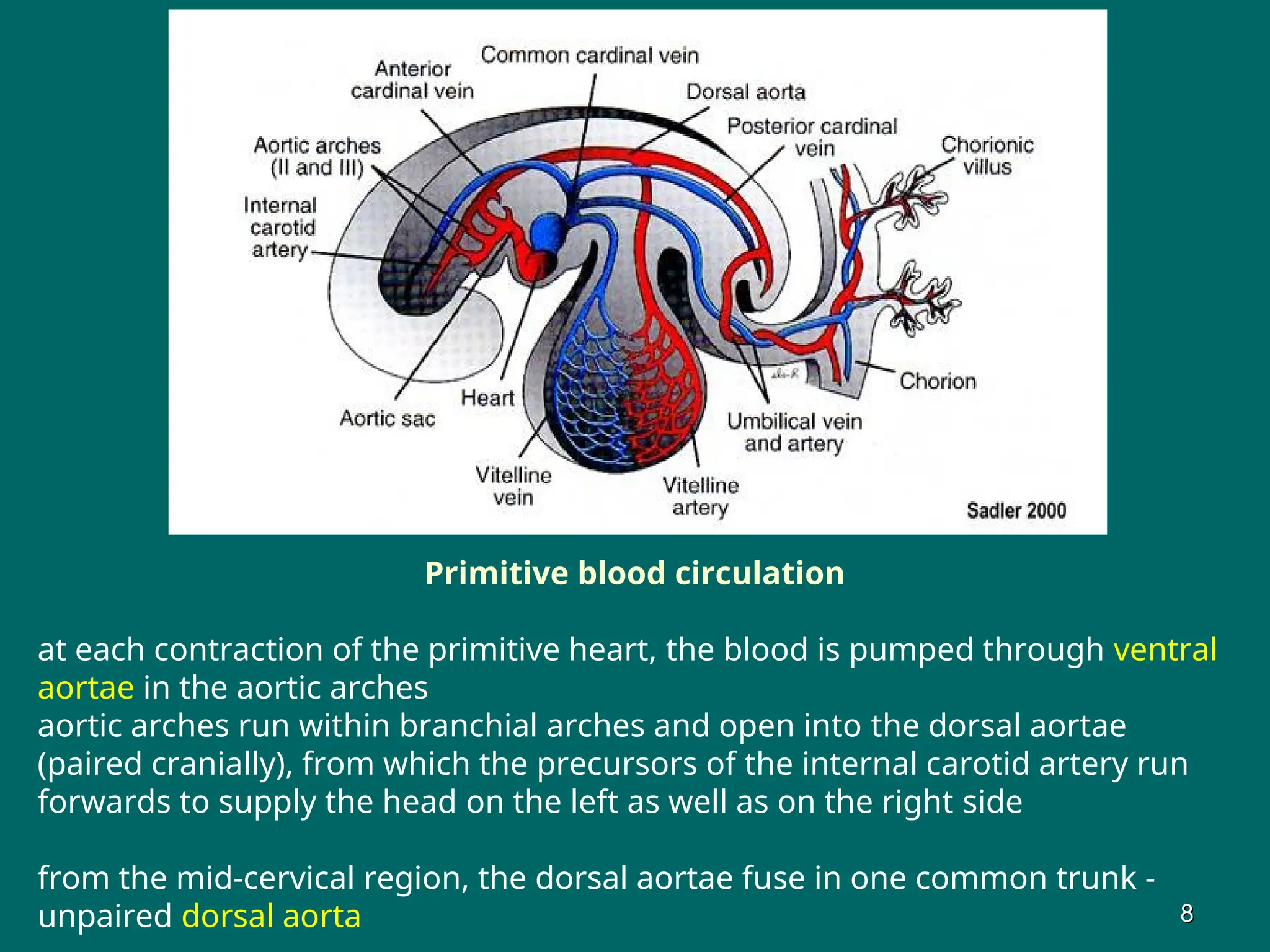
Primitive blood circulation
ateach contraction of the primitive heart, the blood is pumped through ventral
aortae in the aortic arches
aortic arches run within branchial arches and open into the dorsal aortae
(paired cranially), from which the precursors of the internal carotid artery run
forwards to supply the head on the left as well as on the right side
from the mid-cervical region, the dorsal aortae fuse in one common trunk -
unpaired dorsal aorta 8
8
9.
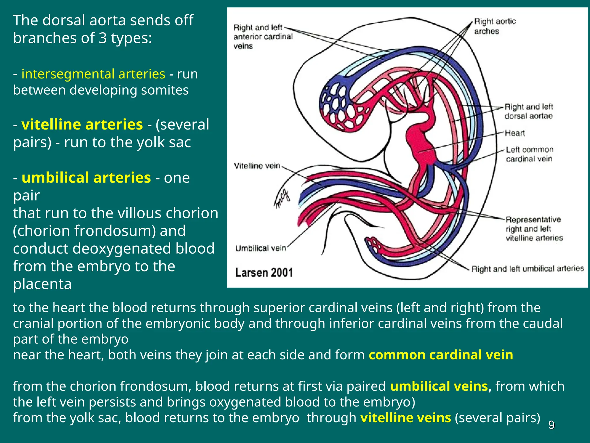
The dorsal aortasends off
branches of 3 types:
- intersegmental arteries - run
between developing somites
- vitelline arteries - (several
pairs) - run to the yolk sac
- umbilical arteries - one
pair
that run to the villous chorion
(chorion frondosum) and
conduct deoxygenated blood
from the embryo to the
placenta
to the heart the blood returns through superior cardinal veins (left and right) from the
cranial portion of the embryonic body and through inferior cardinal veins from the caudal
part of the embryo
near the heart, both veins they join at each side and form common cardinal vein
from the chorion frondosum, blood returns at first via paired umbilical veins, from which
the left vein persists and brings oxygenated blood to the embryo)
from the yolk sac, blood returns to the embryo through vitelline veins (several pairs)
9
9
10.
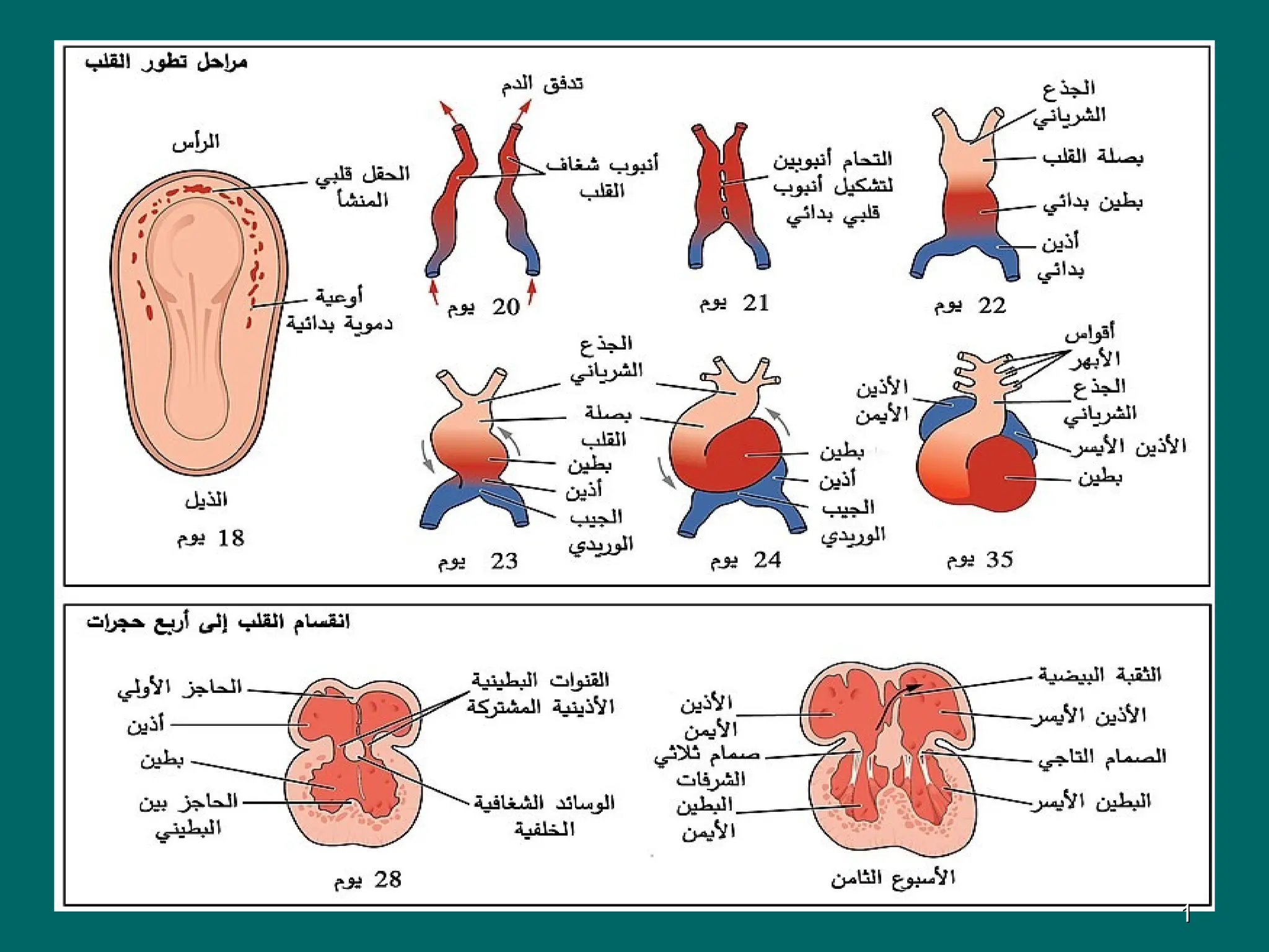
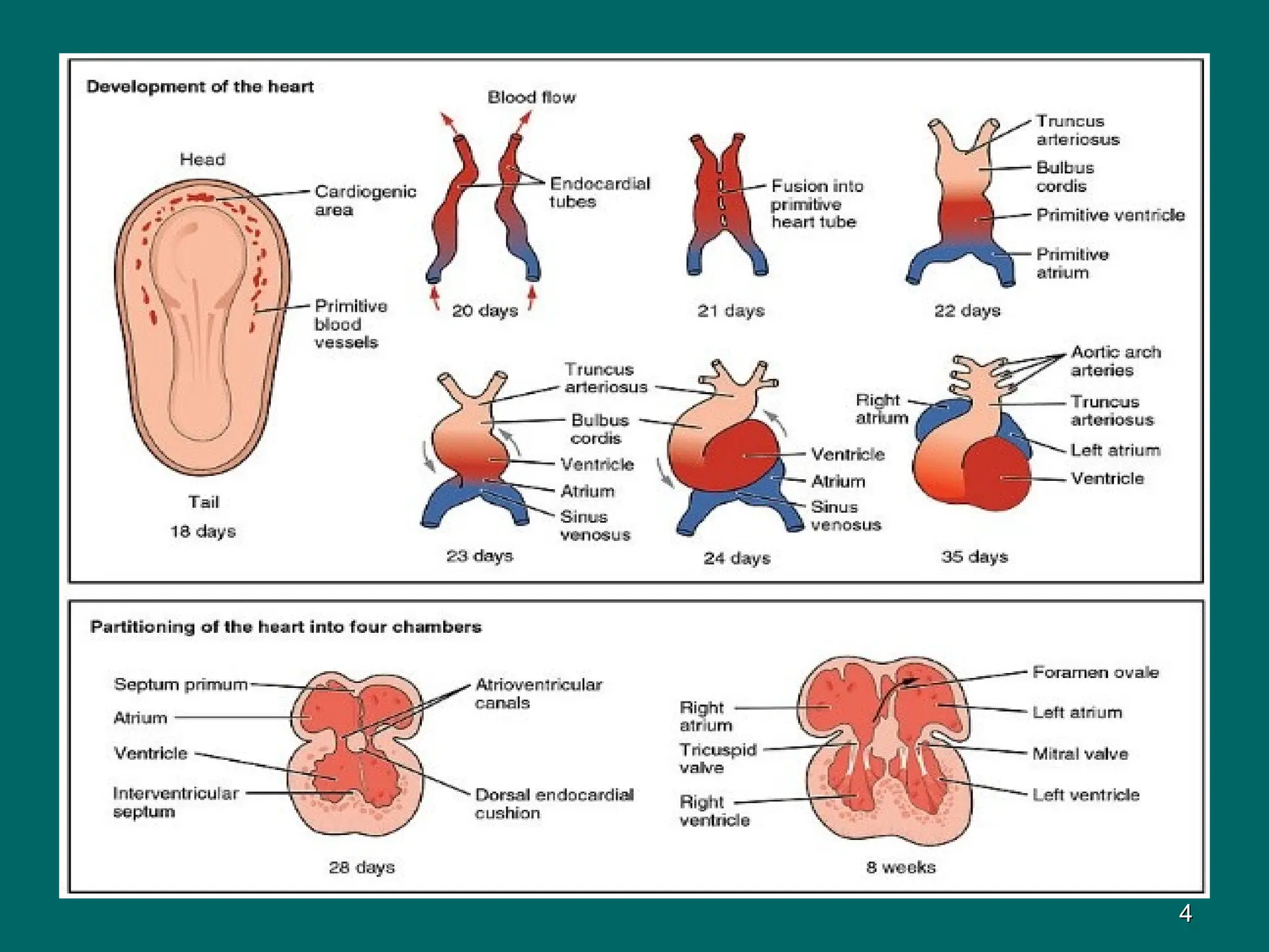
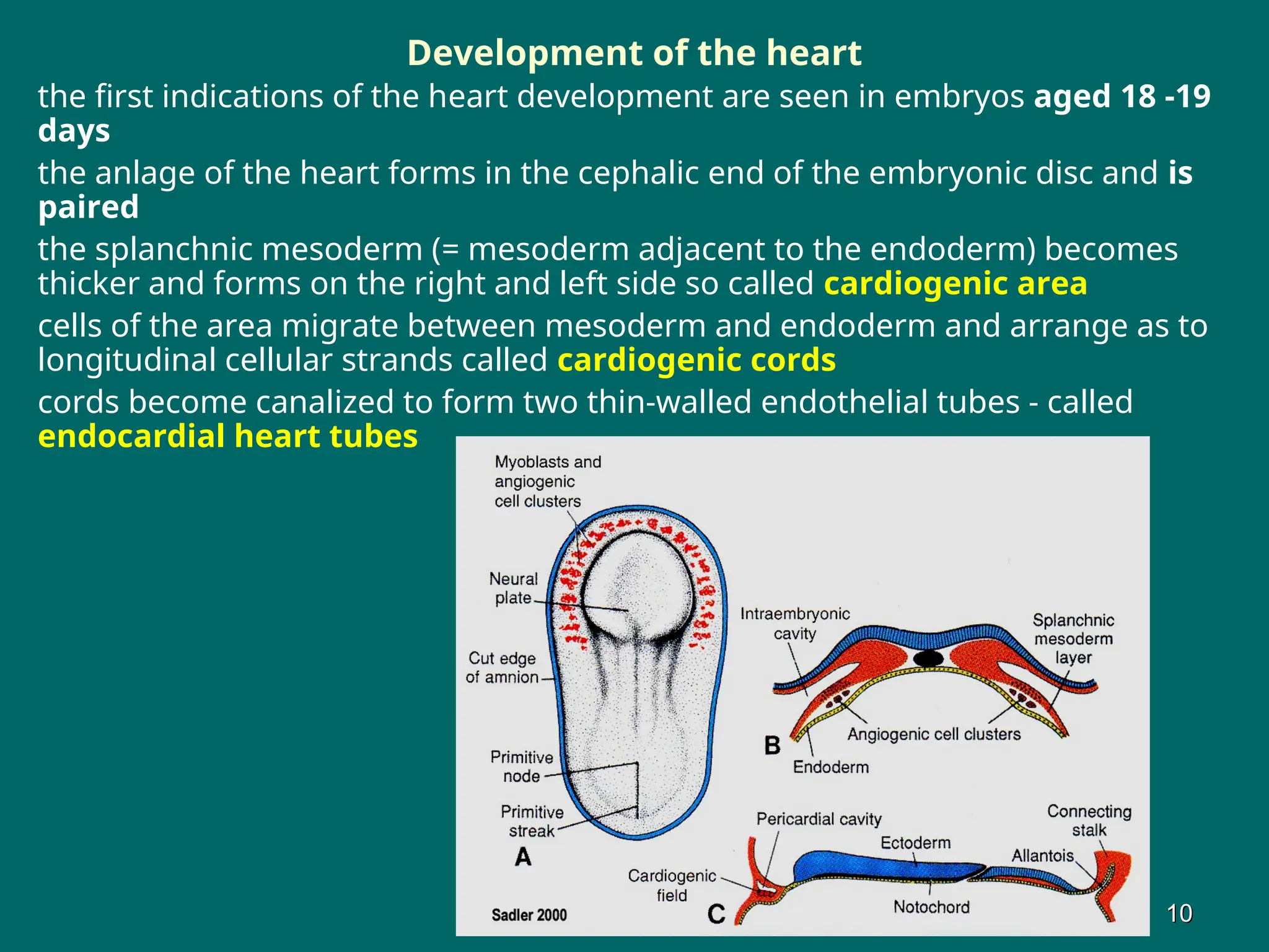
Development of theheart
the first indications of the heart development are seen in embryos aged 18 -19
days
the anlage of the heart forms in the cephalic end of the embryonic disc and is
paired
the splanchnic mesoderm (= mesoderm adjacent to the endoderm) becomes
thicker and forms on the right and left side so called cardiogenic area
cells of the area migrate between mesoderm and endoderm and arrange as to
longitudinal cellular strands called cardiogenic cords
cords become canalized to form two thin-walled endothelial tubes - called
endocardial heart tubes
10
10
11.
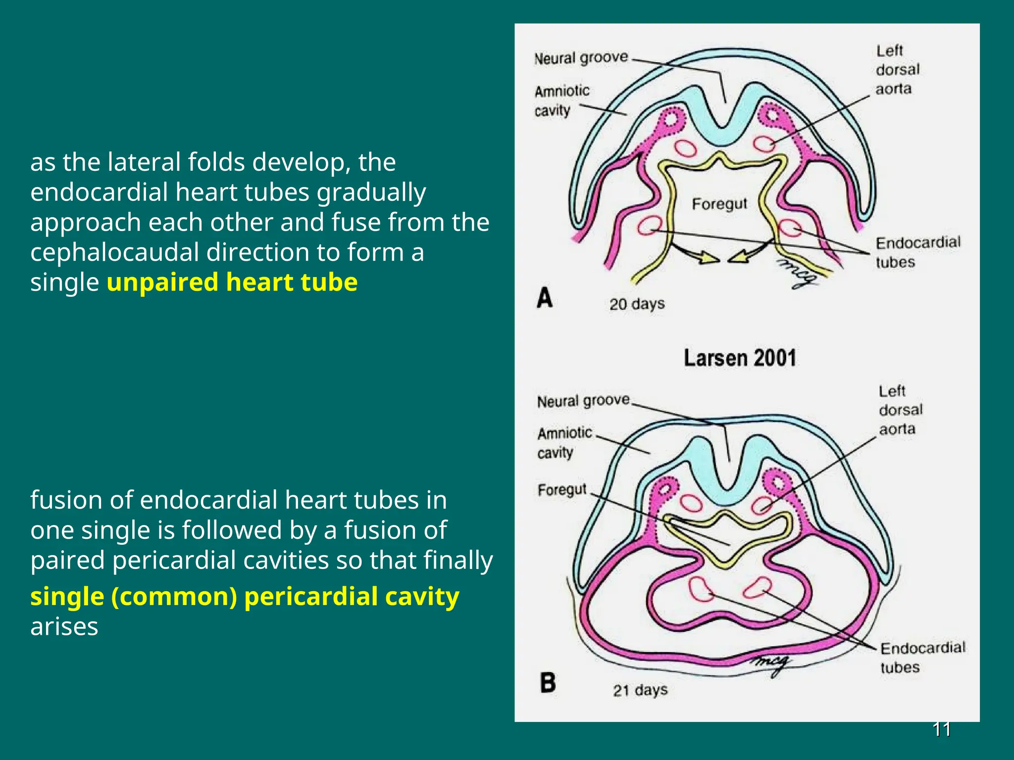
as the lateralfolds develop, the
endocardial heart tubes gradually
approach each other and fuse from the
cephalocaudal direction to form a
single unpaired heart tube
fusion of endocardial heart tubes in
one single is followed by a fusion of
paired pericardial cavities so that finally
single (common) pericardial cavity
arises
11
11
12.
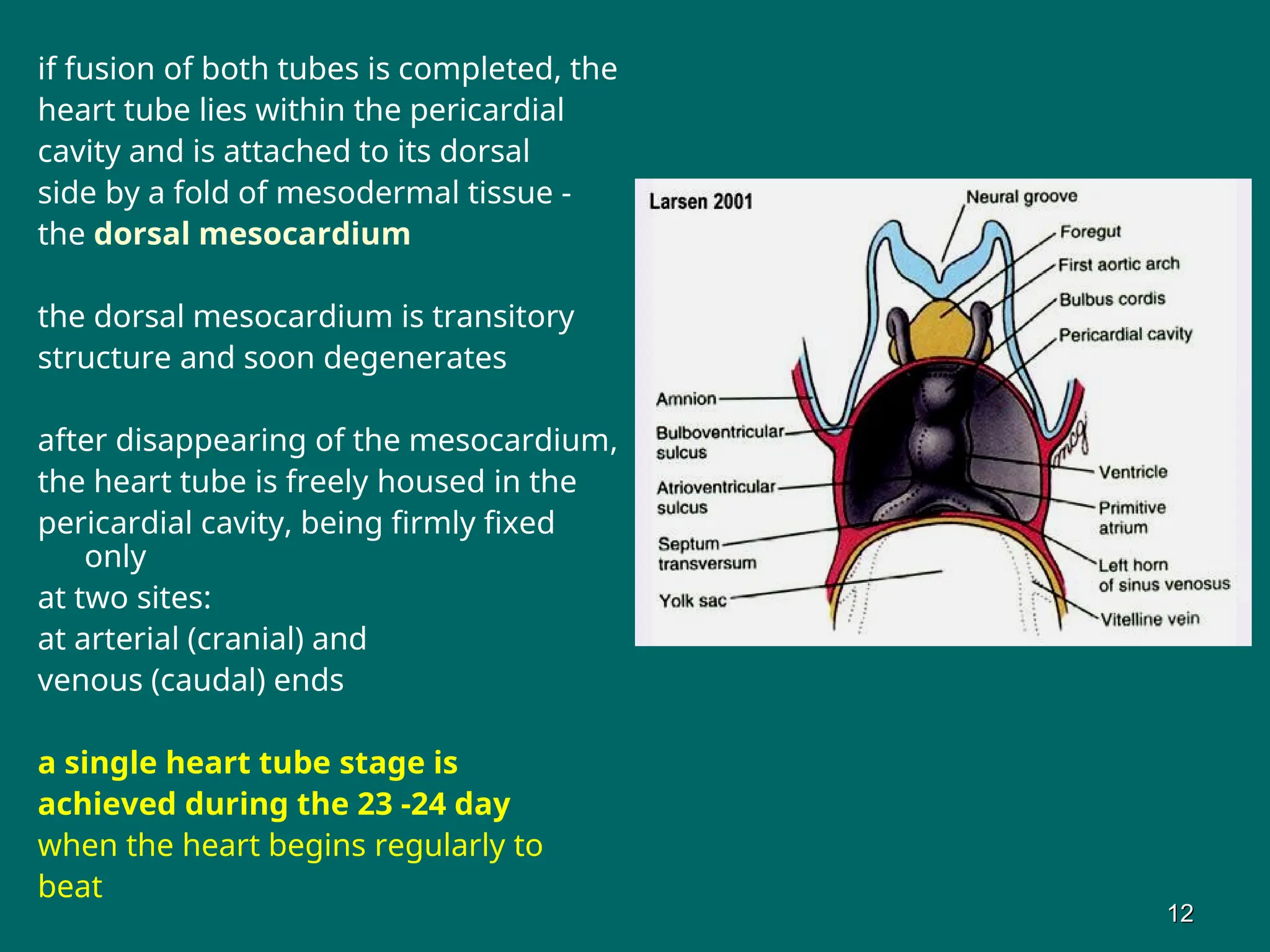
if fusion ofboth tubes is completed, the
heart tube lies within the pericardial
cavity and is attached to its dorsal
side by a fold of mesodermal tissue -
the dorsal mesocardium
the dorsal mesocardium is transitory
structure and soon degenerates
after disappearing of the mesocardium,
the heart tube is freely housed in the
pericardial cavity, being firmly fixed
only
at two sites:
at arterial (cranial) and
venous (caudal) ends
a single heart tube stage is
achieved during the 23 -24 day
when the heart begins regularly to
beat
12
12
13.
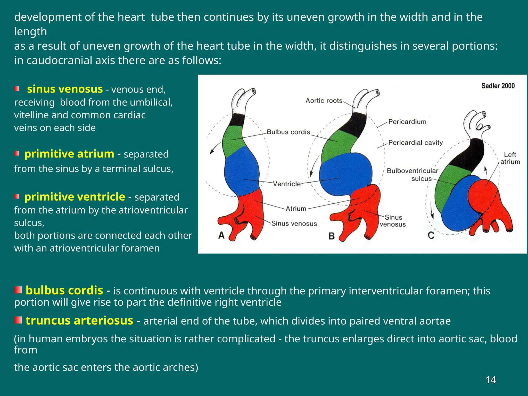
development of theheart tube then continues by its uneven growth in the width and in the
length
as a result of uneven growth of the heart tube in the width, it distinguishes in several portions:
in caudocranial axis there are as follows:
sinus venosus - venous end,
receiving blood from the umbilical,
vitelline and common cardiac
veins on each side
primitive atrium - separated
from the sinus by a terminal sulcus,
primitive ventricle - separated
from the atrium by the atrioventricular
sulcus,
both portions are connected each other
with an atrioventricular foramen
bulbus cordis - is continuous with ventricle through the primary interventricular foramen; this
portion will give rise to part the definitive right ventricle
truncus arteriosus - arterial end of the tube, which divides into paired ventral aortae
(in human embryos the situation is rather complicated - the truncus enlarges direct into aortic sac, blood
from
the aortic sac enters the aortic arches)
14
14
14.
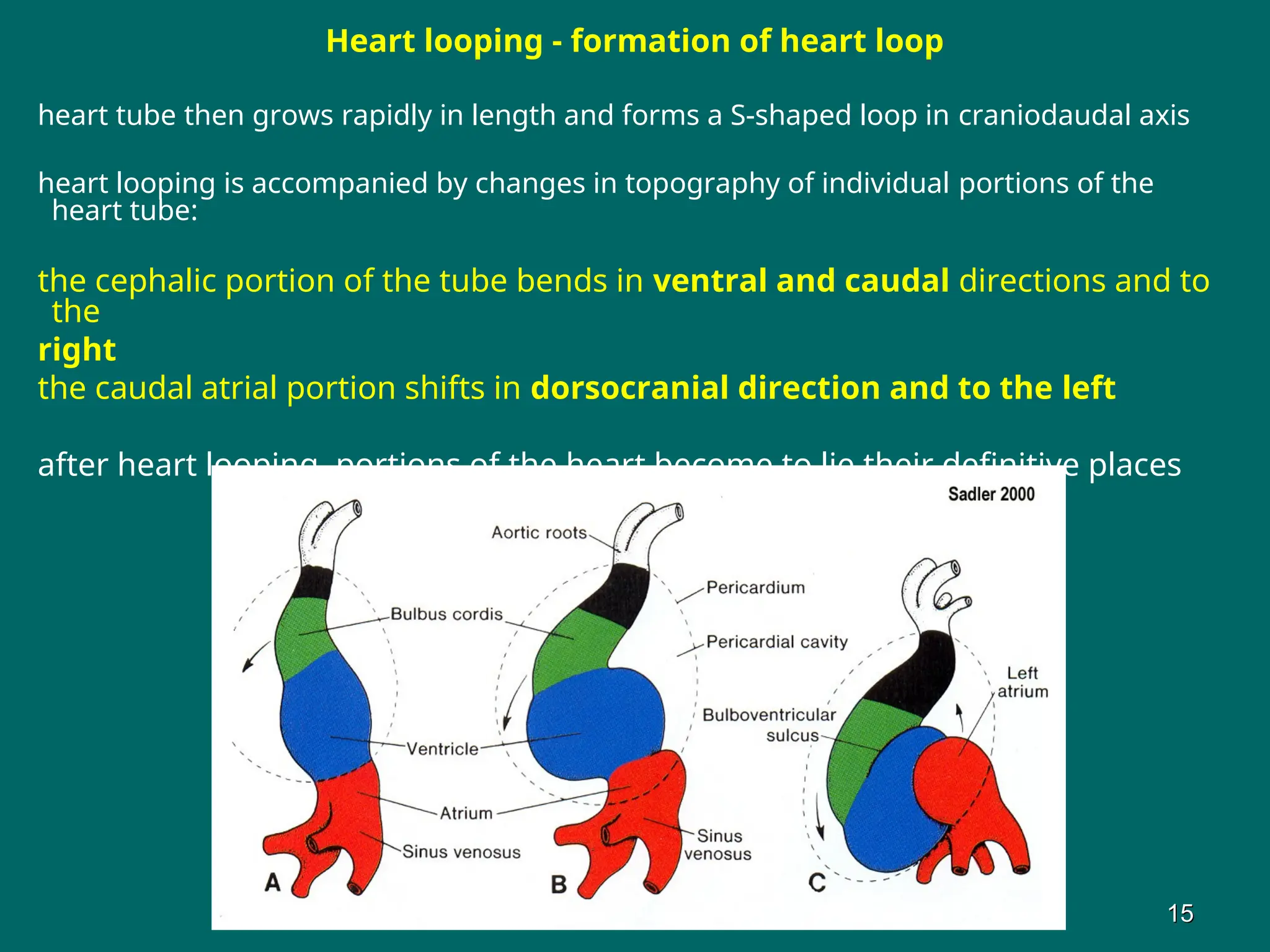
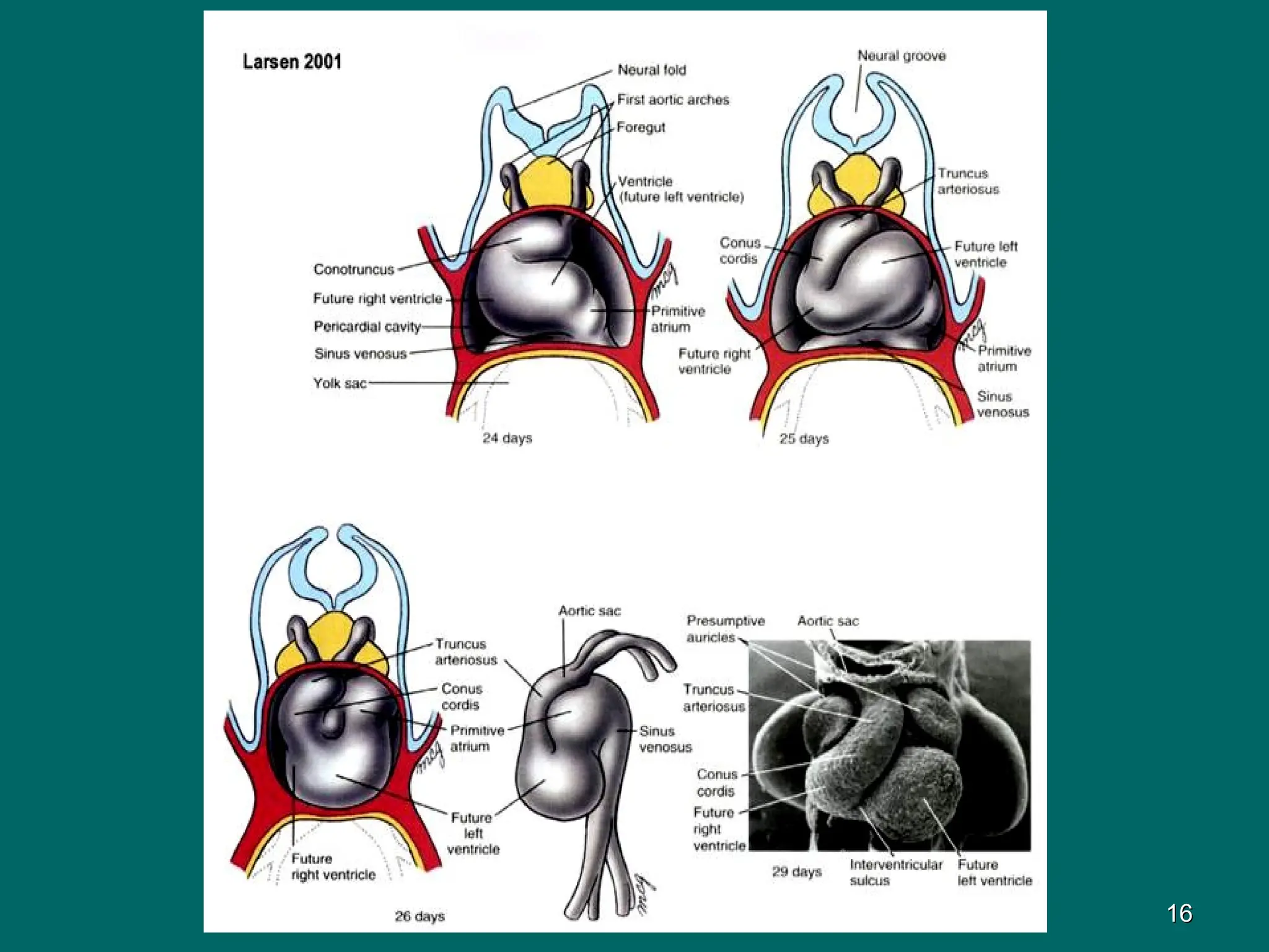
Heart looping -formation of heart loop
heart tube then grows rapidly in length and forms a S-shaped loop in craniodaudal axis
heart looping is accompanied by changes in topography of individual portions of the
heart tube:
the cephalic portion of the tube bends in ventral and caudal directions and to
the
right
the caudal atrial portion shifts in dorsocranial direction and to the left
after heart looping, portions of the heart become to lie their definitive places
15
15
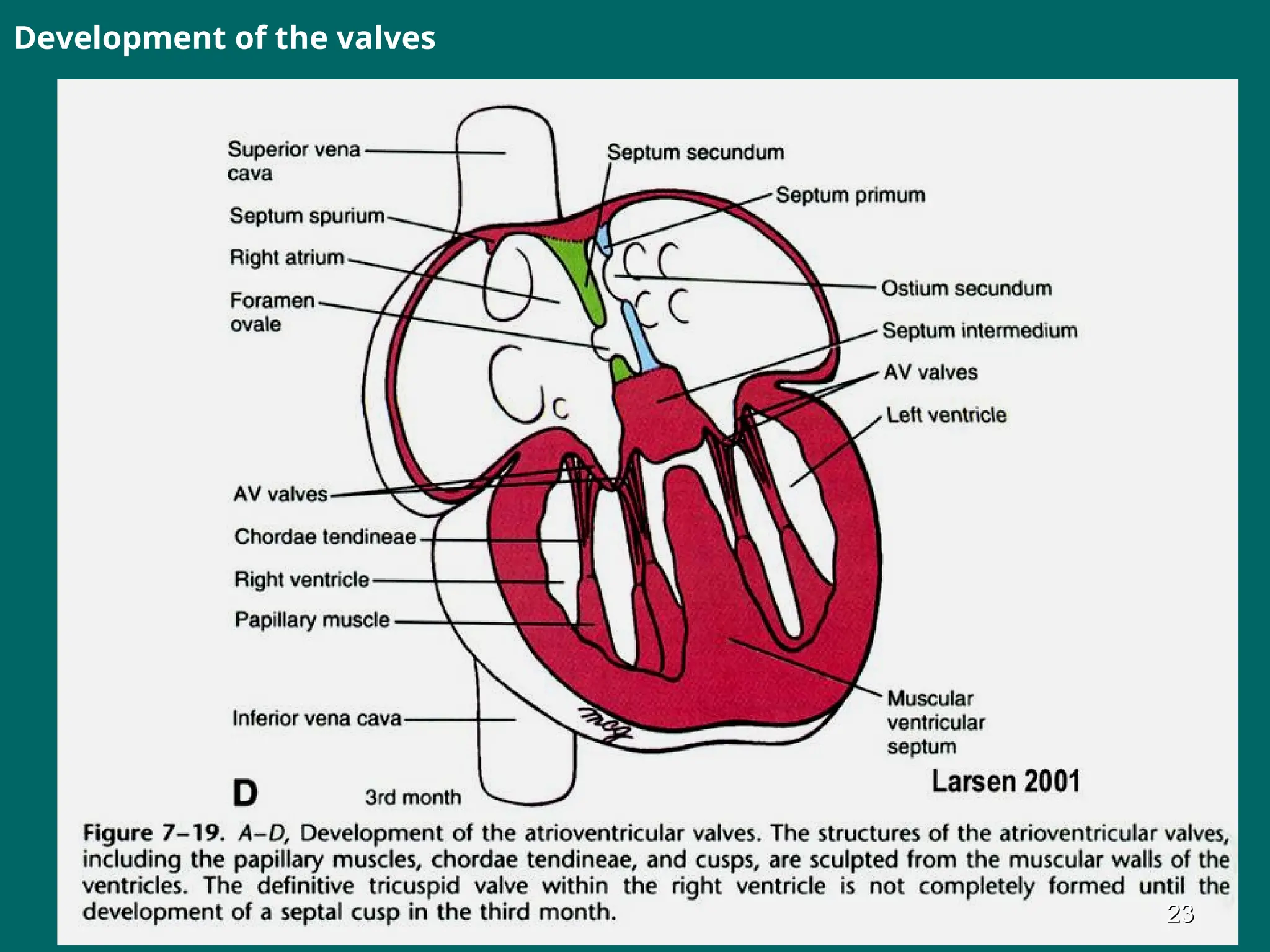
Septation of theheart (formation of cardiac septa)
the septation process = division of the heart into two halves down midline
the process begins in the 5th week and ends in a week later
3 septae take part in division of the heart in the right and left chamber
there are as follows:
interatrial septum
interventricular septum
aorticopulmonary septum
Development of the interatrial septum
the definitive interatrial septum shows a complicated development
septum originates from two septae that fuse each other after birth of the fetus:
the septum primum and
the septum secundum
17
17
17.
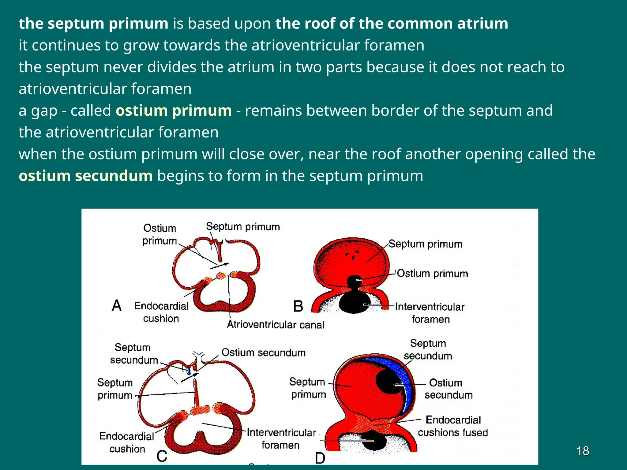
the septum primumis based upon the roof of the common atrium
it continues to grow towards the atrioventricular foramen
the septum never divides the atrium in two parts because it does not reach to
atrioventricular foramen
a gap - called ostium primum - remains between border of the septum and
the atrioventricular foramen
when the ostium primum will close over, near the roof another opening called the
ostium secundum begins to form in the septum primum
18
18
18.
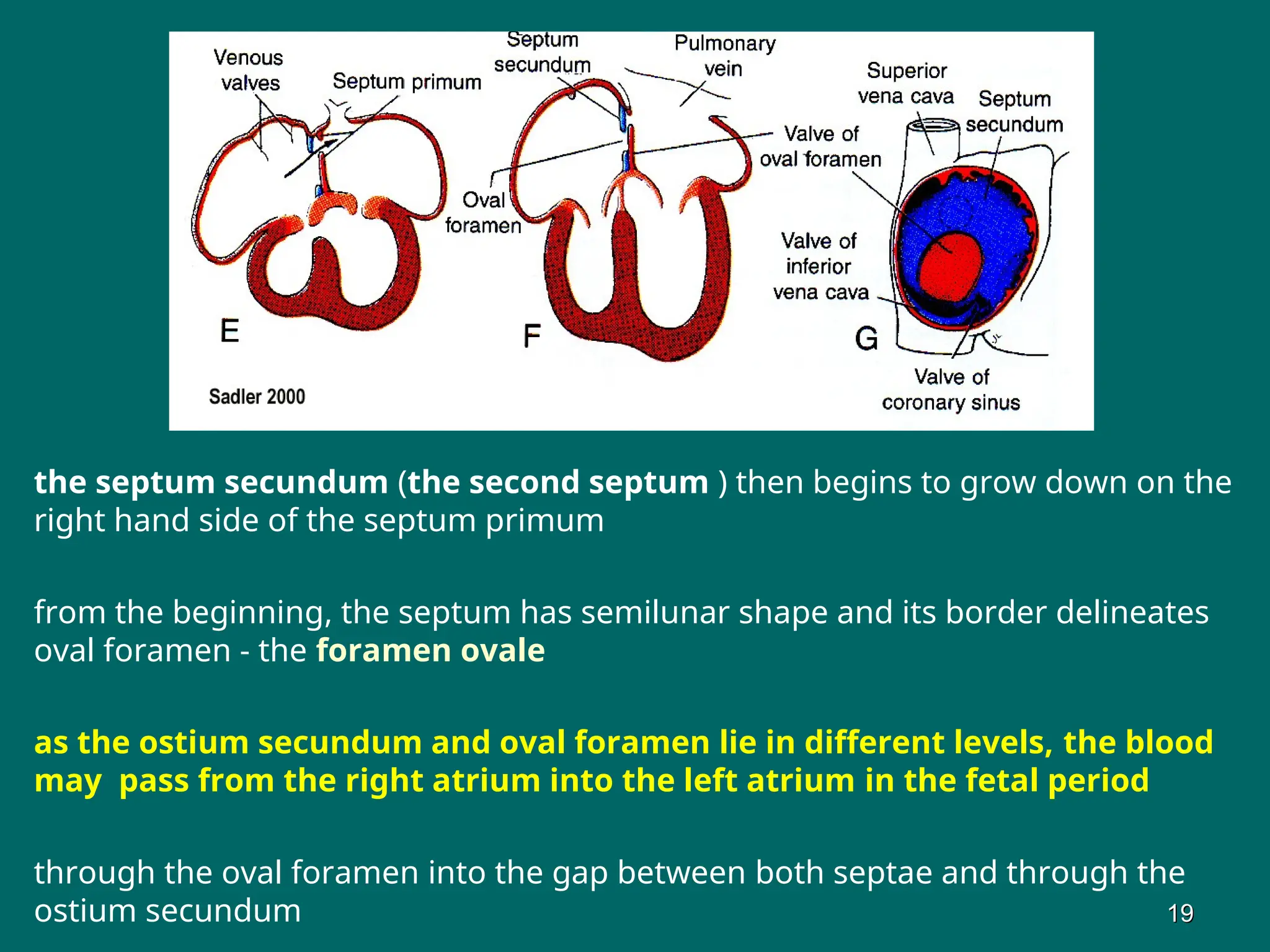
the septum secundum(the second septum ) then begins to grow down on the
right hand side of the septum primum
from the beginning, the septum has semilunar shape and its border delineates
oval foramen - the foramen ovale
as the ostium secundum and oval foramen lie in different levels, the blood
may pass from the right atrium into the left atrium in the fetal period
through the oval foramen into the gap between both septae and through the
ostium secundum 19
19
19.
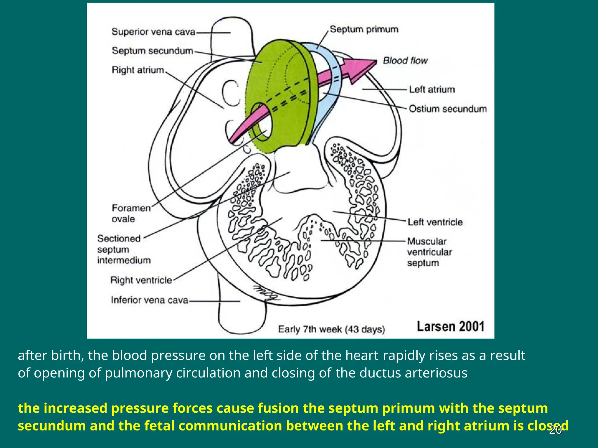
after birth, theblood pressure on the left side of the heart rapidly rises as a result
of opening of pulmonary circulation and closing of the ductus arteriosus
the increased pressure forces cause fusion the septum primum with the septum
secundum and the fetal communication between the left and right atrium is closed
20
20
20.
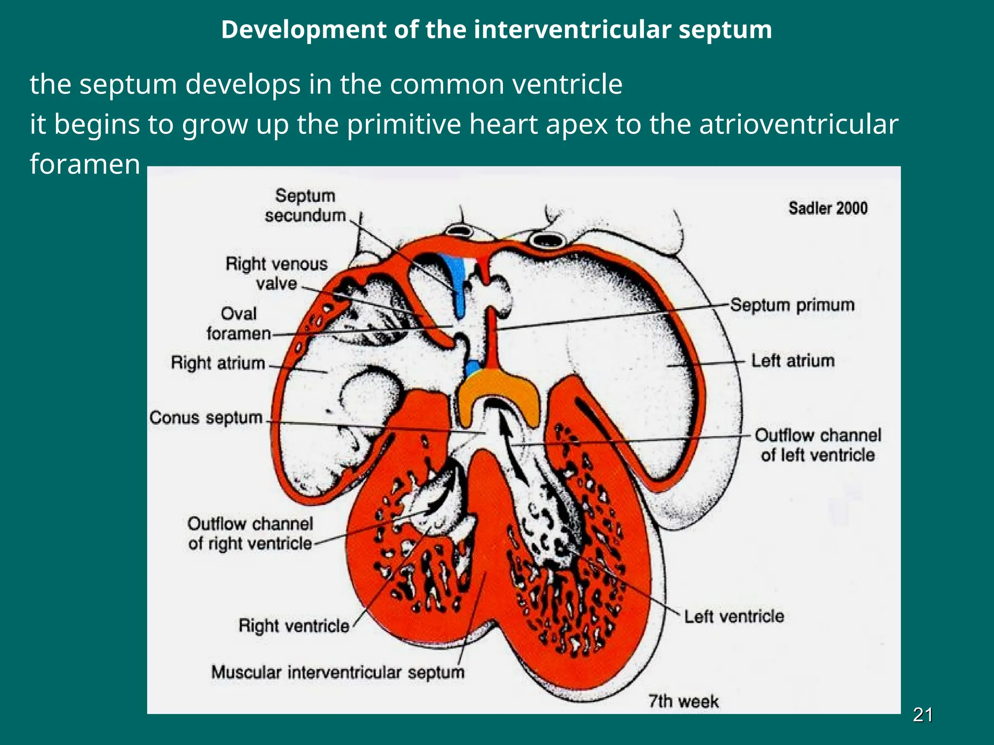
Development of theinterventricular septum
the septum develops in the common ventricle
it begins to grow up the primitive heart apex to the atrioventricular
foramen
21
21
21.
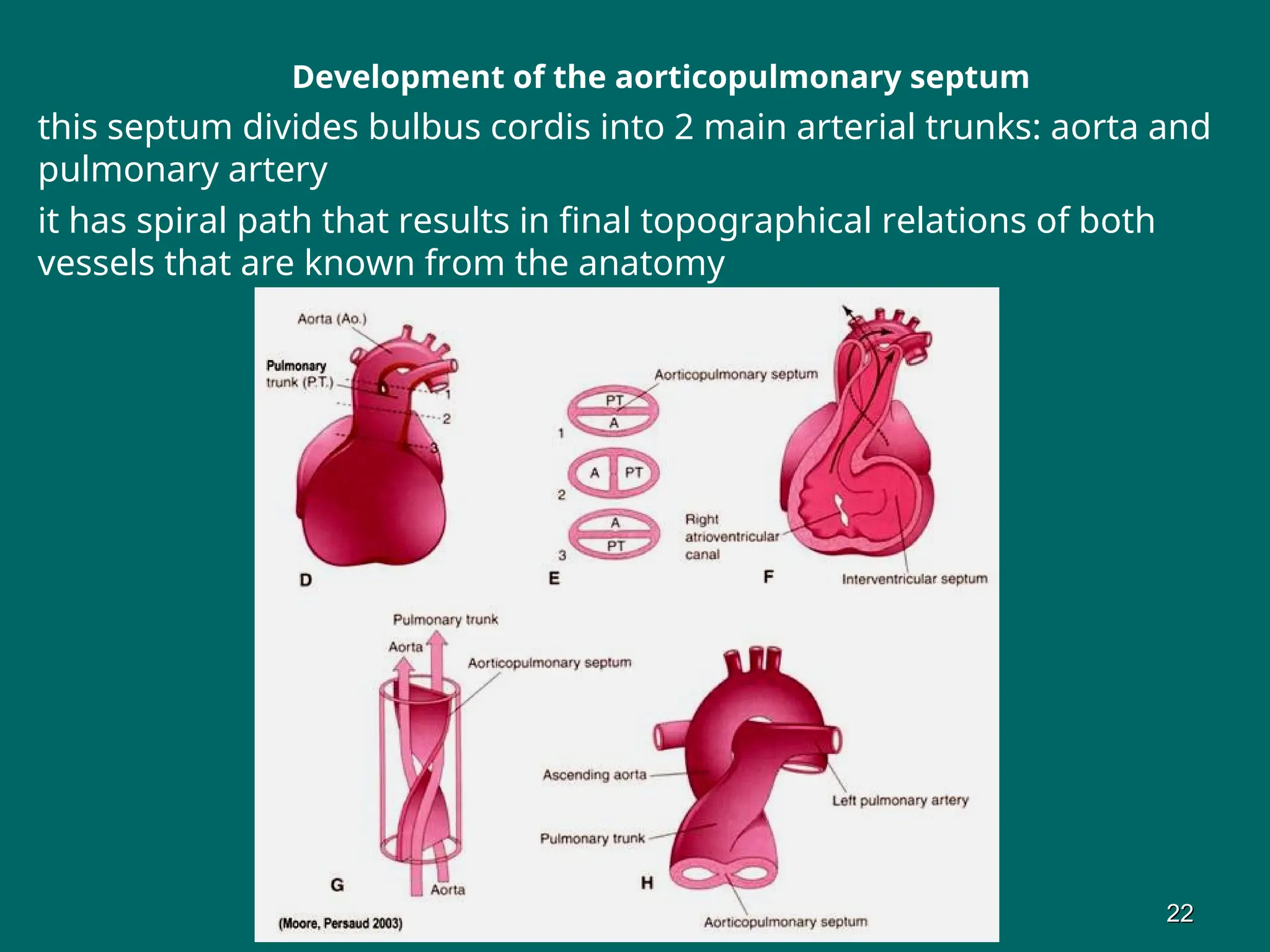
Development of theaorticopulmonary septum
this septum divides bulbus cordis into 2 main arterial trunks: aorta and
pulmonary artery
it has spiral path that results in final topographical relations of both
vessels that are known from the anatomy
22
22
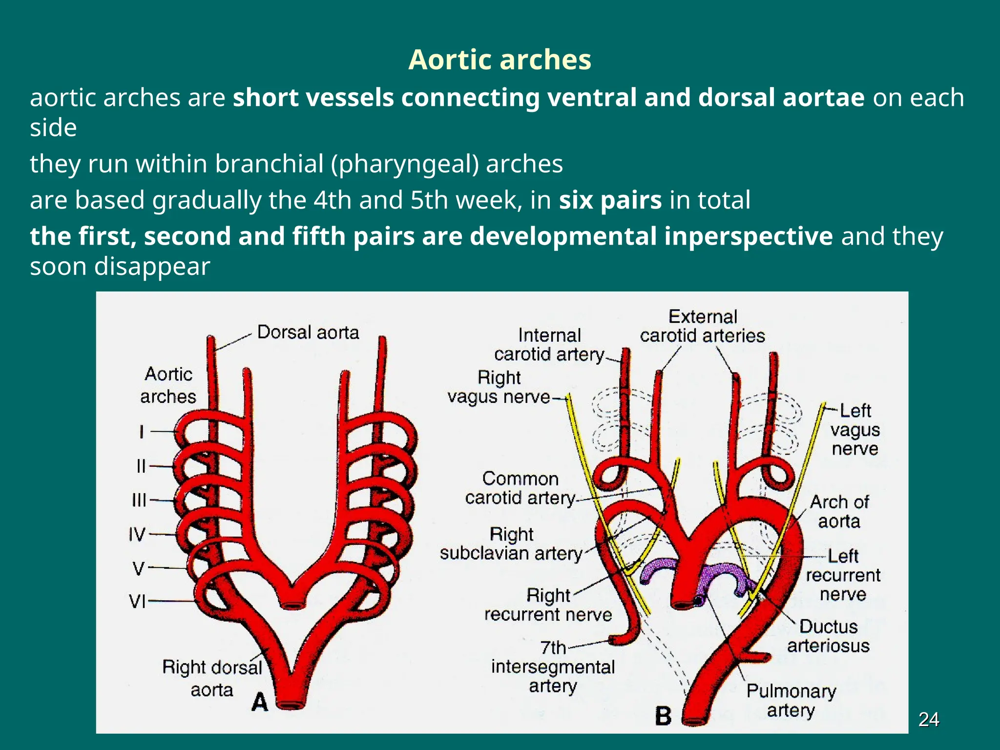
Aortic arches
aortic archesare short vessels connecting ventral and dorsal aortae on each
side
they run within branchial (pharyngeal) arches
are based gradually the 4th and 5th week, in six pairs in total
the first, second and fifth pairs are developmental inperspective and they
soon disappear
24
24
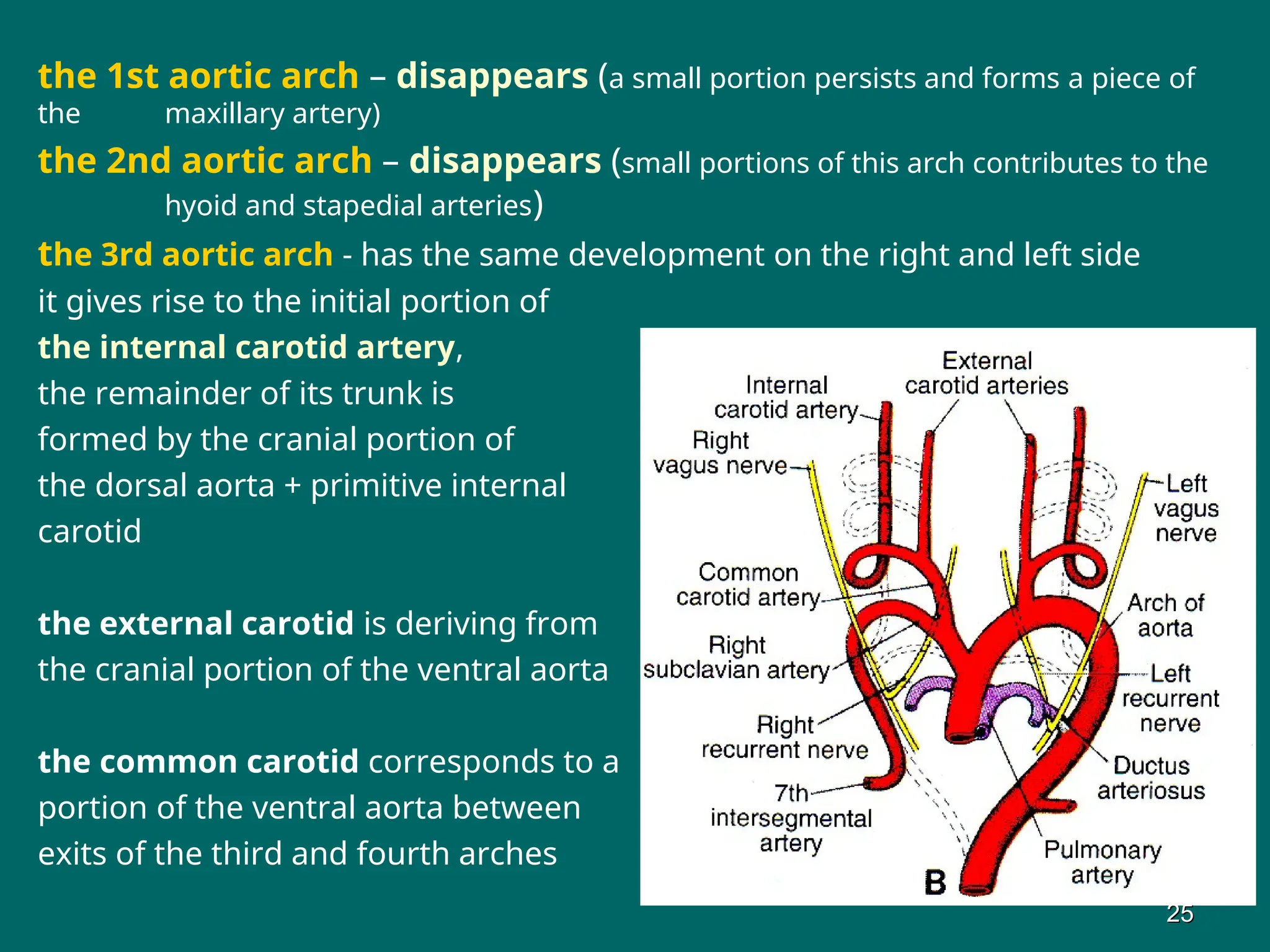
24.
the 1st aorticarch – disappears (a small portion persists and forms a piece of
the maxillary artery)
the 2nd aortic arch – disappears (small portions of this arch contributes to the
hyoid and stapedial arteries)
the 3rd aortic arch - has the same development on the right and left side
it gives rise to the initial portion of
the internal carotid artery,
the remainder of its trunk is
formed by the cranial portion of
the dorsal aorta + primitive internal
carotid
the external carotid is deriving from
the cranial portion of the ventral aorta
the common carotid corresponds to a
portion of the ventral aorta between
exits of the third and fourth arches
25
25
25.
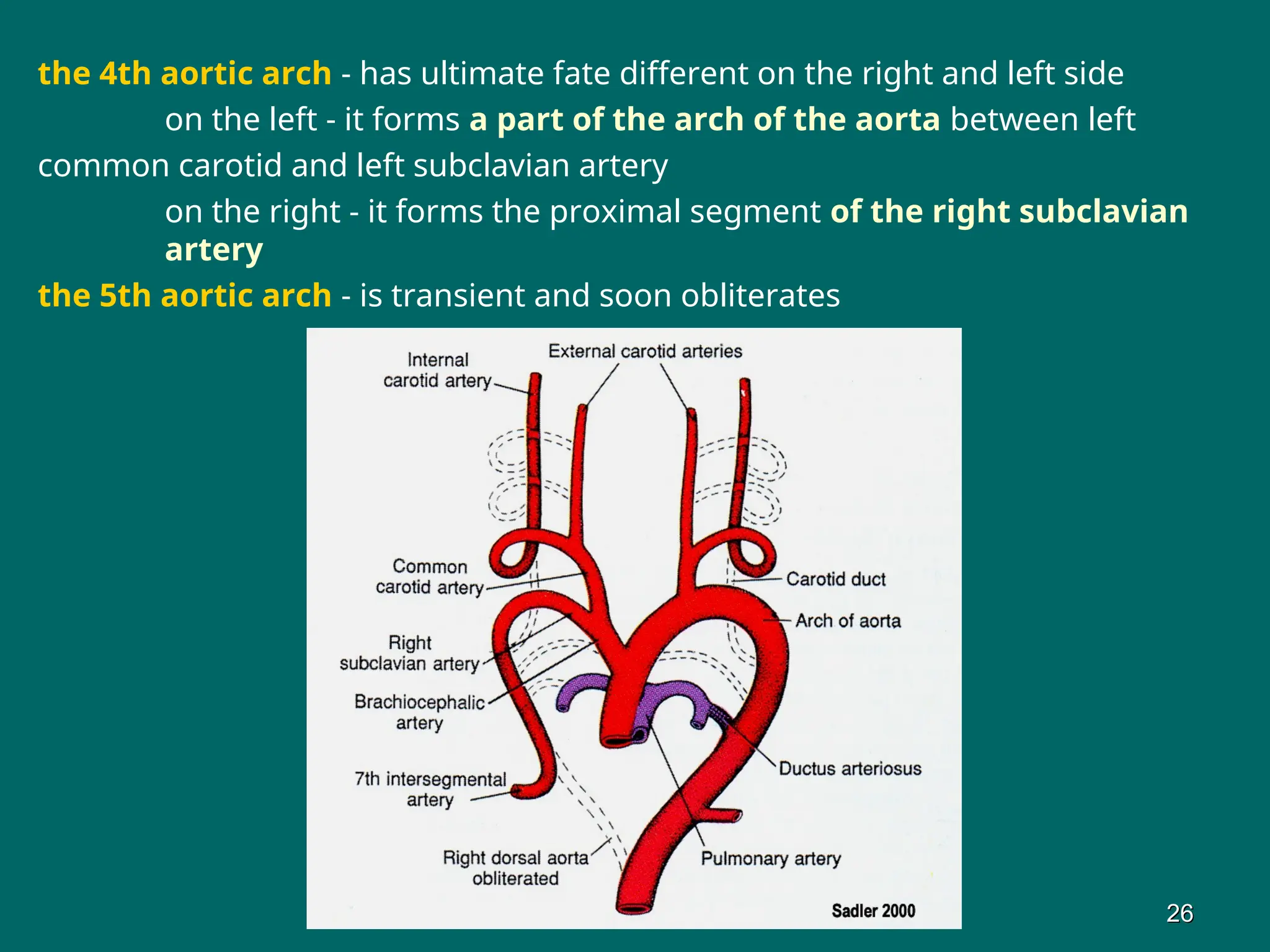
the 4th aorticarch - has ultimate fate different on the right and left side
on the left - it forms a part of the arch of the aorta between left
common carotid and left subclavian artery
on the right - it forms the proximal segment of the right subclavian
artery
the 5th aortic arch - is transient and soon obliterates
26
26
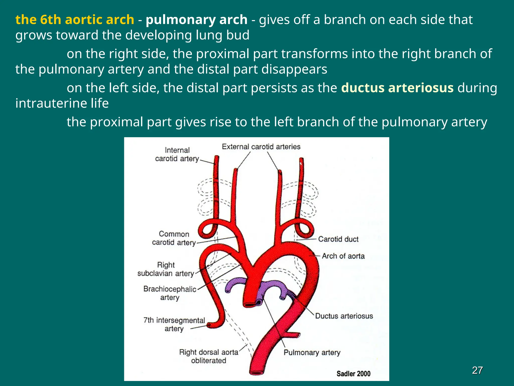
26.
the 6th aorticarch - pulmonary arch - gives off a branch on each side that
grows toward the developing lung bud
on the right side, the proximal part transforms into the right branch of
the pulmonary artery and the distal part disappears
on the left side, the distal part persists as the ductus arteriosus during
intrauterine life
the proximal part gives rise to the left branch of the pulmonary artery
27
27
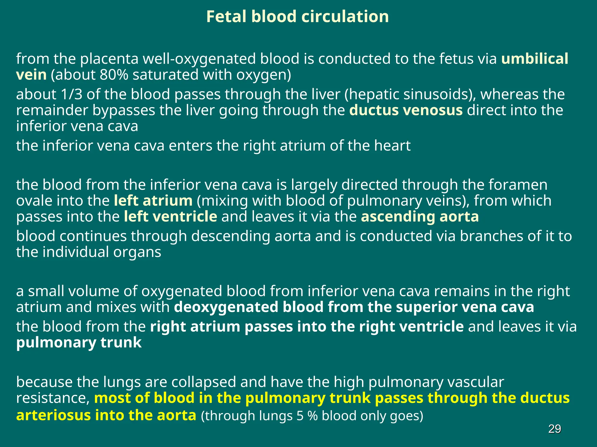
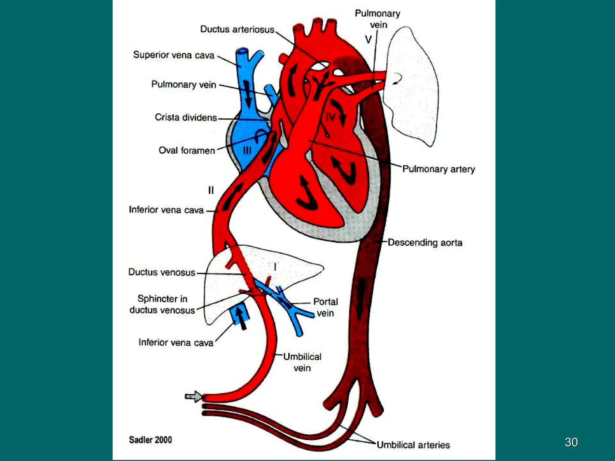
Fetal blood circulation
fromthe placenta well-oxygenated blood is conducted to the fetus via umbilical
vein (about 80% saturated with oxygen)
about 1/3 of the blood passes through the liver (hepatic sinusoids), whereas the
remainder bypasses the liver going through the ductus venosus direct into the
inferior vena cava
the inferior vena cava enters the right atrium of the heart
the blood from the inferior vena cava is largely directed through the foramen
ovale into the left atrium (mixing with blood of pulmonary veins), from which
passes into the left ventricle and leaves it via the ascending aorta
blood continues through descending aorta and is conducted via branches of it to
the individual organs
a small volume of oxygenated blood from inferior vena cava remains in the right
atrium and mixes with deoxygenated blood from the superior vena cava
the blood from the right atrium passes into the right ventricle and leaves it via
pulmonary trunk
because the lungs are collapsed and have the high pulmonary vascular
resistance, most of blood in the pulmonary trunk passes through the ductus
arteriosus into the aorta (through lungs 5 % blood only goes)
29
29
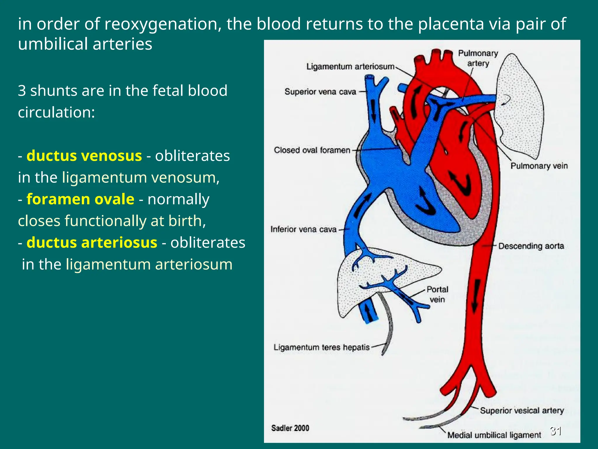
in order ofreoxygenation, the blood returns to the placenta via pair of
umbilical arteries
3 shunts are in the fetal blood
circulation:
- ductus venosus - obliterates
in the ligamentum venosum,
- foramen ovale - normally
closes functionally at birth,
- ductus arteriosus - obliterates
in the ligamentum arteriosum
31
31
31.
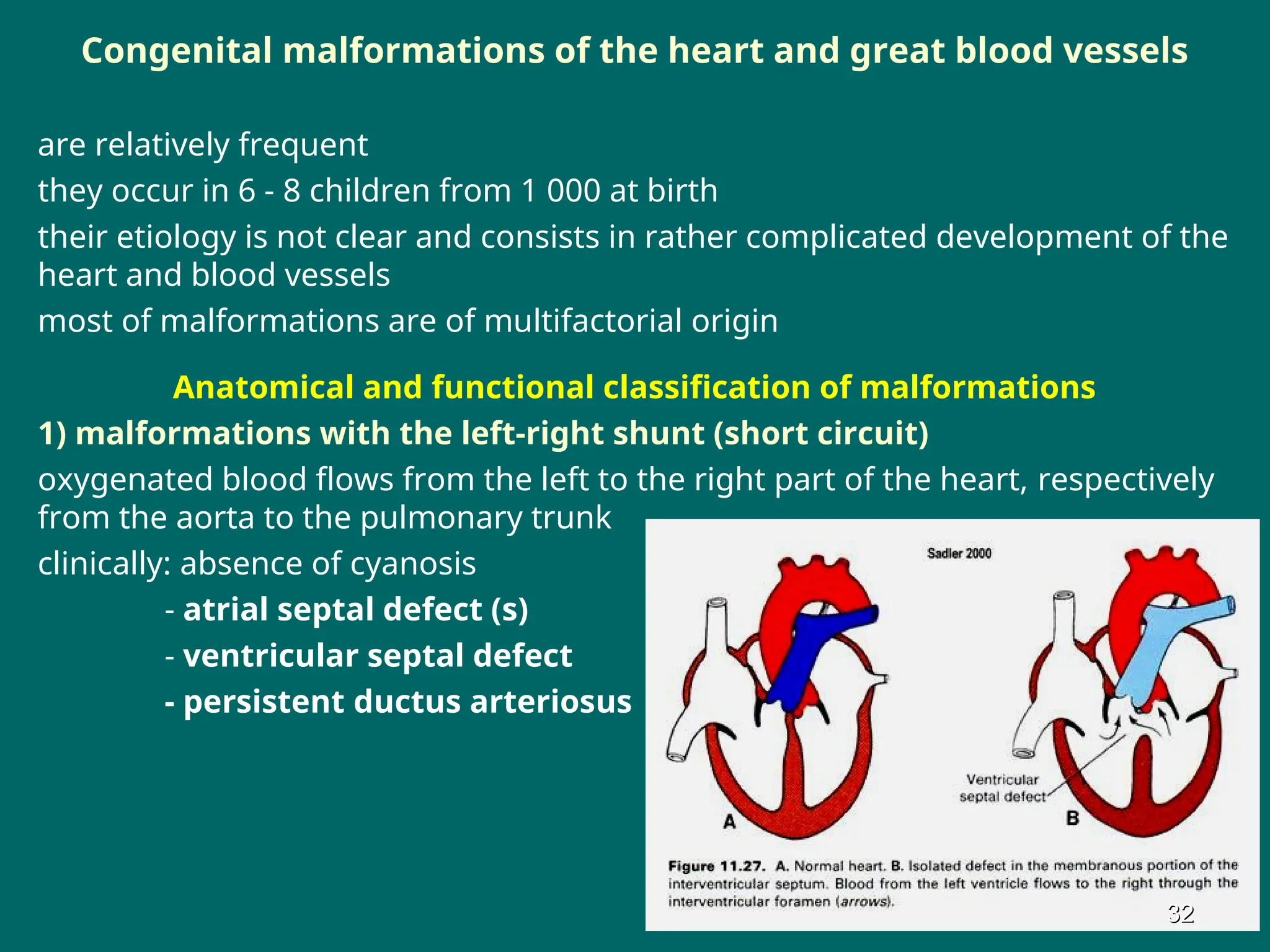
Congenital malformations ofthe heart and great blood vessels
are relatively frequent
they occur in 6 - 8 children from 1 000 at birth
their etiology is not clear and consists in rather complicated development of the
heart and blood vessels
most of malformations are of multifactorial origin
Anatomical and functional classification of malformations
1) malformations with the left-right shunt (short circuit)
oxygenated blood flows from the left to the right part of the heart, respectively
from the aorta to the pulmonary trunk
clinically: absence of cyanosis
- atrial septal defect (s)
- ventricular septal defect
- persistent ductus arteriosus
32
32
32.
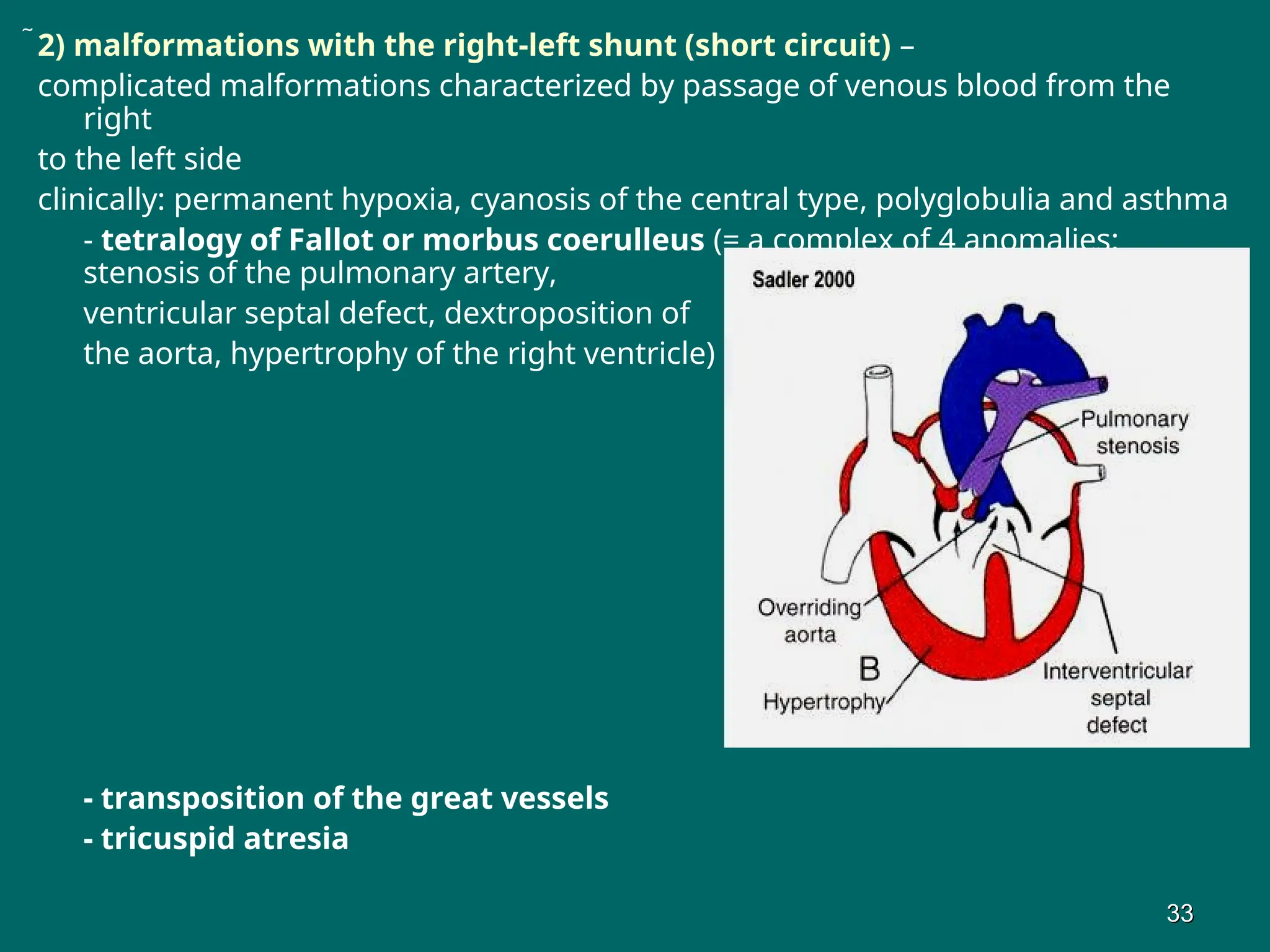
2) malformations withthe right-left shunt (short circuit) –
complicated malformations characterized by passage of venous blood from the
right
to the left side
clinically: permanent hypoxia, cyanosis of the central type, polyglobulia and asthma
- tetralogy of Fallot or morbus coerulleus (= a complex of 4 anomalies:
stenosis of the pulmonary artery,
ventricular septal defect, dextroposition of
the aorta, hypertrophy of the right ventricle)
- transposition of the great vessels
- tricuspid atresia
33
33
33.
3) malformations withoutshunts (short circuits) - the pulmonary and
systemic circulations are separated
blood volumes on the right and the left sides are equal
the group includes:
- aortic valvular stenosis or atresia
- coarctation of the aorta
- double aortic arch
- right aortic arch
- valvular stenosis of the pulmonary artery
4) abnormalities in heart position:
- dextrocardia - the heart lies on the right side
- ectopia cordis - the heart is located on the surface of the
chest
Sequency of CM of the heart and great vessels:
- persistent ductus arteriosus
- ventricular septal defect
- tetralogy of Fallot
- atrial septal defect (s)
- stenosis of pulmonary trunk 34
34