SlideShare a Scribd company logo
1 of 70
DEVELOPMENT
OF MAXILLA
BY – Dr. Vipul Gupta
MDS 1ST YR
1
CONTENTS
EMBRYONIC DEVELOPMENT
ANATOMY OF MAXILLA
GROWTH AND DEVELOPMENT
CRANIOFACIAL GROWTH DISORDERS
REFERENCES
2
PRE-NATAL
GORWTH OF
MAXILLA
3
PRE NATAL EMBROYOLOGY
OF MAXILLA
• Around 4th week of IUL , a prominent bulge
appears on ventral aspect of embryo
corresponding to developing brain.
• Below this bulge a shallow depression which
correspond to the primitive mouth appears
called STOMODEUM.
• The floor of the stomodeum is formed by
BUCCOPHARNGEAL MEMEBRANE that
separate the stomodeum from the foregut.
4
• By around the 4th week of intra-uterine life ,5 branchial arches
form in the region of future head and neck .Each of these arches
give rise to muscles , connective tissue, vasculature, skeletal
components and neural components of the future face.
• The 1st branchial arch is called the MANDIBULAR ARCH and
plays important role in development of naso-maxillary region.
5
• The mesoderm covering the developing forebrain proliferates and
forms a downward projection that overlaps the upper part of
stomodeum.
• This downward projection is known as FRONTO-NASAL
PROCESS.
6
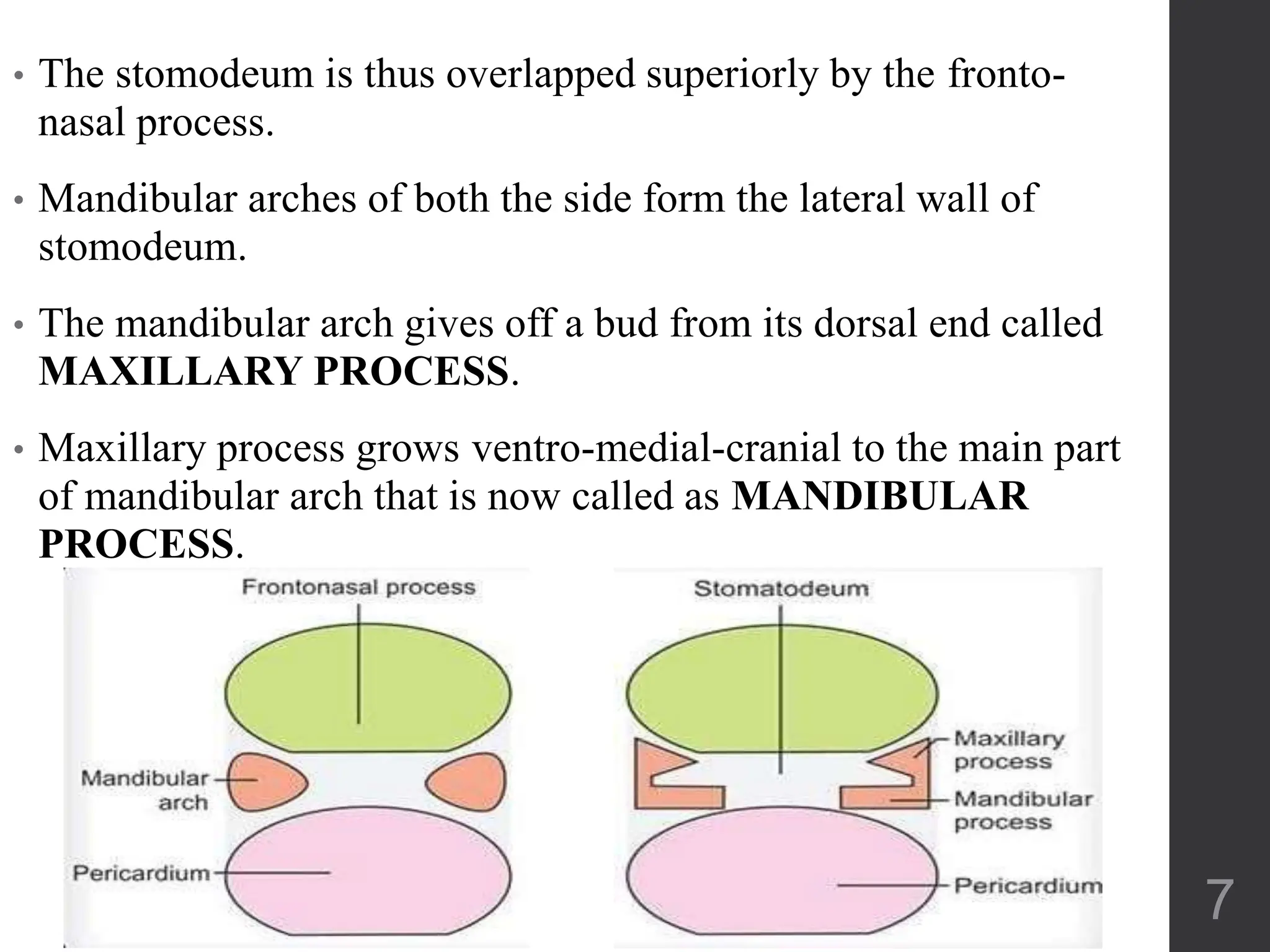
• The stomodeum is thus overlapped superiorly by the fronto-
nasal process.
• Mandibular arches of both the side form the lateral wall of
stomodeum.
• The mandibular arch gives off a bud from its dorsal end called
MAXILLARY PROCESS.
• Maxillary process grows ventro-medial-cranial to the main part
of mandibular arch that is now called as MANDIBULAR
PROCESS.
7
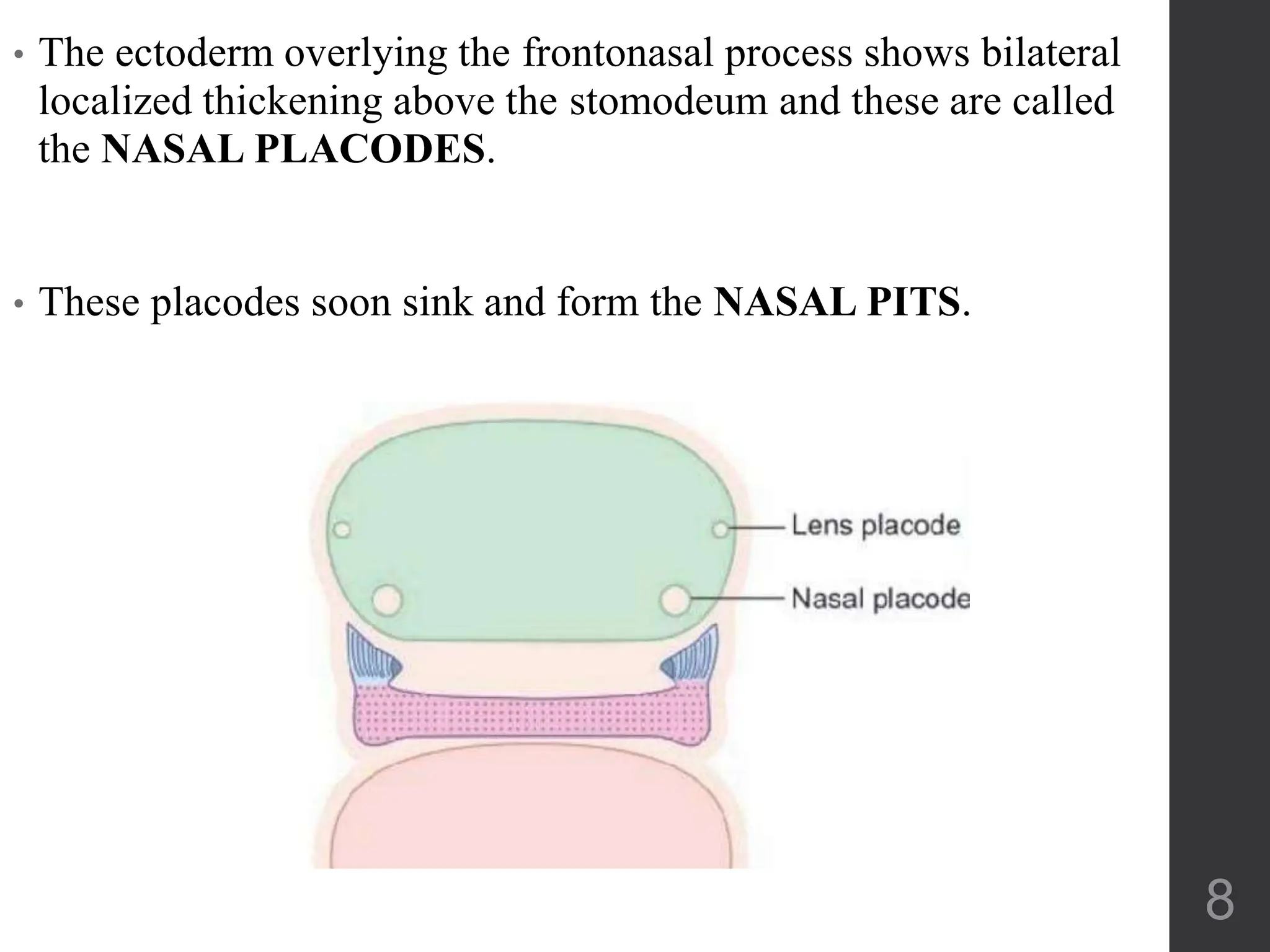
• The ectoderm overlying the frontonasal process shows bilateral
localized thickening above the stomodeum and these are called
the NASAL PLACODES.
• These placodes soon sink and form the NASAL PITS.
8
• The formation of these nasal pits divides the fronto-nasal process into 2
parts-
MESIAL NASAL PROCESS
LATERAL NASAL PROCESS
9
• As the maxillary process undergoes growth fronto-nasal process
becomes narrow so that two nasal pits come closer.
• The line of fusion of maxillary process and medial nasal process
correspond to the naso-lacrimal duct.
• Nasomedial processes grow quickly pushing the frontal
prominence , then fuse with the maxillary process to complete
the arch of upper jaw.
10
DEVELOPMENT OF PALATE
• The palate is formed by contribution of:
• 1) Maxillary process
• 2) Palatal shelves given off by maxillary
process
3) Fronto-nasal process
The fronto-nasal process gives rise to the
premaxillary region while the palatal shelves
form the rest of the palate
11
• As the palatal shelves grow
medially their union is prevented
by the presence of tongue .
• Thus initally the developing
palatal shelves grow vertically
downward towards the floor of
the mouth .
• Sometimes during the 7th week of
IUL, a transformation in position
of palatal shelves occurs.
• They change from vertical to
horizontal position.
12
• This transformation is believed to take place within hours . Various
reasons are given to explain how this transformation occurs.
• A) Alteration in biochemical and physical consistency of the
connective tissue of palatal shelves.
• B) Appearance of an intrinsic shelf area.
• C) Rapid differential mitotic activity.
• D) Alteration in vasculature and blood supply to the palatal shelves.
• E) Withdrawal of embryonic face from against the heart prominence
results in slight jaw opening.
• This results in withdrawal of tongue from between the palatal shelves
and aids in elevation of palatal shelves from vertical to horizontal
positions.
13
• The two palatal shelves , by 8th weeks of IUL, are in close
approximation with each other.
• Intially the two palatal shelves are covered by an epithelial
lining.
• As they join, the epithelial cells degenerate.
• The connective tissue of the palatal shelves intermingle with each
other resulting in the fusion.
14
• The entire palate does not contact and fuse at the same time.
• Intially contact occurs in the central region of the secondary
palate posterior to the premaxilla.
• From this point , closure occurs both anteriorly and posteriorly.
• The mesial edges of the palatal processes fuse with the free lower
end of the nasal septum and thus separates the two nasal cavities
from each other and the oral cavity.
15
• 8th week IUL
• 1) Stomodeum enlarge
• 2) Tongue drops
• 3) Vertically inclined palatal shelves-horizontal
• 4) Shelves contact each other in midline
• 5) By 12th week , fusion of palatal processes is
complete
16
ANATOMY OF MAXILLA
• BODY- large and pyramidal
in shape
• FOUR PROCESSES-
frontal
zygomatic
alveolar
palatine
• It houses the largest sinus of the face: maxillary sinus
17
• The maxillary bone is the second largest bone of the face, first
being the mandible.
• Body of maxilla is like hollow pyramid.
• Base of pyramid is formed by nasal surface and apex is directed
towards zygomatic process.
18
BORDERS OF MAXILLA
• Superiorly: frontal bone
• Posteriorly: sphenoid, palatine,
lacrimal, ethmoid bones
• Medially: nasal bone, vomer
• Inferior: nasal concha
• Laterally: zygomatic bone
19
POST NATAL
GROWTH OF
MAXILLA
20
APPOSITION OF
BONE
SURFACE
REMODELLING
21
SURFACE REMODELLING
• Shape of bone can be changed through removal
(resorption) of bone in one area and addition
(apposition) of bone in another.
• This balance of apposition and resorption, with
new bone being formed in some areas while old
bone is removed in others known as remodelling.
22
Cranial
base
growth
Growth at
sutures
23
• The maxilla develops post natally entirely by
intramembranous ossification
• The growth pattern of the face requires that it grow
"out from under the cranium,"
• It means that the maxilla must move through growth a
considerable distance downward and forward relative
to the cranium and cranial base
24
• As the maxilla grows downward and forward, its
front surfaces are remodelled, and bone is removed
from most of the anterior surface.
• That almost the entire anterior surface of the maxilla
is an area of resorption, not apposition
25
DISPLACEMENT
• PRIMARY DISPLACEMENT: The process of
physical carry, takes place in conjunction with a
bone’s own enlargement.
• SECONDARY DISPLACEMENT: The
movement of bone and its soft tissues is not
directly related to its own enlargement. It is a
fundamental part of the overall process of
craniofacial enlargement.
26
DOWNWARD MAXILLARY
DISPLACEMENT
• New bone is added at the frontomaxillary, zygotemporal,
zygosphenoidal, zygomaxillary, ethmomaxillary, ethmofrontal,
nasomaxillary, nasofrontal, frontolacrimal, palatine, and vomerine
sutures.
• These multiple sutural deposits
accompany displacement.
• The process of displacement
produces the "space" within which remodeling enlargement
occurs.
27
• The displacement of the bones is produced by the expanding
soft tissues.
• The bones of the ethmomaxillary region are displaced
downward.
28
Sutural bone growth takes place
at the same time in response to
It.
Thus enlarging the bones as the
soft tissues continue to develop.
This places all the bones in new
positions in conjunction with the generalized expansion
of the soft tissue matrix and maintains continuous
sutural contact.
29
NASOMAXIlLARY
REMODELING
30
• Extensive remodeling occurs throughout the nasomaxillary
complex as the entire region undergoes inferior (and anterior)
displacement .
31
THE BIOMECHANICAL FORCE
UNDERLYING MAXILLARY
DISPLACEMENT
• “Nasal septum" theory-
developed by Scott
• It developed from the criticisms of
the "sutural theory”
• Cartilage is a special tissue
uniquely structured to provide the
capacity for growth in a field of
compression.
32
• Where as the cartilaginous nasal septum
itself contributes only a small amount of
actual endochondral growth.
• The basis for the “septal” theory is that
the pressure-accommodating expansion of
the cartilage in the nasal septum provides
a source for the physical force that
displaces
(pushes) the whole maxilla anteriorly and
inferiorly.
33
• This sets up fields of tension in all the
maxillary sutures.
• The bones then secondarily, but virtually
simultaneously, enlarge at their sutures in
response to the tension created by the
displacement process.
34
• A notable advance was made with the development of
the functional matrix concept, by Moss.
• According to this theory, the course and extent of bone
growth are secondarily dependent upon the growth and
the functioning of pacemaking soft tissues.
• So, the growth expansion of the facial muscles, the
subcutaneous and submucosal connective tissues, the
oral and nasal epithelia lining the spaces, the vessels
and nerves, and so on, all combine to move the facial
bones passively along with them as they grow.
35
MAXILLARY TUBEROSITY
AND ARCH LENGTENING
• Remodelling of maxillary tuberosity produces horizontal
lenghtening of the bony maxillary arch.
• The backward-facing periosteal surface of the tuberosity
receives continued deposits of new bone as long as
growth in this part of the face continues
36
• The arch also widens, and the lateral
surface is, similarly, depository.
• The endosteal side of the cortex within the
interior of the tuberosity (the maxillary
sinus) is resorptive.
• The cortex thus moves (relocates)
progressively posteriorly and also, to a
lesser extent, in a lateral direction.
37
• After closure of mid- palatine suture, increase in the
arch width is due to remodelling of the alveolar
process.
• The whole maxilla undergoes a simultaneous process
of primary displacement in an anterior and inferior
direction as it grows and lengthens posteriorly
38
THE MAXILLARY
TUBEROSITY AND THE KEY
RIDGE
In the growth of the bony maxillary arch, area A in the fig. is
moving in three directions by bone deposition on the external
surface
39
• A major change in surface contour occurs
along the vertical crest just below the
malar protuberance.
• This crest is called the "key ridge.“
• A reversal occurs here.
• most of the external surface of the
maxillary arch is resorptive
40
The Lacrimal Suture: A Key Growth
Mediator
• The lacrimal bone is a diminutive flake of a bony
island with its entire perimeter bounded by sutural
connective tissue contacts separating it from the many
other surrounding bones.
• The sutural system of the lacrimal bone provides for
the "slippage" of the multiple bones along sutural
interfaces as they all enlarge differentially.
• This is made possible by collagenous linkage
adjustments within the sutural connective tissue 41
• The lacrimal sutures make it possible for
the maxilla to "slide" downward along its
orbital contacts. This allows the whole
maxilla to become displaced inferiorly.
• It is a key midfacial growth event, even
though all the other bones of the orbit and
nasal region develop quite
differently and at different times,
amounts, and directions.
42
THE NASAL AIRWAY
• The lining surfaces of the bony walls
and floor of the nasal chambers are
predominantly resorptive .
• This produces a lateral and anterior
expansion of the nasal chambers and
a downward relocation of the palate;
the oral side of the bony palate is
depository.
43
• The breadth of the nasal bridge in the region just
below the frontonasal sutures does not markedly
increase from early childhood to adulthood
44
PALATAL REMODELLING
• Even though the external (labial) side of
the whole anterior part of the maxillary
arch is resorptive, bone is being added
onto the inside of the arch
• The arch nonetheless increases in width
• The palate becomes wider
• (follows V- Principle)
45
46
THE CHEEKBONE AND
ZYGOMATIC ARCH
• The posterior side of the malar
protuberance within the temporal fossa is
depository.
• Together with a resorptive anterior
surface, the cheekbone relocates
posteriorly as it enlarges.
47
• The zygomatic arch moves laterally by
resorption on the medial side within the
temporal fossa and by deposition on the
lateral side
• This enlarges the temporal fossa and
keeps the cheekbone proportionately
broad in relation to face and jaw size
and the masticatory musculature.
• It also moves the arches bilaterally, thus
increasing the space between for overall
head and brain enlargement.
48
• As the malar region grows and becomes relocated
posteriorly, the contiguous nasal region is
enlarging in an opposite, anterior direction.
• This draws out and greatly expands the contour
between them, resulting in a progressively more
protrusive-appearing nose and an
anteroposteriorly much deeper face
49
• This is a major topographic maturational change in the
childhood-to-adult face.
• The facial contours become opened, the protrusions more
prominent, and the depths all increased
50
ORBITAL GROWTH
• The remodeling changes of the orbit
are complex.
• This is because many separate bones
comprise its enclosing walls, including
• Most of the lining roof and the floor are
depository.
51
As the frontal lobe of the cerebrum expands forward and
downward (until about 5 to 7 years of age), the orbital roof
remodels anteriorly and inferiorly by resorption on the
endocranial side and deposition on the orbital side.
52
The orbit grows by the V principle
The cone-shaped orbital cavity
moves (relocation by remodeling)
in a direction toward its wide
opening; deposits on the inside
thus enlarge, rather than reduce,
the volume.
53
Deposition takes place on the
intraorbital (superior) side of
the orbital floor and resorption
on the maxillary (inferior) sinus
side.
This sustains the orbital floor in
proper position with respect to
the eyeball above it
54
DEVELOPMENTAL
ANOMALIES
AFECTING MAXILLA
55
Defective fusion of the various
components of the palate gives
rise to clefts in the palate.
Cleft lip and palate occur when
mesenchymal connective
tissues from different
embryologic structures fail to
meet and merge with each
other.
21
•The common form of
cleft lip is a result of
failure of fusion of the
medial nasal process
with the maxillary
process.
•Cleft lip may be
unilateral or bilateral
and may extend into
the alveolar process.
• there may be
unilateral cleft lip with
palate
57
Cleft Lip
•Cleft palate is the result of failure of the lateral palatine
shelves to fuse with each other, with the nasal septum, or
with the primary palate.
•Failure of mesodermal proliferation of resistant
epithelial covering and retention of epithelial bridges
can cause cleft palate.
•Clefts of the palate result in anomalous
communications between the mouth and the nose.
• These may be unilateral or bilateral.
58
CLEFT PALATE
59
Figs 11.20A to E: Varieties of cleft palate. (A) Complete cleft with bilateral
harelip; (B) Unilateral cleft palate and cleft of upper lip. The left maxillary
process has fused with the premaxilla, but not with the right maxillary process.
The cleft is accompanied by unilateral harelip; (C) Midline cleft of hard palate
and soft palate; (D) Cleft of soft palate; (E) Bifid uvula
60
VARIOUS SYNDROMES ASSOCIATED
WITH [CL(P)]
Stickler syndrome Mutations in the specific
collagen gene palatal clefting
Waarnderburg’s syndrome I Mutation in
homeodomain- containing protein (pa x3)
• Mutation in sonic hedgehog (shh)
Holoprosencephalic disorders
• Characterized by severe midline defect(failure of
nose to develop)
•van der Woude syndrome is an
autosomal dominant syndrome
typically consisting of a cleft lip or
cleft palate and distinctive pits of the
lower lips.
•These variable manifestations include lip pits
alone, missing teeth or isolated cleft lip, and
palate of varying degrees of severity.
•The van der Woude syndrome can be caused by
deletions in chromosome band 1q32 , and linkage
analysis has confirmed this chromosomal locus as
the disease gene site.
van der Woude Syndrome
61
NON- SYNDROMIC CLEFT
LIP/PALATE
Mutation in no. of different genes familial CL
and CP
1. Transforming Growth Factor- alpha(TGF-α)
2. Retinoic Acid Receptor- alpha(RAR-α)
3. MSX1 Gene
4. BCL3
62
The migration of neural crest cells is important for the derivation of
facial structures
DRUG induced impairement of migration:
•exposure to Thalidomide- congenital defects
•Anti-acne drug Isotretinoin- affects the neural crest cells
Dangerous as it affects the embryo before the mother knows she is
pregnant
•Retinoic acid plays crucial role in ontogenesis of midface
Loss of RAR genes affect post migratory activity of crest cells
63
SMOKING AND CLEFT LIP/ PALATE
• An initial step in development of primary palate is
forward movement of lateral nasal process which
positions it so that it can contact median nasal
process.
• The associated with probably
interferes with this movement
64
Altered development of cells
derived
from neural crest
TREACHER COLLINS
SYNDROME
Characterised by a generalised
lack of mesenchymal tissue.
Now known to be due to
mutation in a specific gene
(TCOF1, POLR1C, POLR1D)
65
CROUZON’S SYNDROME
(Craniosynostosis)
Results due to early closure of the superior and
posterior sutures of maxilla along the wall of orbit
Characterised by:
• Frog face
• Underdeveloped midface
• Eyes that seem to bulge out
• Hypertelorism
66
CLINICAL IMPLICATION
• Because distal movement of the maxillary first molar
is often part of an orthodontic treatment plan, the
maxillary tuberosity is important.
• Every mechanical option designed to move the
maxillary first molar distally exploits the growth
potential of the tuberosity.
• In Rapid Maxillary Expansion (RME), remodelling
of maxilla follows clinically induced displacement.
67
•Maxilla is formed by Ist brachial arch and
ectomesenchyme of neural crest cells. Any factor
interfering with the formation underdeveloped
maxilla. E.g Craniofacial dysostosis, cleidocranial
dysplasia, achondroplasia, downs syndrome
•Underdeveloped maxilla mid-face deficiency
•If cleft lip and palate forms narrow maxilla with high
arched palate tendency for posterior and anterior
crossbites speech, mastication, hearing is impared
68
•Headgear is used for restricting the maxillary growth
and reverse pull head gear or facemask with class III
functional appliance for correction of retrognathic
maxilla
•If growth is completed orthognathic surgical
correction
69
REFERENCES
1. Essentials of facial growth-
Enlows
2. Human Anatomy-I.B SINGH
3. Contemporary Orthodontics-
William R. Profitt
4. Textbook of orthodontics; Bishara
70

More Related Content

Similar to DEVELOPMENT OF MAXILLA with prenatal and postnatal development.pptx

16. FACE AND ASSOCIATED STRUCTURES-1.pptx
16. FACE AND ASSOCIATED STRUCTURES-1.pptx16. FACE AND ASSOCIATED STRUCTURES-1.pptx
16. FACE AND ASSOCIATED STRUCTURES-1.pptx
StanleyOdira
 

Similar to DEVELOPMENT OF MAXILLA with prenatal and postnatal development.pptx (20)

Growth & Development of Maxilla
Growth & Development of MaxillaGrowth & Development of Maxilla
Growth & Development of Maxilla
 
Development of maxilla and palate
Development of maxilla and palateDevelopment of maxilla and palate
Development of maxilla and palate
 
Growth of maxilla /certified fixed orthodontic courses by Indian dental acad...
Growth of maxilla  /certified fixed orthodontic courses by Indian dental acad...Growth of maxilla  /certified fixed orthodontic courses by Indian dental acad...
Growth of maxilla /certified fixed orthodontic courses by Indian dental acad...
 
Developement of maxilla and mandible.
Developement of maxilla and mandible.Developement of maxilla and mandible.
Developement of maxilla and mandible.
 
growth and development of maxilla
growth and development of maxillagrowth and development of maxilla
growth and development of maxilla
 
Growth & Development of Mandible
Growth & Development of MandibleGrowth & Development of Mandible
Growth & Development of Mandible
 
16. FACE AND ASSOCIATED STRUCTURES-1.pptx
16. FACE AND ASSOCIATED STRUCTURES-1.pptx16. FACE AND ASSOCIATED STRUCTURES-1.pptx
16. FACE AND ASSOCIATED STRUCTURES-1.pptx
 
Seminar on HUMAN EMBRYOLOGY for postgraduates
Seminar on HUMAN EMBRYOLOGY for postgraduatesSeminar on HUMAN EMBRYOLOGY for postgraduates
Seminar on HUMAN EMBRYOLOGY for postgraduates
 
SURGICAL AND APPLIED ANATOMY OF MAXILLA.
SURGICAL AND APPLIED ANATOMY OF MAXILLA.SURGICAL AND APPLIED ANATOMY OF MAXILLA.
SURGICAL AND APPLIED ANATOMY OF MAXILLA.
 
Prenatal and postnatal growth & development of maxilla and palate presented b...
Prenatal and postnatal growth & development of maxilla and palate presented b...Prenatal and postnatal growth & development of maxilla and palate presented b...
Prenatal and postnatal growth & development of maxilla and palate presented b...
 
GROWTH AND DEVELOPMENT OF MANDIBLE - DR. SAUMYA PAUL
GROWTH AND DEVELOPMENT OF MANDIBLE - DR. SAUMYA PAULGROWTH AND DEVELOPMENT OF MANDIBLE - DR. SAUMYA PAUL
GROWTH AND DEVELOPMENT OF MANDIBLE - DR. SAUMYA PAUL
 
Growth and development of mandible
Growth and development of mandibleGrowth and development of mandible
Growth and development of mandible
 
enlow counterpart principle with reference articles
enlow counterpart principle with reference articlesenlow counterpart principle with reference articles
enlow counterpart principle with reference articles
 
Development of palate
Development of palateDevelopment of palate
Development of palate
 
Maxxilla seminar ih
Maxxilla seminar  ihMaxxilla seminar  ih
Maxxilla seminar ih
 
Development of maxilla and palate
Development of maxilla and palateDevelopment of maxilla and palate
Development of maxilla and palate
 
Oral Biology
Oral Biology Oral Biology
Oral Biology
 
Growth and Development of Mandible
Growth and Development of MandibleGrowth and Development of Mandible
Growth and Development of Mandible
 
Growth of skull and jaws part 1
Growth of skull and jaws part 1Growth of skull and jaws part 1
Growth of skull and jaws part 1
 
Post-natal Growth of Maxilla and Mandible
Post-natal Growth of Maxilla and MandiblePost-natal Growth of Maxilla and Mandible
Post-natal Growth of Maxilla and Mandible
 

Recently uploaded

Histology of Epithelium - Dr Muhammad Ali Rabbani - Medicose Academics
Histology of Epithelium - Dr Muhammad Ali Rabbani - Medicose AcademicsHistology of Epithelium - Dr Muhammad Ali Rabbani - Medicose Academics
Histology of Epithelium - Dr Muhammad Ali Rabbani - Medicose Academics
MedicoseAcademics
 
VIII.1 Nursing Interventions to Promote Healthy Psychological responses, SELF...
VIII.1 Nursing Interventions to Promote Healthy Psychological responses, SELF...VIII.1 Nursing Interventions to Promote Healthy Psychological responses, SELF...
VIII.1 Nursing Interventions to Promote Healthy Psychological responses, SELF...
JRRolfNeuqelet
 
Cytoskeleton and Cell Inclusions - Dr Muhammad Ali Rabbani - Medicose Academics
Cytoskeleton and Cell Inclusions - Dr Muhammad Ali Rabbani - Medicose AcademicsCytoskeleton and Cell Inclusions - Dr Muhammad Ali Rabbani - Medicose Academics
Cytoskeleton and Cell Inclusions - Dr Muhammad Ali Rabbani - Medicose Academics
MedicoseAcademics
 
CAD CAM DENTURES IN PROSTHODONTICS : Dental advancements
CAD CAM DENTURES IN PROSTHODONTICS : Dental advancementsCAD CAM DENTURES IN PROSTHODONTICS : Dental advancements
CAD CAM DENTURES IN PROSTHODONTICS : Dental advancements
Naveen Gokul Dr
 

Recently uploaded (20)

Vesu + ℂall Girls Serviℂe Surat (Adult Only) 8849756361 Esℂort Serviℂe 24x7 C...
Vesu + ℂall Girls Serviℂe Surat (Adult Only) 8849756361 Esℂort Serviℂe 24x7 C...Vesu + ℂall Girls Serviℂe Surat (Adult Only) 8849756361 Esℂort Serviℂe 24x7 C...
Vesu + ℂall Girls Serviℂe Surat (Adult Only) 8849756361 Esℂort Serviℂe 24x7 C...
 
NDCT Rules, 2019: An Overview | New Drugs and Clinical Trial Rules 2019
NDCT Rules, 2019: An Overview | New Drugs and Clinical Trial Rules 2019NDCT Rules, 2019: An Overview | New Drugs and Clinical Trial Rules 2019
NDCT Rules, 2019: An Overview | New Drugs and Clinical Trial Rules 2019
 
Histology of Epithelium - Dr Muhammad Ali Rabbani - Medicose Academics
Histology of Epithelium - Dr Muhammad Ali Rabbani - Medicose AcademicsHistology of Epithelium - Dr Muhammad Ali Rabbani - Medicose Academics
Histology of Epithelium - Dr Muhammad Ali Rabbani - Medicose Academics
 
Quality control tests of suppository ...
Quality control tests  of suppository ...Quality control tests  of suppository ...
Quality control tests of suppository ...
 
Renal Replacement Therapy in Acute Kidney Injury -time modality -Dr Ayman Se...
Renal Replacement Therapy in Acute Kidney Injury -time  modality -Dr Ayman Se...Renal Replacement Therapy in Acute Kidney Injury -time  modality -Dr Ayman Se...
Renal Replacement Therapy in Acute Kidney Injury -time modality -Dr Ayman Se...
 
Young & Hot Surat ℂall Girls Vesu 8527049040 WhatsApp AnyTime Best Surat ℂall...
Young & Hot Surat ℂall Girls Vesu 8527049040 WhatsApp AnyTime Best Surat ℂall...Young & Hot Surat ℂall Girls Vesu 8527049040 WhatsApp AnyTime Best Surat ℂall...
Young & Hot Surat ℂall Girls Vesu 8527049040 WhatsApp AnyTime Best Surat ℂall...
 
Sell 5cladba adbb JWH-018 5FADB in stock
Sell 5cladba adbb JWH-018 5FADB in stockSell 5cladba adbb JWH-018 5FADB in stock
Sell 5cladba adbb JWH-018 5FADB in stock
 
Stereochemistry & Asymmetric Synthesis.pptx
Stereochemistry & Asymmetric Synthesis.pptxStereochemistry & Asymmetric Synthesis.pptx
Stereochemistry & Asymmetric Synthesis.pptx
 
Tips and tricks to pass the cardiovascular station for PACES exam
Tips and tricks to pass the cardiovascular station for PACES examTips and tricks to pass the cardiovascular station for PACES exam
Tips and tricks to pass the cardiovascular station for PACES exam
 
VIII.1 Nursing Interventions to Promote Healthy Psychological responses, SELF...
VIII.1 Nursing Interventions to Promote Healthy Psychological responses, SELF...VIII.1 Nursing Interventions to Promote Healthy Psychological responses, SELF...
VIII.1 Nursing Interventions to Promote Healthy Psychological responses, SELF...
 
VIP ℂall Girls Kandivali west Mumbai 8250077686 WhatsApp: Me All Time Serviℂe...
VIP ℂall Girls Kandivali west Mumbai 8250077686 WhatsApp: Me All Time Serviℂe...VIP ℂall Girls Kandivali west Mumbai 8250077686 WhatsApp: Me All Time Serviℂe...
VIP ℂall Girls Kandivali west Mumbai 8250077686 WhatsApp: Me All Time Serviℂe...
 
^In Pietermaritzburg Hager Werken Embalming +27789155305 Compound Powder in ...
^In Pietermaritzburg  Hager Werken Embalming +27789155305 Compound Powder in ...^In Pietermaritzburg  Hager Werken Embalming +27789155305 Compound Powder in ...
^In Pietermaritzburg Hager Werken Embalming +27789155305 Compound Powder in ...
 
DR. Neha Mehta Best Psychologist.in India
DR. Neha Mehta Best Psychologist.in IndiaDR. Neha Mehta Best Psychologist.in India
DR. Neha Mehta Best Psychologist.in India
 
Cytoskeleton and Cell Inclusions - Dr Muhammad Ali Rabbani - Medicose Academics
Cytoskeleton and Cell Inclusions - Dr Muhammad Ali Rabbani - Medicose AcademicsCytoskeleton and Cell Inclusions - Dr Muhammad Ali Rabbani - Medicose Academics
Cytoskeleton and Cell Inclusions - Dr Muhammad Ali Rabbani - Medicose Academics
 
How to buy 5cladba precursor raw 5cl-adb-a raw material
How to buy 5cladba precursor raw 5cl-adb-a raw materialHow to buy 5cladba precursor raw 5cl-adb-a raw material
How to buy 5cladba precursor raw 5cl-adb-a raw material
 
Young & Hot ℂall Girls Salem 8250077686 WhatsApp Number Best Rates of Surat ℂ...
Young & Hot ℂall Girls Salem 8250077686 WhatsApp Number Best Rates of Surat ℂ...Young & Hot ℂall Girls Salem 8250077686 WhatsApp Number Best Rates of Surat ℂ...
Young & Hot ℂall Girls Salem 8250077686 WhatsApp Number Best Rates of Surat ℂ...
 
CAD CAM DENTURES IN PROSTHODONTICS : Dental advancements
CAD CAM DENTURES IN PROSTHODONTICS : Dental advancementsCAD CAM DENTURES IN PROSTHODONTICS : Dental advancements
CAD CAM DENTURES IN PROSTHODONTICS : Dental advancements
 
Treatment Choices for Slip Disc at Gokuldas Hospital
Treatment Choices for Slip Disc at Gokuldas HospitalTreatment Choices for Slip Disc at Gokuldas Hospital
Treatment Choices for Slip Disc at Gokuldas Hospital
 
Kamrej + ℂall Girls Serviℂe Surat (Adult Only) 8849756361 Esℂort Serviℂe 24x7...
Kamrej + ℂall Girls Serviℂe Surat (Adult Only) 8849756361 Esℂort Serviℂe 24x7...Kamrej + ℂall Girls Serviℂe Surat (Adult Only) 8849756361 Esℂort Serviℂe 24x7...
Kamrej + ℂall Girls Serviℂe Surat (Adult Only) 8849756361 Esℂort Serviℂe 24x7...
 
Unlocking Holistic Wellness: Addressing Depression, Mental Well-Being, and St...
Unlocking Holistic Wellness: Addressing Depression, Mental Well-Being, and St...Unlocking Holistic Wellness: Addressing Depression, Mental Well-Being, and St...
Unlocking Holistic Wellness: Addressing Depression, Mental Well-Being, and St...
 

DEVELOPMENT OF MAXILLA with prenatal and postnatal development.pptx

  • 1. DEVELOPMENT OF MAXILLA BY – Dr. Vipul Gupta MDS 1ST YR 1
  • 2. CONTENTS EMBRYONIC DEVELOPMENT ANATOMY OF MAXILLA GROWTH AND DEVELOPMENT CRANIOFACIAL GROWTH DISORDERS REFERENCES 2
  • 4. PRE NATAL EMBROYOLOGY OF MAXILLA • Around 4th week of IUL , a prominent bulge appears on ventral aspect of embryo corresponding to developing brain. • Below this bulge a shallow depression which correspond to the primitive mouth appears called STOMODEUM. • The floor of the stomodeum is formed by BUCCOPHARNGEAL MEMEBRANE that separate the stomodeum from the foregut. 4
  • 5. • By around the 4th week of intra-uterine life ,5 branchial arches form in the region of future head and neck .Each of these arches give rise to muscles , connective tissue, vasculature, skeletal components and neural components of the future face. • The 1st branchial arch is called the MANDIBULAR ARCH and plays important role in development of naso-maxillary region. 5
  • 6. • The mesoderm covering the developing forebrain proliferates and forms a downward projection that overlaps the upper part of stomodeum. • This downward projection is known as FRONTO-NASAL PROCESS. 6
  • 7. • The stomodeum is thus overlapped superiorly by the fronto- nasal process. • Mandibular arches of both the side form the lateral wall of stomodeum. • The mandibular arch gives off a bud from its dorsal end called MAXILLARY PROCESS. • Maxillary process grows ventro-medial-cranial to the main part of mandibular arch that is now called as MANDIBULAR PROCESS. 7
  • 8. • The ectoderm overlying the frontonasal process shows bilateral localized thickening above the stomodeum and these are called the NASAL PLACODES. • These placodes soon sink and form the NASAL PITS. 8
  • 9. • The formation of these nasal pits divides the fronto-nasal process into 2 parts- MESIAL NASAL PROCESS LATERAL NASAL PROCESS 9
  • 10. • As the maxillary process undergoes growth fronto-nasal process becomes narrow so that two nasal pits come closer. • The line of fusion of maxillary process and medial nasal process correspond to the naso-lacrimal duct. • Nasomedial processes grow quickly pushing the frontal prominence , then fuse with the maxillary process to complete the arch of upper jaw. 10
  • 11. DEVELOPMENT OF PALATE • The palate is formed by contribution of: • 1) Maxillary process • 2) Palatal shelves given off by maxillary process 3) Fronto-nasal process The fronto-nasal process gives rise to the premaxillary region while the palatal shelves form the rest of the palate 11
  • 12. • As the palatal shelves grow medially their union is prevented by the presence of tongue . • Thus initally the developing palatal shelves grow vertically downward towards the floor of the mouth . • Sometimes during the 7th week of IUL, a transformation in position of palatal shelves occurs. • They change from vertical to horizontal position. 12
  • 13. • This transformation is believed to take place within hours . Various reasons are given to explain how this transformation occurs. • A) Alteration in biochemical and physical consistency of the connective tissue of palatal shelves. • B) Appearance of an intrinsic shelf area. • C) Rapid differential mitotic activity. • D) Alteration in vasculature and blood supply to the palatal shelves. • E) Withdrawal of embryonic face from against the heart prominence results in slight jaw opening. • This results in withdrawal of tongue from between the palatal shelves and aids in elevation of palatal shelves from vertical to horizontal positions. 13
  • 14. • The two palatal shelves , by 8th weeks of IUL, are in close approximation with each other. • Intially the two palatal shelves are covered by an epithelial lining. • As they join, the epithelial cells degenerate. • The connective tissue of the palatal shelves intermingle with each other resulting in the fusion. 14
  • 15. • The entire palate does not contact and fuse at the same time. • Intially contact occurs in the central region of the secondary palate posterior to the premaxilla. • From this point , closure occurs both anteriorly and posteriorly. • The mesial edges of the palatal processes fuse with the free lower end of the nasal septum and thus separates the two nasal cavities from each other and the oral cavity. 15
  • 16. • 8th week IUL • 1) Stomodeum enlarge • 2) Tongue drops • 3) Vertically inclined palatal shelves-horizontal • 4) Shelves contact each other in midline • 5) By 12th week , fusion of palatal processes is complete 16
  • 17. ANATOMY OF MAXILLA • BODY- large and pyramidal in shape • FOUR PROCESSES- frontal zygomatic alveolar palatine • It houses the largest sinus of the face: maxillary sinus 17
  • 18. • The maxillary bone is the second largest bone of the face, first being the mandible. • Body of maxilla is like hollow pyramid. • Base of pyramid is formed by nasal surface and apex is directed towards zygomatic process. 18
  • 19. BORDERS OF MAXILLA • Superiorly: frontal bone • Posteriorly: sphenoid, palatine, lacrimal, ethmoid bones • Medially: nasal bone, vomer • Inferior: nasal concha • Laterally: zygomatic bone 19
  • 22. SURFACE REMODELLING • Shape of bone can be changed through removal (resorption) of bone in one area and addition (apposition) of bone in another. • This balance of apposition and resorption, with new bone being formed in some areas while old bone is removed in others known as remodelling. 22
  • 24. • The maxilla develops post natally entirely by intramembranous ossification • The growth pattern of the face requires that it grow "out from under the cranium," • It means that the maxilla must move through growth a considerable distance downward and forward relative to the cranium and cranial base 24
  • 25. • As the maxilla grows downward and forward, its front surfaces are remodelled, and bone is removed from most of the anterior surface. • That almost the entire anterior surface of the maxilla is an area of resorption, not apposition 25
  • 26. DISPLACEMENT • PRIMARY DISPLACEMENT: The process of physical carry, takes place in conjunction with a bone’s own enlargement. • SECONDARY DISPLACEMENT: The movement of bone and its soft tissues is not directly related to its own enlargement. It is a fundamental part of the overall process of craniofacial enlargement. 26
  • 27. DOWNWARD MAXILLARY DISPLACEMENT • New bone is added at the frontomaxillary, zygotemporal, zygosphenoidal, zygomaxillary, ethmomaxillary, ethmofrontal, nasomaxillary, nasofrontal, frontolacrimal, palatine, and vomerine sutures. • These multiple sutural deposits accompany displacement. • The process of displacement produces the "space" within which remodeling enlargement occurs. 27
  • 28. • The displacement of the bones is produced by the expanding soft tissues. • The bones of the ethmomaxillary region are displaced downward. 28
  • 29. Sutural bone growth takes place at the same time in response to It. Thus enlarging the bones as the soft tissues continue to develop. This places all the bones in new positions in conjunction with the generalized expansion of the soft tissue matrix and maintains continuous sutural contact. 29
  • 31. • Extensive remodeling occurs throughout the nasomaxillary complex as the entire region undergoes inferior (and anterior) displacement . 31
  • 32. THE BIOMECHANICAL FORCE UNDERLYING MAXILLARY DISPLACEMENT • “Nasal septum" theory- developed by Scott • It developed from the criticisms of the "sutural theory” • Cartilage is a special tissue uniquely structured to provide the capacity for growth in a field of compression. 32
  • 33. • Where as the cartilaginous nasal septum itself contributes only a small amount of actual endochondral growth. • The basis for the “septal” theory is that the pressure-accommodating expansion of the cartilage in the nasal septum provides a source for the physical force that displaces (pushes) the whole maxilla anteriorly and inferiorly. 33
  • 34. • This sets up fields of tension in all the maxillary sutures. • The bones then secondarily, but virtually simultaneously, enlarge at their sutures in response to the tension created by the displacement process. 34
  • 35. • A notable advance was made with the development of the functional matrix concept, by Moss. • According to this theory, the course and extent of bone growth are secondarily dependent upon the growth and the functioning of pacemaking soft tissues. • So, the growth expansion of the facial muscles, the subcutaneous and submucosal connective tissues, the oral and nasal epithelia lining the spaces, the vessels and nerves, and so on, all combine to move the facial bones passively along with them as they grow. 35
  • 36. MAXILLARY TUBEROSITY AND ARCH LENGTENING • Remodelling of maxillary tuberosity produces horizontal lenghtening of the bony maxillary arch. • The backward-facing periosteal surface of the tuberosity receives continued deposits of new bone as long as growth in this part of the face continues 36
  • 37. • The arch also widens, and the lateral surface is, similarly, depository. • The endosteal side of the cortex within the interior of the tuberosity (the maxillary sinus) is resorptive. • The cortex thus moves (relocates) progressively posteriorly and also, to a lesser extent, in a lateral direction. 37
  • 38. • After closure of mid- palatine suture, increase in the arch width is due to remodelling of the alveolar process. • The whole maxilla undergoes a simultaneous process of primary displacement in an anterior and inferior direction as it grows and lengthens posteriorly 38
  • 39. THE MAXILLARY TUBEROSITY AND THE KEY RIDGE In the growth of the bony maxillary arch, area A in the fig. is moving in three directions by bone deposition on the external surface 39
  • 40. • A major change in surface contour occurs along the vertical crest just below the malar protuberance. • This crest is called the "key ridge.“ • A reversal occurs here. • most of the external surface of the maxillary arch is resorptive 40
  • 41. The Lacrimal Suture: A Key Growth Mediator • The lacrimal bone is a diminutive flake of a bony island with its entire perimeter bounded by sutural connective tissue contacts separating it from the many other surrounding bones. • The sutural system of the lacrimal bone provides for the "slippage" of the multiple bones along sutural interfaces as they all enlarge differentially. • This is made possible by collagenous linkage adjustments within the sutural connective tissue 41
  • 42. • The lacrimal sutures make it possible for the maxilla to "slide" downward along its orbital contacts. This allows the whole maxilla to become displaced inferiorly. • It is a key midfacial growth event, even though all the other bones of the orbit and nasal region develop quite differently and at different times, amounts, and directions. 42
  • 43. THE NASAL AIRWAY • The lining surfaces of the bony walls and floor of the nasal chambers are predominantly resorptive . • This produces a lateral and anterior expansion of the nasal chambers and a downward relocation of the palate; the oral side of the bony palate is depository. 43
  • 44. • The breadth of the nasal bridge in the region just below the frontonasal sutures does not markedly increase from early childhood to adulthood 44
  • 45. PALATAL REMODELLING • Even though the external (labial) side of the whole anterior part of the maxillary arch is resorptive, bone is being added onto the inside of the arch • The arch nonetheless increases in width • The palate becomes wider • (follows V- Principle) 45
  • 46. 46
  • 47. THE CHEEKBONE AND ZYGOMATIC ARCH • The posterior side of the malar protuberance within the temporal fossa is depository. • Together with a resorptive anterior surface, the cheekbone relocates posteriorly as it enlarges. 47
  • 48. • The zygomatic arch moves laterally by resorption on the medial side within the temporal fossa and by deposition on the lateral side • This enlarges the temporal fossa and keeps the cheekbone proportionately broad in relation to face and jaw size and the masticatory musculature. • It also moves the arches bilaterally, thus increasing the space between for overall head and brain enlargement. 48
  • 49. • As the malar region grows and becomes relocated posteriorly, the contiguous nasal region is enlarging in an opposite, anterior direction. • This draws out and greatly expands the contour between them, resulting in a progressively more protrusive-appearing nose and an anteroposteriorly much deeper face 49
  • 50. • This is a major topographic maturational change in the childhood-to-adult face. • The facial contours become opened, the protrusions more prominent, and the depths all increased 50
  • 51. ORBITAL GROWTH • The remodeling changes of the orbit are complex. • This is because many separate bones comprise its enclosing walls, including • Most of the lining roof and the floor are depository. 51
  • 52. As the frontal lobe of the cerebrum expands forward and downward (until about 5 to 7 years of age), the orbital roof remodels anteriorly and inferiorly by resorption on the endocranial side and deposition on the orbital side. 52
  • 53. The orbit grows by the V principle The cone-shaped orbital cavity moves (relocation by remodeling) in a direction toward its wide opening; deposits on the inside thus enlarge, rather than reduce, the volume. 53
  • 54. Deposition takes place on the intraorbital (superior) side of the orbital floor and resorption on the maxillary (inferior) sinus side. This sustains the orbital floor in proper position with respect to the eyeball above it 54
  • 56. Defective fusion of the various components of the palate gives rise to clefts in the palate. Cleft lip and palate occur when mesenchymal connective tissues from different embryologic structures fail to meet and merge with each other. 21
  • 57. •The common form of cleft lip is a result of failure of fusion of the medial nasal process with the maxillary process. •Cleft lip may be unilateral or bilateral and may extend into the alveolar process. • there may be unilateral cleft lip with palate 57 Cleft Lip
  • 58. •Cleft palate is the result of failure of the lateral palatine shelves to fuse with each other, with the nasal septum, or with the primary palate. •Failure of mesodermal proliferation of resistant epithelial covering and retention of epithelial bridges can cause cleft palate. •Clefts of the palate result in anomalous communications between the mouth and the nose. • These may be unilateral or bilateral. 58 CLEFT PALATE
  • 59. 59 Figs 11.20A to E: Varieties of cleft palate. (A) Complete cleft with bilateral harelip; (B) Unilateral cleft palate and cleft of upper lip. The left maxillary process has fused with the premaxilla, but not with the right maxillary process. The cleft is accompanied by unilateral harelip; (C) Midline cleft of hard palate and soft palate; (D) Cleft of soft palate; (E) Bifid uvula
  • 60. 60 VARIOUS SYNDROMES ASSOCIATED WITH [CL(P)] Stickler syndrome Mutations in the specific collagen gene palatal clefting Waarnderburg’s syndrome I Mutation in homeodomain- containing protein (pa x3) • Mutation in sonic hedgehog (shh) Holoprosencephalic disorders • Characterized by severe midline defect(failure of nose to develop)
  • 61. •van der Woude syndrome is an autosomal dominant syndrome typically consisting of a cleft lip or cleft palate and distinctive pits of the lower lips. •These variable manifestations include lip pits alone, missing teeth or isolated cleft lip, and palate of varying degrees of severity. •The van der Woude syndrome can be caused by deletions in chromosome band 1q32 , and linkage analysis has confirmed this chromosomal locus as the disease gene site. van der Woude Syndrome 61
  • 62. NON- SYNDROMIC CLEFT LIP/PALATE Mutation in no. of different genes familial CL and CP 1. Transforming Growth Factor- alpha(TGF-α) 2. Retinoic Acid Receptor- alpha(RAR-α) 3. MSX1 Gene 4. BCL3 62
  • 63. The migration of neural crest cells is important for the derivation of facial structures DRUG induced impairement of migration: •exposure to Thalidomide- congenital defects •Anti-acne drug Isotretinoin- affects the neural crest cells Dangerous as it affects the embryo before the mother knows she is pregnant •Retinoic acid plays crucial role in ontogenesis of midface Loss of RAR genes affect post migratory activity of crest cells 63
  • 64. SMOKING AND CLEFT LIP/ PALATE • An initial step in development of primary palate is forward movement of lateral nasal process which positions it so that it can contact median nasal process. • The associated with probably interferes with this movement 64
  • 65. Altered development of cells derived from neural crest TREACHER COLLINS SYNDROME Characterised by a generalised lack of mesenchymal tissue. Now known to be due to mutation in a specific gene (TCOF1, POLR1C, POLR1D) 65
  • 66. CROUZON’S SYNDROME (Craniosynostosis) Results due to early closure of the superior and posterior sutures of maxilla along the wall of orbit Characterised by: • Frog face • Underdeveloped midface • Eyes that seem to bulge out • Hypertelorism 66
  • 67. CLINICAL IMPLICATION • Because distal movement of the maxillary first molar is often part of an orthodontic treatment plan, the maxillary tuberosity is important. • Every mechanical option designed to move the maxillary first molar distally exploits the growth potential of the tuberosity. • In Rapid Maxillary Expansion (RME), remodelling of maxilla follows clinically induced displacement. 67
  • 68. •Maxilla is formed by Ist brachial arch and ectomesenchyme of neural crest cells. Any factor interfering with the formation underdeveloped maxilla. E.g Craniofacial dysostosis, cleidocranial dysplasia, achondroplasia, downs syndrome •Underdeveloped maxilla mid-face deficiency •If cleft lip and palate forms narrow maxilla with high arched palate tendency for posterior and anterior crossbites speech, mastication, hearing is impared 68
  • 69. •Headgear is used for restricting the maxillary growth and reverse pull head gear or facemask with class III functional appliance for correction of retrognathic maxilla •If growth is completed orthognathic surgical correction 69
  • 70. REFERENCES 1. Essentials of facial growth- Enlows 2. Human Anatomy-I.B SINGH 3. Contemporary Orthodontics- William R. Profitt 4. Textbook of orthodontics; Bishara 70

Editor's Notes

  1. Thus at this stage the primitive mouth or stomodeum is overlaped from above by frontal process below by mandibular process and on either side by maxillary process.
  2. Intramembranous ossification – bone develops directly fro sheet of mesenchymal connective tissue Endrochondeal ossifaction – bone develops by replacing the hylaine cartilage
  3. Diagram showing summary of maxillary remodeliing Arrow entering – show resorption of bone Arrow emerging – show deposition of bone
  4. The lining cortical surfaces of the maxillary sinuses are all resorptive, except the medial nasal wall, which is depository because it remodels laterally to accommodate nasal expansion
  5. pressure-accommodating expansion of the cartilage in the nasal septum provides a source for the physical force that displaces (pushes) the whole maxilla anteriorly and inferiorly.
  6. The suture is essentially a tension-adapted tissue. The presence of any unusual pressure on a suture triggers bone resorption, not deposition
  7. The suture is essentially a tension-adapted tissue. The presence of any unusual pressure on a suture triggers bone resorption, not deposition
  8. Reveral refers to couple of bone resorption and bone formation by generating osteogenic enviroment at remodelling sites Key ridge also corrspond to point a and is concave LABIAL surfaces faces upward rather than downward
  9. The lacrimal bone and its suture is a developmental hub providing key traffic controls.
  10. that bone deposition occurs on the inner side of the V; resorption takes place on the outside surface. The V thereby moves from position A to B and, at the same time, increases in overall dimensions. The direction of movement is toward the wide end of the V.
  11. Malar region grows posteriorly and the nasal region grows in anterior direction
  12. Malar region grows posteriorly and the nasal region grows in anterior direction
  13. Figs 11.20A to E: Varieties of cleft palate. (A) Complete cleft with bilateral harelip; (B) Unilateral cleft palate and cleft of upper lip. The left maxillary process has fused with the premaxilla, but not with the right maxillary process. The cleft is accompanied by unilateral harelip; (C) Midline cleft of hard palate and soft palate; (D) Cleft of soft palate; (E) Bifid uvula
  14. Stickler syndrome also caled as hereditary progressive arthoopthalmopath – distintive facial appearance , hearning loss and joint problems Waanderburg syndrome –hearing loss , changes in colouring of hair skin and eyes Holoprosencephalic – failure of embroynic forebrain to sufficiently divide into double lobes of cerebral hemishpere
  15. Thalidomide afects developing neral pathways causes missing limb Isotretionin artifical vitamin a Retinoic acid metabolite of vitamin a1
  16. Symtoms incude downward slting eyes , very small chin and jaw , small and flatenned cheekbone
  17. Caonacve facial profile , genetic syndrome
  18. Craniofacial dysostosis – crouzon syndeome – skull fuse abnormally Cleidocranial dyslpasia – delayed loss of primary teeth and delayed appeaence of secondary teeh , peg liked teeth Achondroplasia – macrocephly,short upper arm and thigh , limited range motion of elbow Downs syndrome – genetic chromose no. 21 flat masal bridge , short starure , protuding tongue