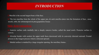
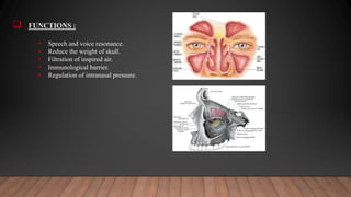
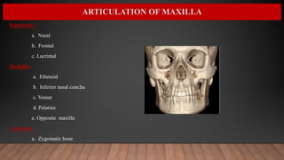
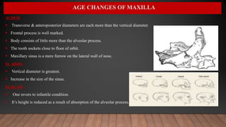
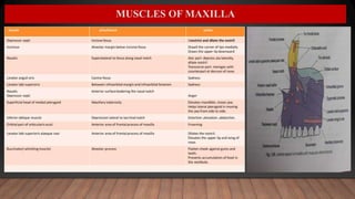
SEMINAR ON MAXILLA
DEPARTMENT OF ORAL AND MAXILLOFACIAL SURGERY
MAHARANA PRATAP COLLEGE OF DENTISTRY AND RESEARCH CENTRE (GWALIOR)
PRESENTED BY:
DR.MRINALINI SINGH
PG 1ST YEAR
CONTENTS
 Introduction
 Development
 Anatomy
 Articulation of maxilla
 Ossification
 Muscles attachment of maxilla
 Blood supply and innervation
 Age changes in maxilla
 Applied anatomy of maxilla
 References
INTRODUCTION
Maxilla is the second largest bone of the face.
 The two maxillae form the whole of the upper jaw, & each maxilla enters into the formation of face , nose,
mouth, orbit, the infratemporal & pterygopalatine fossae.
Side determination-
 Anterior surface ends medially into a deeply cancave border, called the nasal notch. Posterior surface is
convex.
 Alveolar border with sockets for upper teeth faces downward with its convexity directed outward. Frontal
process is the longest process which is directed upward.
 Medial surface is marked by a large irregular opening, the maxillary hiatus..
 PRENATAL GROWTH OF MAXILLA :
 Around the 4th week of intrauterine life , a prominent bulge appears on the ventral aspect
of the embryo corresponding to the developing brain.
 Below the bulge a shallow depression which corresponds to the primitive mouth appears
called STOMODAEUM.
 The floor of the stomodaeum is formed by buccopharyngeal membrane which seperates the
stomodeum from the foregut.
DEVELOPMENT OF MAXILLA
 By around the 4th week of IUL , 5 branchial arches form in the region of future head and
neck.
 Each of these arches gives rise to muscles, connective tissue, vasculature, skeletal
components and neural components of the future face.
 The first branchial arch plays an important role in the development of the naso-maxillary region.
 The mesoderm covering the developing forebrain proliferates and forms a downward
projection thar overlaps the upper part of stomodaeum . This downward projection is called
FRONTO-NASAL PROCESS.
• The stomodaeum is thus overlapped superiorly by the frontonasal process.
• The mandibular arches of both the sides form the lateral walls of the stomodaeum.
• The mandibular arch gives off a bud from its dorsal end called the MAXILLARY PROCESS.
• The maxillary process grows ventro -medio-cranial to the main part of the mandibular arch
which is now called the MANDIBULAR PROCESS.
• Thus at this stage the primitive mouth or stomodaeum is overlapped from above by the
frontal process, below by the mandibular process and on either side by the maxillary process.
• The ectoderm overlying the frontal process shows bilateral localized thickenings above
stomodaeum. These are called the NASAL PLACODES.
• These placodes soon sinks from the nasal pits.
• The formation of these nasal pits divide the fronto-nasal process in to 2 parts:
A. The medial nasal process
B. Lateral nasal process
• The 2 mandibular processess grow medially and fuse to form the lower lip and lower jaw.
• As the maxillary process becomes narrow so that the 2 nasal pits come closer.
• The line of fusion of maxillary process and the medial nasal process correspond
to naso-lacrimal duct.
The palate is formed by the contribution of :
• Maxillary process
• Palatal shelves given off by the maxillary process
• Frontonasal process.
 DEVELOPMENT OF PALATE
PRIMARY PALATE :
• By the fusion of the maxillary and medial nasal process in the roof of stomodaeum the primitive palate ( or primary palate)
is formed, and the olfactory pits extend backward above it.
SECONDARY PALATE :
• The development of the secondary palate commences in the 6th week of IUL. It is characterized by the formation of two
palatal shelves on the maxillary prominences.
• As the palatal shelves grow medially , their union is prevented by the presence of tongue.
• Initially the developing palatal shelves grow vertically toward the floor of mouth.
• During 7th week of IUL , a transformation in the the position of the palatine shelf occurs.
• They change from a vertical to a horizontal position.
• The two palatal shelves by 8 weeks of IUL are in close approximation to each other.
• Initially the 2 palatal shelves are covered by an epithelial lining. As they join the epithelial cells degenerate.
• Initially the contact occurs in the central region of the secondary palate posterior to the premaxilla.
• From this point, closure occurs both anteriorly and posteriorly.
 OSSIFICATION OF PALATE
 Ossification of the palate occurs from the 8th week of IUL.
 Intramembranous type of ossification.
 The palate ossifies from a single centre derived from the maxilla.
 The most posterior part of the palate does not ossify. This forms soft palate.
 The mid palatal suture ossifies by 12-14 yrs.
 POSTNATAL PERIOD
The growth of naso-maxillary complex is produces by the following mechanism:
a. Displacement
b. Growth at suture
c. Surface remodelling
 DISPLACEMENT:
• It is the movement of the whole bone as a unit.
• Displacement can be of 2 types:
A. Primary
B. Secondary
 GROWTH AT SUTURES
• The maxilla is connected to the cranium and cranial base by a number of sutures. These sutures
include:
• Fronto-nasal suture
• Fronto-maxillary suture
• Zygomatico-maxillary suture
• Zygomatico-temporal suture
• Pterygopalatine suture
 SURFACE REMODELING:
• a. Increase in size
• b. Change in shape
• c. Change functional relationship
BONE DEPOSITION AND RESORPTION :
• Bone changes in shape and size by 2 basic mechanisms bone deposition and bone resorption. The bone deposition
and resorption together is called BONE REMODELING.
• The changes that bone depostion and resorption can produce are:
• Change in size
• Change in shape
• Change in proportion
• Change in relationship of the bone with adajacent structures.
 EXPANDING “V” PRINCIPLE OF MAXILLA
As the maxilla descends, transversely, additive growth on the free ends
increases the distance between them.
The buccal segment move outward and downward, as the maxilla itself is
moving downward and forward, following the principle of expanding.
Each maxilla has
A) BODY
The body of maxilla is pyramidal in shape, with its base directed medially at the nasal surface , the apex directed laterally at
the zygomatic process.
It has four surfaces & encloses a large cavity; the maxillary sinus ,its surfaces are:
• Anterior or facial
• Posterior & infratemporal
• Superior or orbital &
• Medial or nasal
B) PROCESESS
• Frontal
• Zygomatic
• Alveolar
• Palatine
ANATOMY OF MAXILLA
SURFACES OF MAXILLA
ANTERIOR/ FACIAL SURFACE :
Anterior surface is directed forwards & laterally & displays inferior elevation overlying the root of teeth.
POSTERIOR / INFRATEMPORAL SURFACE:
Posterior surface is convex & directed backwards and laterally.
SUPERIOR / ORBITAL SURFACE:
Superior surface is smooth , triangular & slightly
concave,& forms a greater part of the floor of the orbit.
The superior surface presents:
• Infraorbital groove and canal
• Canalis sinosus
• Inferior oblique muscle
MEDIAL NASAL SURFACE:
Form lateral wall of nose
MAXILLARY SINUS (ANTRUM OF HIGHMORE)
 Maxillary sinus is first sinus to develop & is a large cavity in the body of maxilla.
 Pyramidal in shape
 Size is variable.
Average measurments are:
 Height-3.7cm.
 Width-2.5cm.
 Anteroposterior depth-3.7cm
 The sinus opens into the middle meatus of nose usually by two
openings one of which is closed by mucous membrane.
 FUNCTIONS :
 Speech and voice resonance.
 Reduce the weight of skull.
 Filtration of inspired air.
 Immunological barrier.
 Regulation of intranasal pressure.
PROCESS OF MAXILLA
1-Frontal process: it projects upwards & backwards to articulate above with the nasal margin
of frontal bone, in front with nasal bone, & behind with the lacrimal bone.
2-Zygomatic process: is a pyramidal projection where anterior, infratemporal & orbital surface
converge.
3-Alveolar process: it forms half of the alveolar arch, & bears socket for the root of upper teeth.
In adults, there are 8 sockets; canine socket is deepest; molar sockets are widest & divided into
three minor socket by septa; the incisor & second premolar socket is sometimes divided into two.
4- Palatine process: Palatine process is a thick horizontal plate projecting medially from the lowest
part of the nasal surface. It forms a large part of the roof of mouth & the floor of nasal cavity.
ARTICULATION OF MAXILLA
Superiorly :
a. Nasal
b. Frontal
c. Lacrimal
Medially:
a. Ethmoid
b. Inferior nasal concha
c. Vomer
d. Palatine
e. Opposite maxilla
Laterally:
a. Zygomatic bone
• Maxilla ossifies in membrane from three centres, one for maxilla proper and two for os incisivum or premaxilla.
• The centre for maxilla proper appears above the canine fossa during 6th week of IUL.
• Of the two premaxillary centres, the main centre appears above the incisive fossa during 7th week of IUL.
• The second centres appear at the ventral margin of nasal septum during 10th week of IUL and soon fuses with
palatal process of maxilla.
• Premaxilla begins to fuse with alveolar process almost immediately after the ossification begins, the evidence of
premaxilla as a separate bone may persist until the middle decades.
OSSIFICATION OF MAXILLA
AGE CHANGES OF MAXILLA
At birth
 Transverse & anteroposterior diameters are each more than the vertical diameter.
 Frontal process is well marked.
 Body consists of little more than the alveolar process.
 The tooth sockets close to floor of orbit.
 Maxillary sinus is a mere furrow on the lateral wall of nose.
In adults
 Vertical diameter is greatest.
 Increase in the size of the sinus.
In the old
 One revers to infantile condition.
 It’s height is reduced as a result of absorption of the alveolar process.
MUSCLES OF MAXILLA
BLOOD SUPPLY OF MAXILLA
MAXILLARY ARTERY IS DIVIDED INTO 3 PARTS:
1ST PART (MANDIBULAR PART)
1. Deep auricular artery
2. Anterior tympanic branch
3. Middle meningeal artery
4. Accessory meningeal artery
5. Inferior alveolar artery
2ND PART (PTERYGOID PART)
1. Deep temporal
2. Pterygoid
3. Massetric
4. Buccal
3RD PART (PTERYGOPALATINE PART)
1. Posterior superior alveolar artery
2. Infraorbital
3. Greater palatine
4. Pharyngeal
5. Artery of pterygoid canal
6. Sphenopalatine
NERVE SUPPLY
Branches :
IN THE CRANIAL CAVITY
• Meningeal branch
IN THE PTERYGOPALATINE FOSSA
• 2 ganglionic branches
IN THE INFRATEMPORAL FOSSA
• Posterior superior alveolar nerve
• Zygomatic nerve
VEINOUS DRAINAGE
LYMPHATIC DRAINAGE
APPLIED ANATOMY
 MAXILLARY SINUSITIS : is usually secondary to allergy or infection.
Clinical features –
Pain & tenderness which is usually associated with headache.
Pain also radiates to teeth & is often mistaken as dental pain (referred odontalgia).
Radiological features-
SOFT TISSUE INVOLVEMENT :
Management –
Medicinal treatment: Surgical treatment:
Antihistaminic Nasal Antrostomy
Decongestants Caldwell-luc operation
Analgesics & antipyretic
It is a pathological communication between the oral cavity & the maxillary sinus,which is
lined by epithelium.
Management-
1-General management-
Gentle packing of the socket with wet gauze to control the bleeding.
2-Definitive management –
Buccal flap
Palatal flap
Combination of buccal & palatal flap
OROANTRAL FISTULA
 Torus palatinus : is a benign, slowly growing, bony projection of the palatine processes of the maxillae &
occasionally of the horizontal plates of the palatine bones.
Management-
A median palatal incision is made in the mucosa the full length of the torus with two short, obliquely diverging incisions at
the anterior & posterior ends, avoiding the vascular foramina.
HARD TISSUE INVOLVEMENT
Anatomic space infection :
 canine fossa contains varying amounts of connective tissue & fat & is bounded superiorly by the quadratus labii
superioris muscle, anteriorly by the orbicularis oris & posteriorly by the buccinator.
 Infection of this area results in swelling of the upper lip, obliterating the nasolabial fold & of the upper & lower eyelids,
closing the eye.
:
 MAXILLARY SAGITTAL FRACTURE :
 Fracture of a maxilla in a sagittal plane, involving anterior - lateral wall of a maxillary sinus.
 Due to direct blow together at right & left midface.
 ISOLATED ALVEOLAR PROCESS FRACTURE :
 Fracture of any portion of alveolar process.
 Clinically evident by malalignment and displacement of teeth contained within fracture segment.
CONGENITAL DEFECTS ASSOCIATED WITH MAXILLA
MICROGNATHIA : Micrognathia of the maxilla frequently occurs due to a
deficiency in the premaxillary area, and patients with this deformity appear to have the
middle third of the face retracted.
MACROGNATHIA : Macrognathia refers to the condition of abnormally large jaws.
Paget’s disease of bone, in which overgrowth of the cranium and maxilla or occasionally
the mandible occurs.
Leontiasis ossea, a form of fibrous dysplasia in which there is enlargement of the
maxilla.
OBLIQUE CLEFT LIP CLEFT LIP CLEFT PALATE BILATERAL CLEFTLIP
Triangular flap technique
Millard rotational flap technique Straight line repair
Primary : Closure of the lip and
anterior palate.
Closure of the palate
Secondary : Pharyngoplasty
Orthognathic proceddure
SYNDROME ASSOCIATED WITH MAXILLA
• ROMBERG / PARRY ROMBERG SYNDROME : Progressive hemifacial atrophy.
• Atrophy of the skin, subcutaneous tissue, muscles, bones, cartilages, alveolar bone and soft palate on that side
of the face.
• In addition to facial wasting that may include the ipsilateral salivary glands and hemiatrophy of the tongue,
unilateral involvement of the ear, larynx, esophagus, diaphragm, kidney and brain.
APERT SYNDROME / ACROCEPHALOSTNDUCTYLY
Middle third of face is markedly reduded.
Apert’s syndrome = Acrocephaly + low IQ. + Complex syndactyly
Oral manifestations - a high arched palatal vault , Bifid uvula as well as malocclusion.
In addition, multiple odontogenic cysts of the maxilla and mandible .
One additional finding sometimes present, is temporomandibular dysarthrosis
CRUZON SYNDROME / CRANIOFACIAL DYSOSTOSIS :
Midface hypoplasia, underdeveloped maxilla.
Wide face and hypoplastic maxilla producing pseudoprognathism are observed.
MARFAN SYNDROME
REFERENCES :
• B.D. chaurasia’s, Human Anatomy 5th edition Volume 3 head & neck, brain.
• Gray’s Anatomy, 38th edition, Churchill Livingstone.
• Shafer’s oral pathology
• Langman’s medical embryology
seminar on maxilla.pptx

seminar on maxilla.pptx

  • 1.
    SEMINAR ON MAXILLA DEPARTMENTOF ORAL AND MAXILLOFACIAL SURGERY MAHARANA PRATAP COLLEGE OF DENTISTRY AND RESEARCH CENTRE (GWALIOR) PRESENTED BY: DR.MRINALINI SINGH PG 1ST YEAR
  • 2.
    CONTENTS  Introduction  Development Anatomy  Articulation of maxilla  Ossification  Muscles attachment of maxilla  Blood supply and innervation  Age changes in maxilla  Applied anatomy of maxilla  References
  • 3.
    INTRODUCTION Maxilla is thesecond largest bone of the face.  The two maxillae form the whole of the upper jaw, & each maxilla enters into the formation of face , nose, mouth, orbit, the infratemporal & pterygopalatine fossae. Side determination-  Anterior surface ends medially into a deeply cancave border, called the nasal notch. Posterior surface is convex.  Alveolar border with sockets for upper teeth faces downward with its convexity directed outward. Frontal process is the longest process which is directed upward.  Medial surface is marked by a large irregular opening, the maxillary hiatus..
  • 4.
     PRENATAL GROWTHOF MAXILLA :  Around the 4th week of intrauterine life , a prominent bulge appears on the ventral aspect of the embryo corresponding to the developing brain.  Below the bulge a shallow depression which corresponds to the primitive mouth appears called STOMODAEUM.  The floor of the stomodaeum is formed by buccopharyngeal membrane which seperates the stomodeum from the foregut. DEVELOPMENT OF MAXILLA  By around the 4th week of IUL , 5 branchial arches form in the region of future head and neck.  Each of these arches gives rise to muscles, connective tissue, vasculature, skeletal components and neural components of the future face.
  • 5.
     The firstbranchial arch plays an important role in the development of the naso-maxillary region.  The mesoderm covering the developing forebrain proliferates and forms a downward projection thar overlaps the upper part of stomodaeum . This downward projection is called FRONTO-NASAL PROCESS.
  • 6.
    • The stomodaeumis thus overlapped superiorly by the frontonasal process. • The mandibular arches of both the sides form the lateral walls of the stomodaeum. • The mandibular arch gives off a bud from its dorsal end called the MAXILLARY PROCESS. • The maxillary process grows ventro -medio-cranial to the main part of the mandibular arch which is now called the MANDIBULAR PROCESS. • Thus at this stage the primitive mouth or stomodaeum is overlapped from above by the frontal process, below by the mandibular process and on either side by the maxillary process. • The ectoderm overlying the frontal process shows bilateral localized thickenings above stomodaeum. These are called the NASAL PLACODES. • These placodes soon sinks from the nasal pits. • The formation of these nasal pits divide the fronto-nasal process in to 2 parts: A. The medial nasal process B. Lateral nasal process
  • 7.
    • The 2mandibular processess grow medially and fuse to form the lower lip and lower jaw. • As the maxillary process becomes narrow so that the 2 nasal pits come closer. • The line of fusion of maxillary process and the medial nasal process correspond to naso-lacrimal duct. The palate is formed by the contribution of : • Maxillary process • Palatal shelves given off by the maxillary process • Frontonasal process.  DEVELOPMENT OF PALATE
  • 8.
    PRIMARY PALATE : •By the fusion of the maxillary and medial nasal process in the roof of stomodaeum the primitive palate ( or primary palate) is formed, and the olfactory pits extend backward above it. SECONDARY PALATE : • The development of the secondary palate commences in the 6th week of IUL. It is characterized by the formation of two palatal shelves on the maxillary prominences. • As the palatal shelves grow medially , their union is prevented by the presence of tongue. • Initially the developing palatal shelves grow vertically toward the floor of mouth. • During 7th week of IUL , a transformation in the the position of the palatine shelf occurs. • They change from a vertical to a horizontal position.
  • 9.
    • The twopalatal shelves by 8 weeks of IUL are in close approximation to each other. • Initially the 2 palatal shelves are covered by an epithelial lining. As they join the epithelial cells degenerate. • Initially the contact occurs in the central region of the secondary palate posterior to the premaxilla. • From this point, closure occurs both anteriorly and posteriorly.  OSSIFICATION OF PALATE  Ossification of the palate occurs from the 8th week of IUL.  Intramembranous type of ossification.  The palate ossifies from a single centre derived from the maxilla.  The most posterior part of the palate does not ossify. This forms soft palate.  The mid palatal suture ossifies by 12-14 yrs.
  • 10.
     POSTNATAL PERIOD Thegrowth of naso-maxillary complex is produces by the following mechanism: a. Displacement b. Growth at suture c. Surface remodelling  DISPLACEMENT: • It is the movement of the whole bone as a unit. • Displacement can be of 2 types: A. Primary B. Secondary
  • 11.
     GROWTH ATSUTURES • The maxilla is connected to the cranium and cranial base by a number of sutures. These sutures include: • Fronto-nasal suture • Fronto-maxillary suture • Zygomatico-maxillary suture • Zygomatico-temporal suture • Pterygopalatine suture
  • 12.
     SURFACE REMODELING: •a. Increase in size • b. Change in shape • c. Change functional relationship BONE DEPOSITION AND RESORPTION : • Bone changes in shape and size by 2 basic mechanisms bone deposition and bone resorption. The bone deposition and resorption together is called BONE REMODELING. • The changes that bone depostion and resorption can produce are: • Change in size • Change in shape • Change in proportion • Change in relationship of the bone with adajacent structures.
  • 13.
     EXPANDING “V”PRINCIPLE OF MAXILLA As the maxilla descends, transversely, additive growth on the free ends increases the distance between them. The buccal segment move outward and downward, as the maxilla itself is moving downward and forward, following the principle of expanding.
  • 14.
    Each maxilla has A)BODY The body of maxilla is pyramidal in shape, with its base directed medially at the nasal surface , the apex directed laterally at the zygomatic process. It has four surfaces & encloses a large cavity; the maxillary sinus ,its surfaces are: • Anterior or facial • Posterior & infratemporal • Superior or orbital & • Medial or nasal B) PROCESESS • Frontal • Zygomatic • Alveolar • Palatine ANATOMY OF MAXILLA
  • 15.
    SURFACES OF MAXILLA ANTERIOR/FACIAL SURFACE : Anterior surface is directed forwards & laterally & displays inferior elevation overlying the root of teeth. POSTERIOR / INFRATEMPORAL SURFACE: Posterior surface is convex & directed backwards and laterally.
  • 16.
    SUPERIOR / ORBITALSURFACE: Superior surface is smooth , triangular & slightly concave,& forms a greater part of the floor of the orbit. The superior surface presents: • Infraorbital groove and canal • Canalis sinosus • Inferior oblique muscle MEDIAL NASAL SURFACE: Form lateral wall of nose
  • 17.
    MAXILLARY SINUS (ANTRUMOF HIGHMORE)  Maxillary sinus is first sinus to develop & is a large cavity in the body of maxilla.  Pyramidal in shape  Size is variable. Average measurments are:  Height-3.7cm.  Width-2.5cm.  Anteroposterior depth-3.7cm  The sinus opens into the middle meatus of nose usually by two openings one of which is closed by mucous membrane.
  • 18.
     FUNCTIONS : Speech and voice resonance.  Reduce the weight of skull.  Filtration of inspired air.  Immunological barrier.  Regulation of intranasal pressure.
  • 19.
    PROCESS OF MAXILLA 1-Frontalprocess: it projects upwards & backwards to articulate above with the nasal margin of frontal bone, in front with nasal bone, & behind with the lacrimal bone. 2-Zygomatic process: is a pyramidal projection where anterior, infratemporal & orbital surface converge. 3-Alveolar process: it forms half of the alveolar arch, & bears socket for the root of upper teeth. In adults, there are 8 sockets; canine socket is deepest; molar sockets are widest & divided into three minor socket by septa; the incisor & second premolar socket is sometimes divided into two. 4- Palatine process: Palatine process is a thick horizontal plate projecting medially from the lowest part of the nasal surface. It forms a large part of the roof of mouth & the floor of nasal cavity.
  • 21.
    ARTICULATION OF MAXILLA Superiorly: a. Nasal b. Frontal c. Lacrimal Medially: a. Ethmoid b. Inferior nasal concha c. Vomer d. Palatine e. Opposite maxilla Laterally: a. Zygomatic bone
  • 22.
    • Maxilla ossifiesin membrane from three centres, one for maxilla proper and two for os incisivum or premaxilla. • The centre for maxilla proper appears above the canine fossa during 6th week of IUL. • Of the two premaxillary centres, the main centre appears above the incisive fossa during 7th week of IUL. • The second centres appear at the ventral margin of nasal septum during 10th week of IUL and soon fuses with palatal process of maxilla. • Premaxilla begins to fuse with alveolar process almost immediately after the ossification begins, the evidence of premaxilla as a separate bone may persist until the middle decades. OSSIFICATION OF MAXILLA
  • 23.
    AGE CHANGES OFMAXILLA At birth  Transverse & anteroposterior diameters are each more than the vertical diameter.  Frontal process is well marked.  Body consists of little more than the alveolar process.  The tooth sockets close to floor of orbit.  Maxillary sinus is a mere furrow on the lateral wall of nose. In adults  Vertical diameter is greatest.  Increase in the size of the sinus. In the old  One revers to infantile condition.  It’s height is reduced as a result of absorption of the alveolar process.
  • 24.
  • 25.
    BLOOD SUPPLY OFMAXILLA MAXILLARY ARTERY IS DIVIDED INTO 3 PARTS: 1ST PART (MANDIBULAR PART) 1. Deep auricular artery 2. Anterior tympanic branch 3. Middle meningeal artery 4. Accessory meningeal artery 5. Inferior alveolar artery 2ND PART (PTERYGOID PART) 1. Deep temporal 2. Pterygoid 3. Massetric 4. Buccal 3RD PART (PTERYGOPALATINE PART) 1. Posterior superior alveolar artery 2. Infraorbital 3. Greater palatine 4. Pharyngeal 5. Artery of pterygoid canal 6. Sphenopalatine
  • 26.
    NERVE SUPPLY Branches : INTHE CRANIAL CAVITY • Meningeal branch IN THE PTERYGOPALATINE FOSSA • 2 ganglionic branches IN THE INFRATEMPORAL FOSSA • Posterior superior alveolar nerve • Zygomatic nerve
  • 27.
  • 28.
  • 29.
    APPLIED ANATOMY  MAXILLARYSINUSITIS : is usually secondary to allergy or infection. Clinical features – Pain & tenderness which is usually associated with headache. Pain also radiates to teeth & is often mistaken as dental pain (referred odontalgia). Radiological features- SOFT TISSUE INVOLVEMENT :
  • 30.
    Management – Medicinal treatment:Surgical treatment: Antihistaminic Nasal Antrostomy Decongestants Caldwell-luc operation Analgesics & antipyretic
  • 31.
    It is apathological communication between the oral cavity & the maxillary sinus,which is lined by epithelium. Management- 1-General management- Gentle packing of the socket with wet gauze to control the bleeding. 2-Definitive management – Buccal flap Palatal flap Combination of buccal & palatal flap OROANTRAL FISTULA
  • 32.
     Torus palatinus: is a benign, slowly growing, bony projection of the palatine processes of the maxillae & occasionally of the horizontal plates of the palatine bones. Management- A median palatal incision is made in the mucosa the full length of the torus with two short, obliquely diverging incisions at the anterior & posterior ends, avoiding the vascular foramina. HARD TISSUE INVOLVEMENT
  • 33.
    Anatomic space infection:  canine fossa contains varying amounts of connective tissue & fat & is bounded superiorly by the quadratus labii superioris muscle, anteriorly by the orbicularis oris & posteriorly by the buccinator.  Infection of this area results in swelling of the upper lip, obliterating the nasolabial fold & of the upper & lower eyelids, closing the eye.
  • 34.
    :  MAXILLARY SAGITTALFRACTURE :  Fracture of a maxilla in a sagittal plane, involving anterior - lateral wall of a maxillary sinus.  Due to direct blow together at right & left midface.  ISOLATED ALVEOLAR PROCESS FRACTURE :  Fracture of any portion of alveolar process.  Clinically evident by malalignment and displacement of teeth contained within fracture segment.
  • 35.
    CONGENITAL DEFECTS ASSOCIATEDWITH MAXILLA MICROGNATHIA : Micrognathia of the maxilla frequently occurs due to a deficiency in the premaxillary area, and patients with this deformity appear to have the middle third of the face retracted. MACROGNATHIA : Macrognathia refers to the condition of abnormally large jaws. Paget’s disease of bone, in which overgrowth of the cranium and maxilla or occasionally the mandible occurs. Leontiasis ossea, a form of fibrous dysplasia in which there is enlargement of the maxilla.
  • 36.
    OBLIQUE CLEFT LIPCLEFT LIP CLEFT PALATE BILATERAL CLEFTLIP Triangular flap technique Millard rotational flap technique Straight line repair Primary : Closure of the lip and anterior palate. Closure of the palate Secondary : Pharyngoplasty Orthognathic proceddure
  • 37.
    SYNDROME ASSOCIATED WITHMAXILLA • ROMBERG / PARRY ROMBERG SYNDROME : Progressive hemifacial atrophy. • Atrophy of the skin, subcutaneous tissue, muscles, bones, cartilages, alveolar bone and soft palate on that side of the face. • In addition to facial wasting that may include the ipsilateral salivary glands and hemiatrophy of the tongue, unilateral involvement of the ear, larynx, esophagus, diaphragm, kidney and brain. APERT SYNDROME / ACROCEPHALOSTNDUCTYLY Middle third of face is markedly reduded. Apert’s syndrome = Acrocephaly + low IQ. + Complex syndactyly
  • 38.
    Oral manifestations -a high arched palatal vault , Bifid uvula as well as malocclusion. In addition, multiple odontogenic cysts of the maxilla and mandible . One additional finding sometimes present, is temporomandibular dysarthrosis CRUZON SYNDROME / CRANIOFACIAL DYSOSTOSIS : Midface hypoplasia, underdeveloped maxilla. Wide face and hypoplastic maxilla producing pseudoprognathism are observed. MARFAN SYNDROME
  • 39.
    REFERENCES : • B.D.chaurasia’s, Human Anatomy 5th edition Volume 3 head & neck, brain. • Gray’s Anatomy, 38th edition, Churchill Livingstone. • Shafer’s oral pathology • Langman’s medical embryology