Development of theEar
Development of the Ear
For medicine students
For medicine students
2.
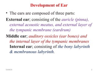
Development of Ear
•The ears are composed of three parts:
External ear; consisting of the auricle (pinna),
external acoustic meatus, and external layer of
the tympanic membrane (eardrum)
Middle ear; auditory ossicles (ear bones) and
the internal layer of the tympanic membranes
Internal ear; consisting of the bony labyrinth
& membranous labyrinth.
03/08/25 2
3.
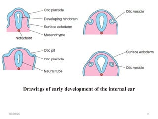
Development of InternalEar
• It is the first part of the ears to develop.
• Early in the fourth week, the otic placode (a
thickening of surface ectoderm) appears on each side
of the hindbrain.
• Each otic placode soon invaginates into the
underlying mesenchyme and form an otic pit.
• The edges of the pit come together and fuse to form
an otic vesicle,
– which is the primordium of the membranous labyrinth.
• The vesicle soon loses its connection with the surface
ectoderm.
03/08/25 3
Development of InternalEar cont…
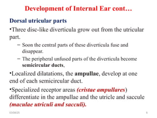
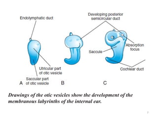
• Two regions of the otic vesicles are
recognizable:
– Dorsal utricular parts, from which the small
endolymphatic ducts, utricles, and semicircular
ducts arise.
– Ventral saccular parts, which give rise to the
saccules and cochlear ducts.
03/08/25 5
6.
Development of InternalEar cont…
Dorsal utricular parts
•Three disc-like diverticula grow out from the utricular
part.
– Soon the central parts of these diverticula fuse and
disappear.
– The peripheral unfused parts of the diverticula become
semicircular ducts,
•Localized dilatations, the ampullae, develop at one
end of each semicircular duct.
•Specialized receptor areas (cristae ampullares)
differentiate in the ampullae and the utricle and saccule
(maculae utriculi and sacculi).
03/08/25 6
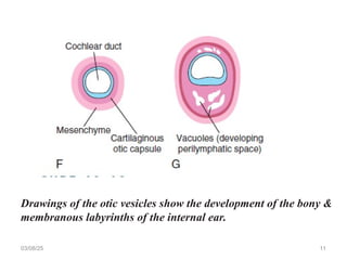
7.
7
Drawings of theotic vesicles show the development of the
membranous labyrinths of the internal ear.
8.
8
Drawings of theotic vesicles show the development of the
membranous labyrinths of the internal ear.
9.
Ventral saccular parts
•Fromthis part, a tubular diverticulum (cochlear duct)
grows and coils to form the membranous cochlea.
•A connection of the cochlea with the saccule (ductus
reuniens) soon forms.
•The spiral organ differentiates from cells in the wall of
the cochlear duct
•Inductive influences from the otic vesicle stimulate the
mesenchyme around it to condense and differentiate
into a cartilaginous otic capsule
03/08/25 9
Development of Internal Ear cont…
10.
• As themembranous labyrinth enlarges, vacuoles
appear in the cartilaginous otic capsule and soon
coalesce to form the perilymphatic space.
– The perilymphatic space, which is related to the cochlear
duct, develops two divisions, the scala tympani and scala
vestibule.
• The cartilaginous otic capsule later ossifies to form
the bony labyrinth of the internal ear.
• The internal ear reaches its adult size and shape by
the middle of the fetal period (20–22 weeks).
10
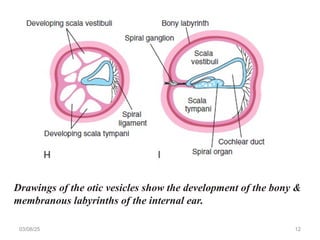
Development of Internal Ear cont…
11.
03/08/25 11
Drawings ofthe otic vesicles show the development of the bony &
membranous labyrinths of the internal ear.
12.
03/08/25 12
Drawings ofthe otic vesicles show the development of the bony &
membranous labyrinths of the internal ear.
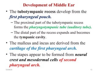
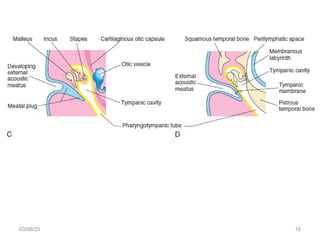
13.
• The tubotympanicrecess develop from the
first pharyngeal pouch.
– The proximal part of the tubotympanic recess
forms the pharyngotympanic tube (auditory tube).
– The distal part of the recess expands and becomes
the tympanic cavity.
• The malleus and incus are derived from the
cartilage of the first pharyngeal arch.
• The stapes appear to be formed from neural
crest and mesodermal cells of second
pharyngeal arch.
03/08/25 13
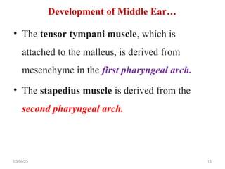
Development of Middle Ear
• The tensortympani muscle, which is
attached to the malleus, is derived from
mesenchyme in the first pharyngeal arch.
• The stapedius muscle is derived from the
second pharyngeal arch.
03/08/25 15
Development of Middle Ear…
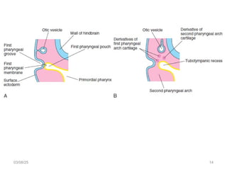
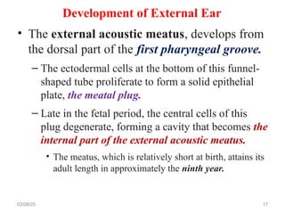
Development of ExternalEar
• The external acoustic meatus, develops from
the dorsal part of the first pharyngeal groove.
– The ectodermal cells at the bottom of this funnel-
shaped tube proliferate to form a solid epithelial
plate, the meatal plug.
– Late in the fetal period, the central cells of this
plug degenerate, forming a cavity that becomes the
internal part of the external acoustic meatus.
• The meatus, which is relatively short at birth, attains its
adult length in approximately the ninth year.
03/08/25 17
18.
Development of ExternalEar cont…
• The primordium of the tympanic membrane is
the first pharyngeal membrane.
– In the embryo, the pharyngeal membrane separates
the first pharyngeal groove from the first
pharyngeal pouch.
• The tympanic membrane develops from three
sources:
– Ectoderm of the first pharyngeal groove
– Endoderm of the tubotympanic recess, a derivative
of the first pharyngeal pouch
– Mesenchyme of the first and second pharyngeal
arches 18
19.
Development of ExternalEar cont…
• The auricle (pinna) develops from
mesenchymal proliferations in the first and
second pharyngeal arches (auricular
hillocks).
– As the auricle grows, the contribution from the first
arch is reduced.
• The auricles begin to develop at the base of the
neck.
• As the mandible develops, the auricles assume
their normal position at the side of the head
19
20.
Congenital Deafness
• Approximately3 in 1000 neonates have
significant hearing loss.
• Most types of congenital deafness are caused by
genetic factors.
• Congenital deafness may be associated with
several other head and neck defects as a part
of the first arch syndrome.
– Abnormalities of the malleus and incus are often
associated with this syndrome.
– A rubella infection (particularly in 7th
& 8th
wks)
can cause defects of the spiral organ & deafness.
20
21.
Microtia
• Microtia (smallor rudimentary auricle) results
from suppressed mesenchymal proliferation.
• This defect often serves as an indicator of
associated birth defects, such as
– atresia of the external acoustic meatus (80% of
cases) and middle ear anomalies.
• The cause can be both genetic & environmental.
03/08/25 21
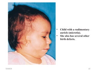
22.
03/08/25 22
• Childwith a rudimentary
auricle (microtia).
• She also has several other
birth defects.
23.
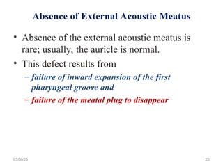
Absence of ExternalAcoustic Meatus
• Absence of the external acoustic meatus is
rare; usually, the auricle is normal.
• This defect results from
– failure of inward expansion of the first
pharyngeal groove and
– failure of the meatal plug to disappear
03/08/25 23
24.
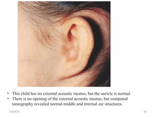
03/08/25 24
• Thischild has no external acoustic meatus, but the auricle is normal.
• There is no opening of the external acoustic meatus, but computed
tomography revealed normal middle and internal ear structures.


![PERI-PROSTHETIC FRACTURE NAIL-PLATE CONSTRUCT [NPC].pptx](https://cdn.slidesharecdn.com/ss_thumbnails/drarunkumardrmohamedashrafperiprostheticfrasturenail-plateconstructnpc-260209164459-7e9d15a1-thumbnail.jpg?width=640&height=640&fit=bounds)


