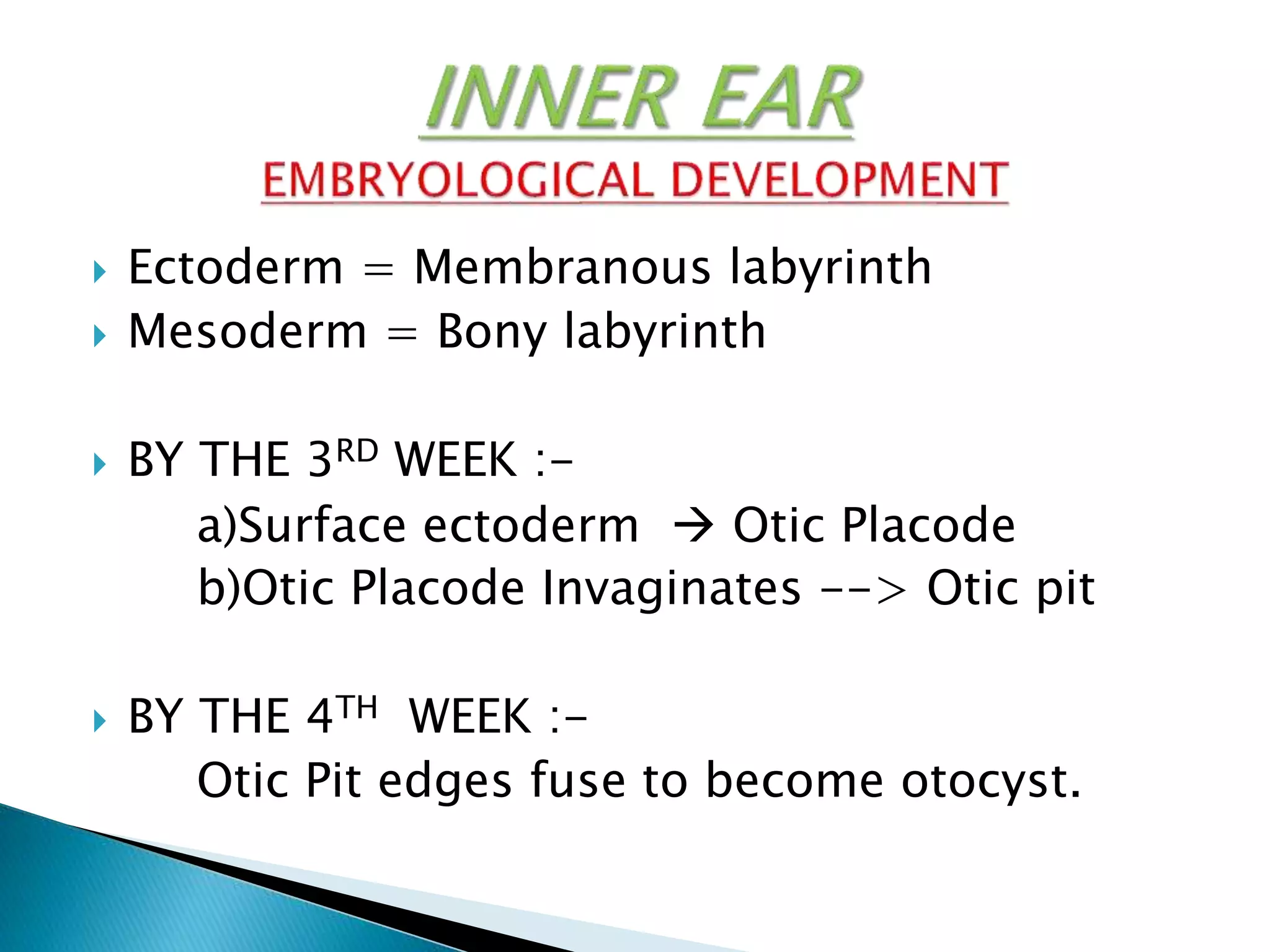
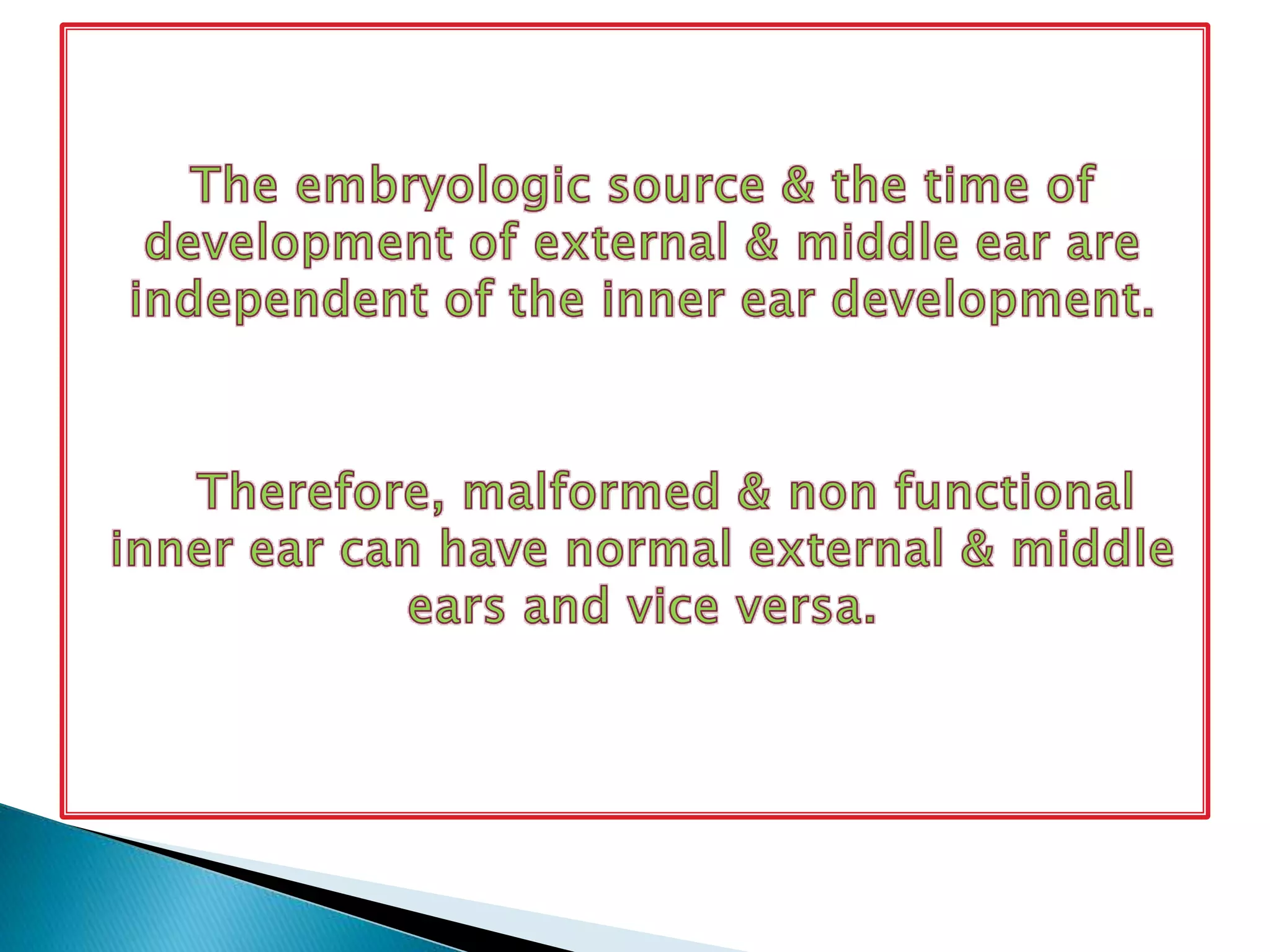
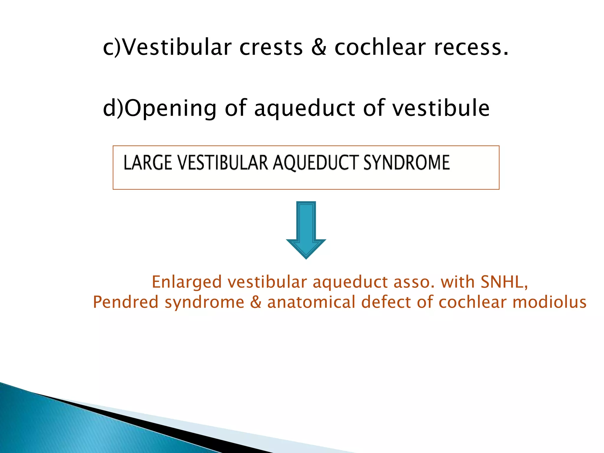
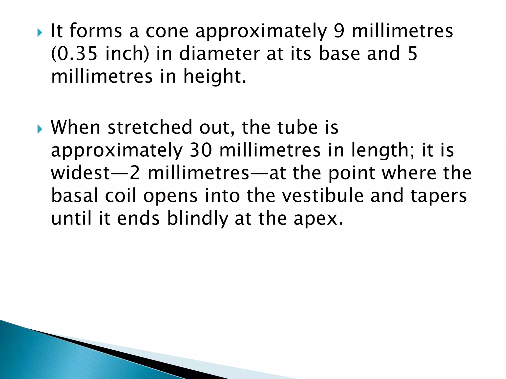
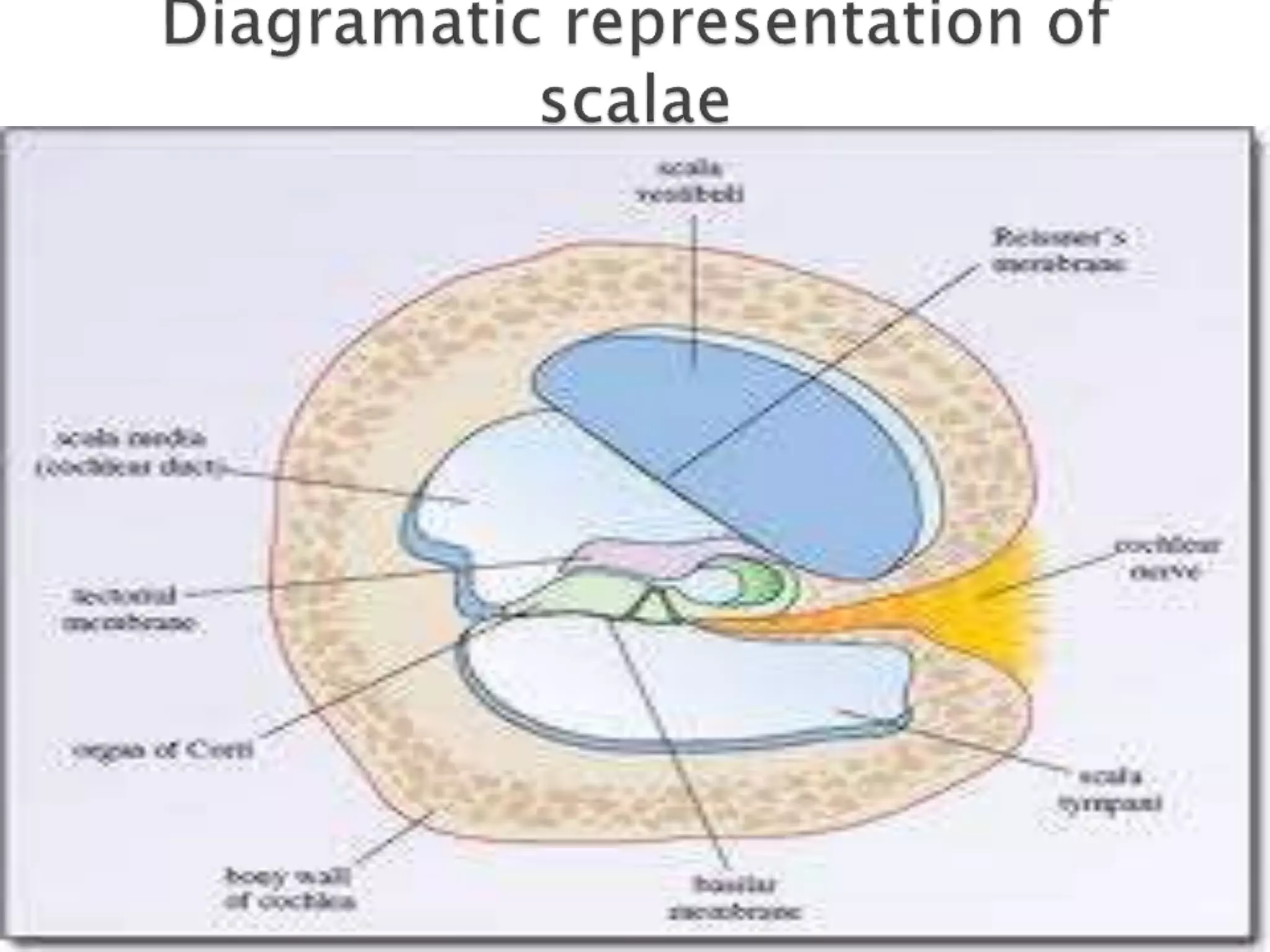
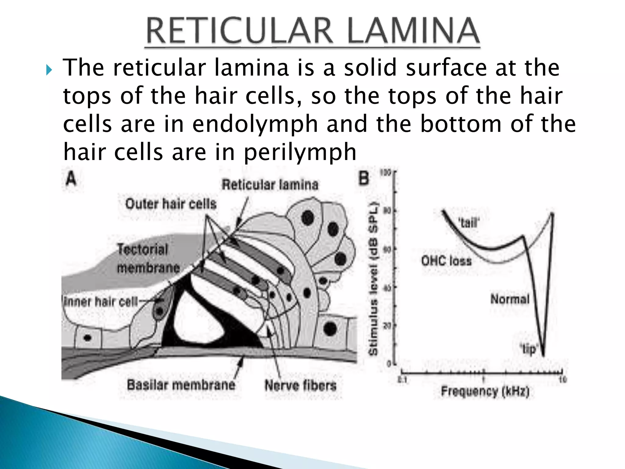
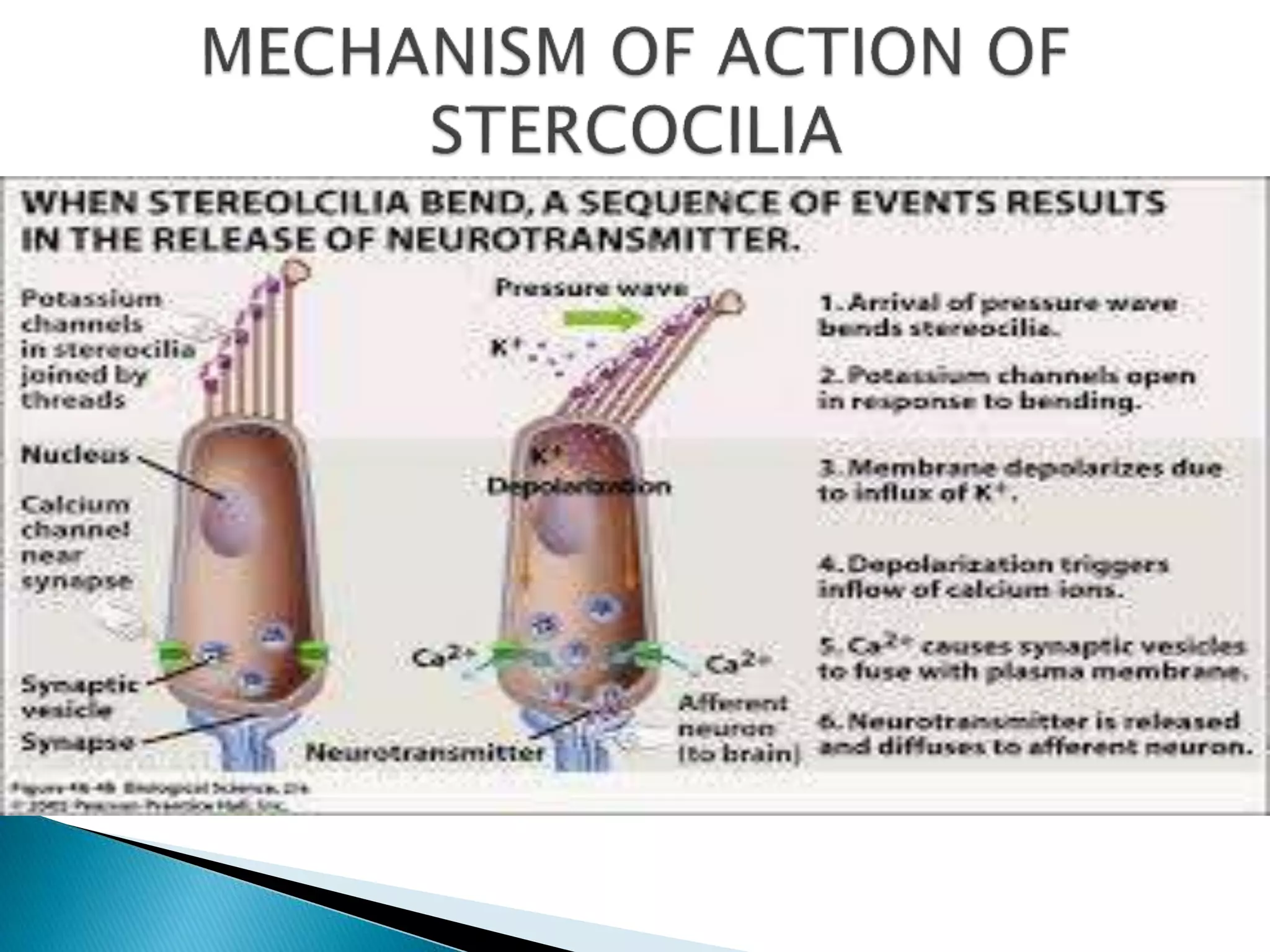
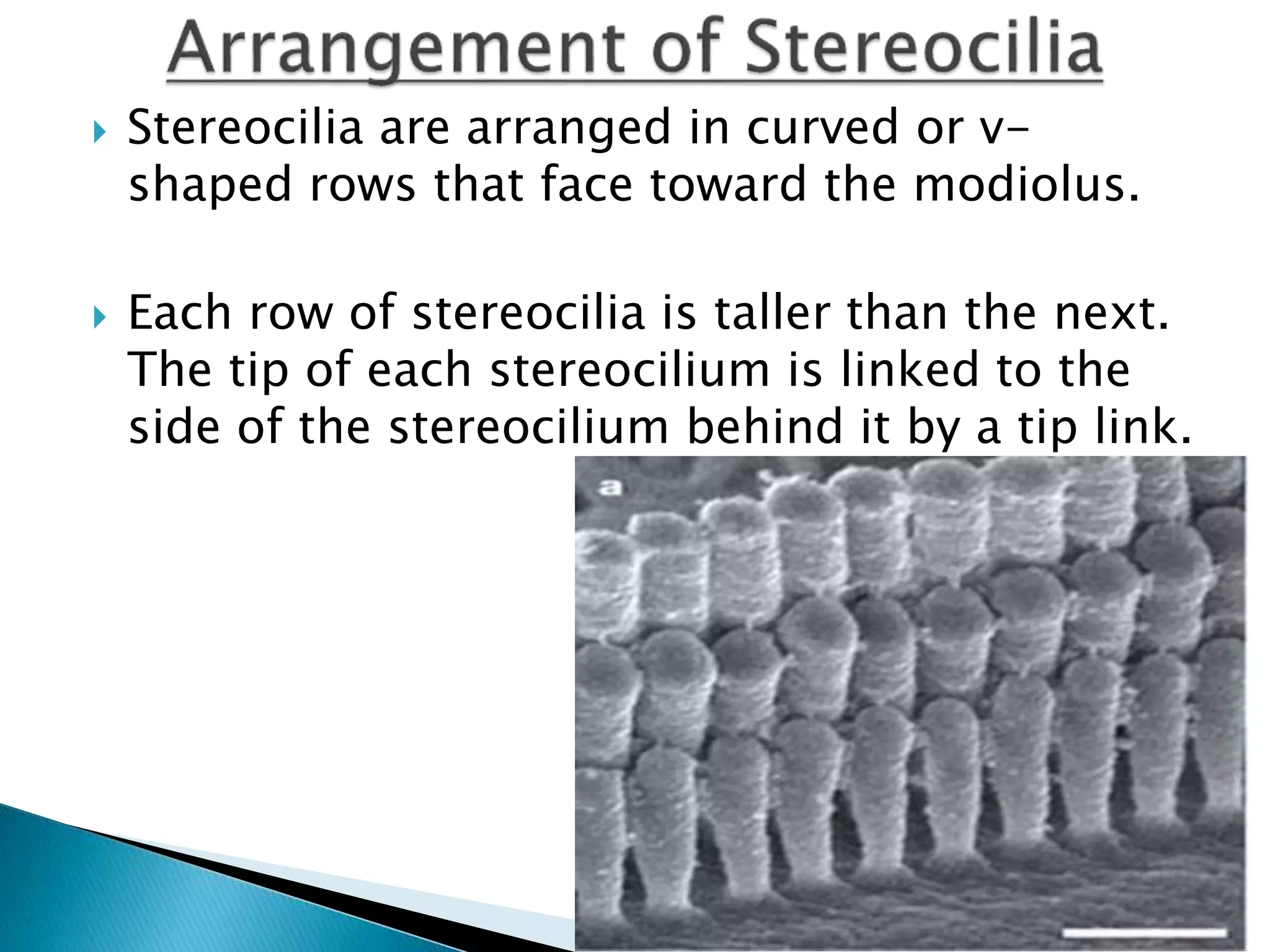
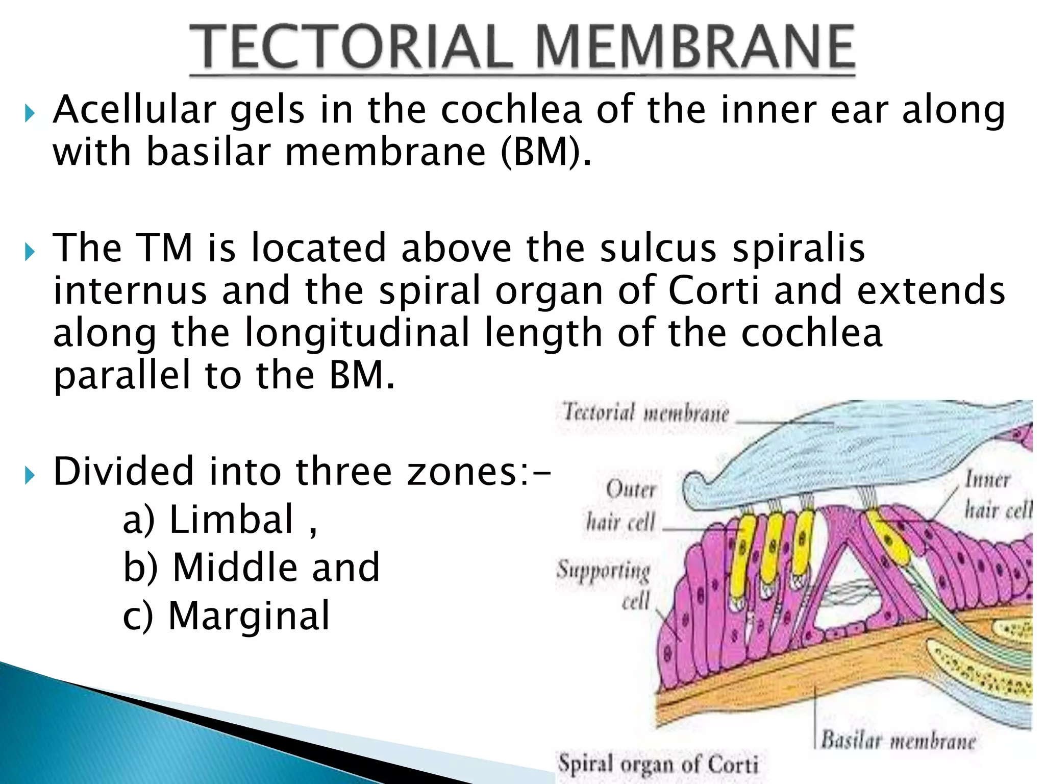
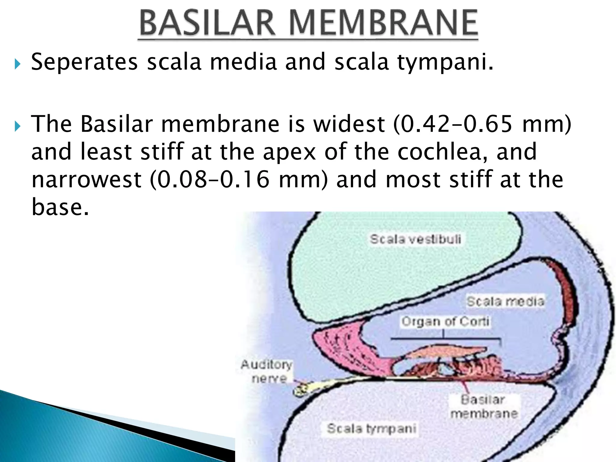
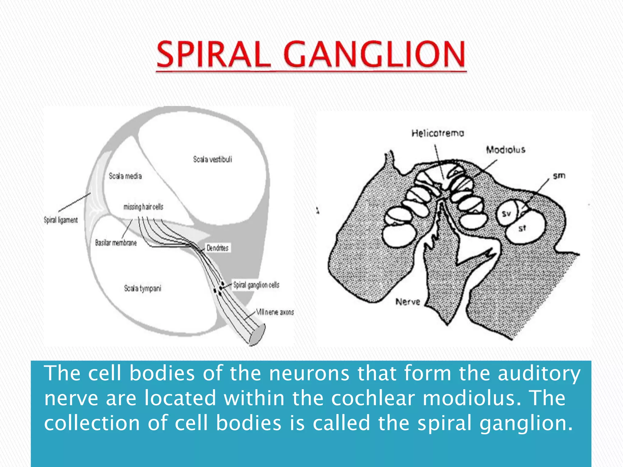
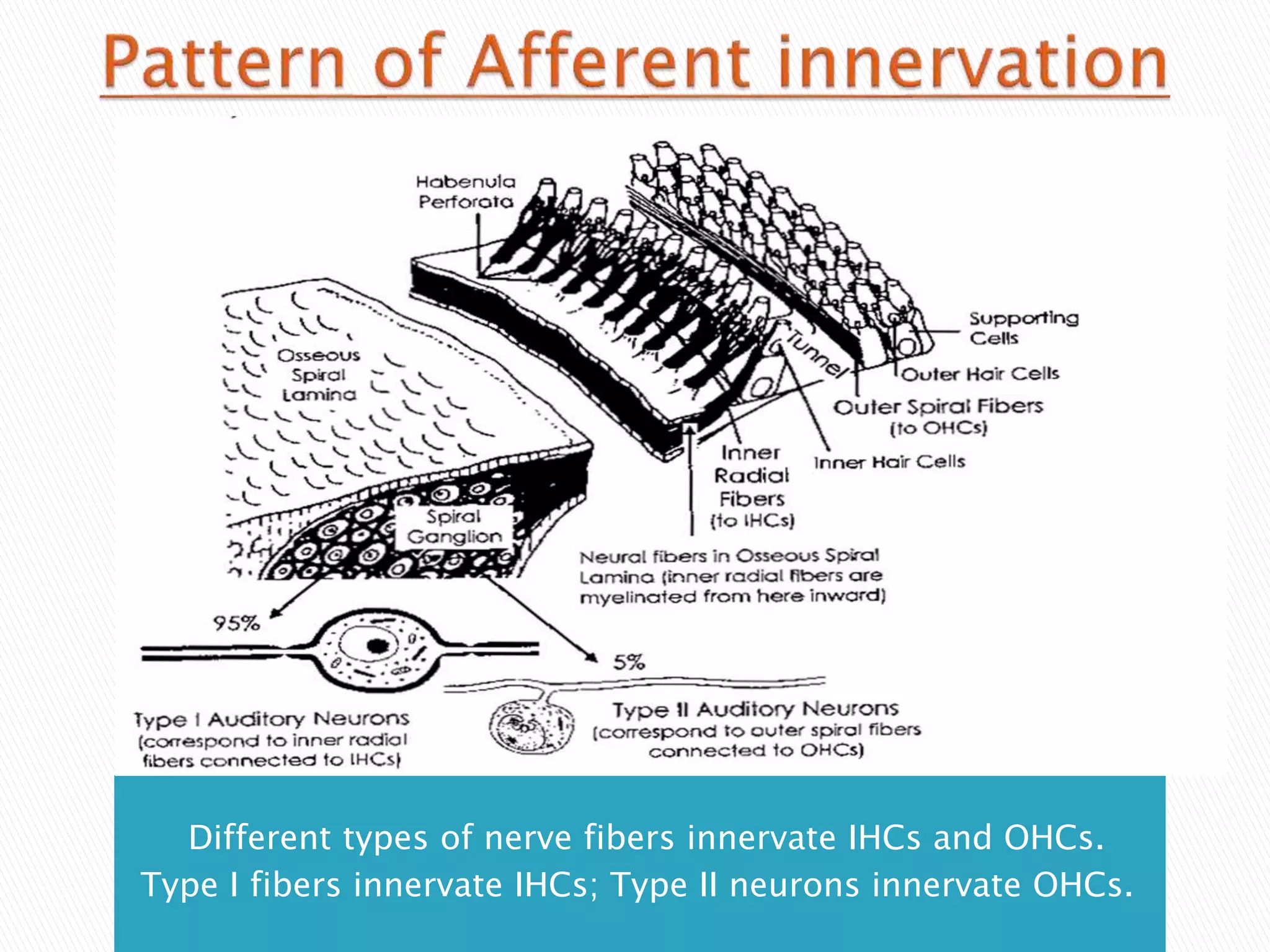
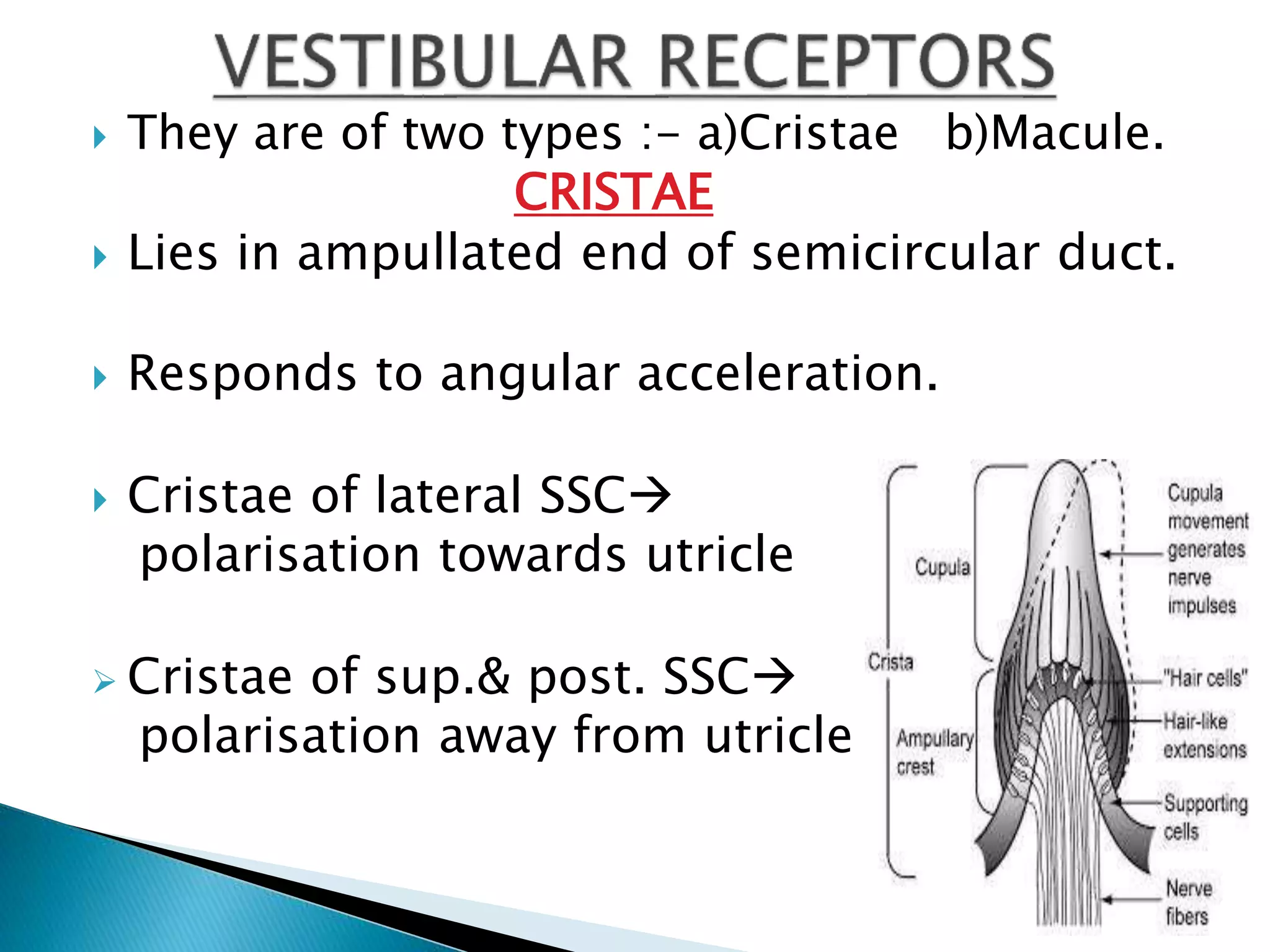
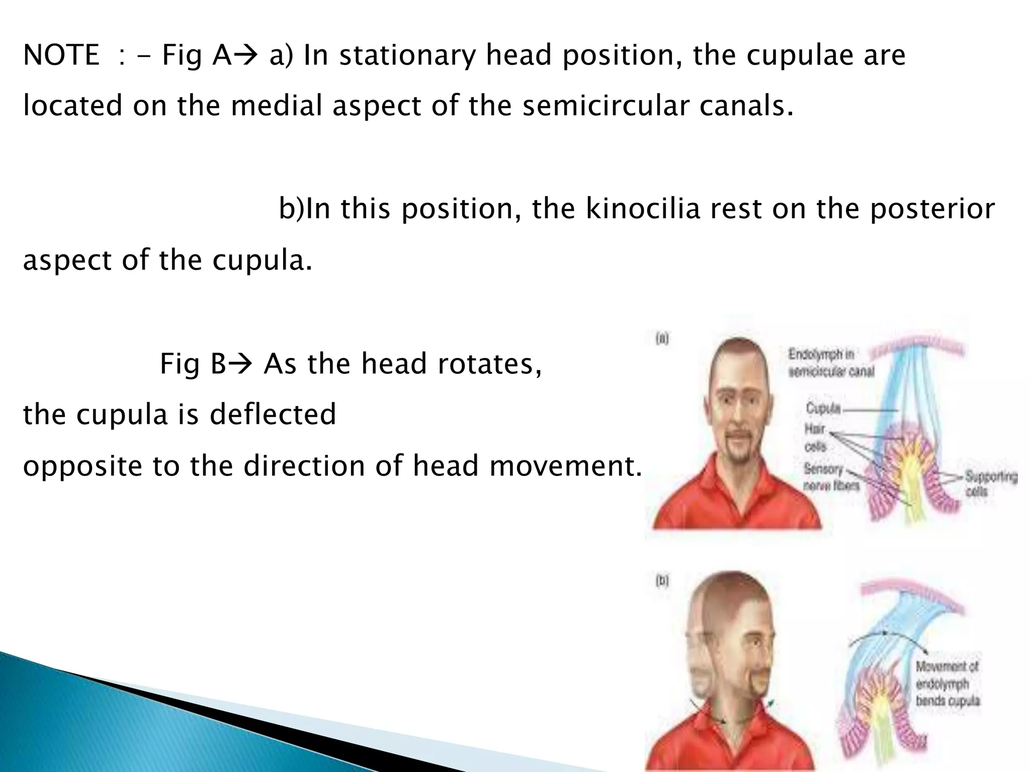
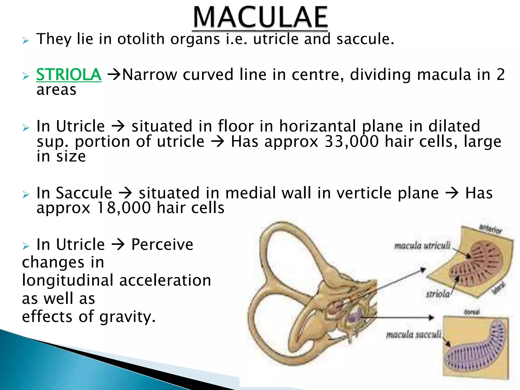
Anatomy of the
Inner Ear
By
Dr. Aditya Tiwari
Brain
 Ectoderm = Membranous labyrinth
 Mesoderm = Bony labyrinth
 BY THE 3RD WEEK :-
a)Surface ectoderm  Otic Placode
b)Otic Placode Invaginates --> Otic pit
 BY THE 4TH WEEK :-
Otic Pit edges fuse to become otocyst.
 Otocyst has 2 portion : -
a)Dorsal Utricular portion
b)Ventral Saccular portion
 Utricular portion :-
3 diverticula for Semicircular Canals
 Saccular portion :-
a)Tubular diverticulum (cochlear duct)
grows in spiral fashion to become membranous
cochlea
b)The organ of Corti differentiates from
cells along the wall of the cochlear duct
 BY THE 6TH WEEK :-
Neuroectoderm --> Spinal and Vestibular
ganglia and corresponding sensory nerves
 Mesoderm around otocyst soon forms a
cartilaginous otic capsule.
 Ossifies by 25 weeks
Bony labyrinth ossifies from 14 ossification centres 
Starts at 16th week of intrauterine life & ends by 20-21
weeks
of gestation.
 Vacuoles containing the perilymph develop
within the Otic capsule.
 The vacuoles enlarge and unite to form the
perilymphatic space.
 Perilymphatic space divides into the scala
tympani and the scala vestibuli.
 The cartilaginous otic capsule ossifies to form
the bony labyrinth of the inner ear
(mesoderm).
 Two compartments:-
а) Bony labyrinth and
b) Membraneous labyrinth.
 BONY LABYRINTH :-
Complex cavity in dense bone (Pars Petrosa)
 Parts of the bony labyrinth: -
a) Vestibulum.
b) Semicircular canals.
C) Cochlea.
 So, the inner ear consists of a “membranous”
labyrinth encased in an “osseous” labyrinth.
 The vestibule and semicircular canals are
concerned with vestibular function (balance).
 The cochlea is concerned with hearing. The
cochlea is a coiled tube.
 It should be noticed that the oval window and
round window open into the vestibule, at the
base of the cochlea.
 Two walls: External and internal.
 External wall has
a)Fenestra Vestibuli.
 Internal wall has:
a) Recessus Ellipticus
b) Recessus Sphericus
c) Recessus Cochlearis
d)Maculae cribrosae superior, medius,
inferior
EXTERNAL WALL INTERNAL WALL
 Lateral wall  oval windowclosed by stapes
footplate & surronded by annular ligament.
 Medial wall :-
a)Sperical recess ant. Situatedlodge
sacculeperforaton of macula cribrosa media
provides passage for fibres of inf vestibular
nerve.
b)Elliptical recesspost. Situatedlodge
utricle perforaton of macula cribrosa
superior(MIKE’S DOT) provides passage for
nerve fibres that supply to uticle & ampulla of
sup & lateral SSC
c)Vestibular crests & cochlear recess.
d)Opening of aqueduct of vestibule
Enlarged vestibular aqueduct asso. with SNHL,
Pendred syndrome & anatomical defect of cochlear modiolus
a) Fenestra vestibuli.
b) Fenestra cochleae.
c) Openings (5) of the semicircular canals
d) Aqueductus vestibuli
 They are 3 in numbers :-
a) Anterior,
b) Posterior and
c) Lateral.
 Have ampulla and crus.
A) Canalis Semicircularis Superior/Anterior –
Frontal.
- Eminentia arcuata on pars petrosa of os
temporale
B) Canalis Semicircularis Lateralis – Horizontal.
- Eminentia canalis semicircularis
lateralis on the medial wall of tymypanic cavity.
C) Canalis semicircularis posterior –Sagittal
 The cochlea is the auditory portion of the inner ear.
 The cochlea (plural is cochleae) is a spiralled,
hollow, conical chamber of bone in the bony
labyrinth
 The cochlea resembles a snail shell and spirals for
about 2 3/4 turns around a bony column
 It is coiled around the
modiolus.
 It forms a cone approximately 9 millimetres
(0.35 inch) in diameter at its base and 5
millimetres in height.
 When stretched out, the tube is
approximately 30 millimetres in length; it is
widest—2 millimetres—at the point where the
basal coil opens into the vestibule and tapers
until it ends blindly at the apex.
Area bounded by the Bony labyrinth anterioly,
Sigmoid sinus posteriorly & the dura or
superior petrosal sinus superiorly.
 In cochlea, waves propagate from the base
(near the middle ear and the oval window) to
the apex (the top or center of the spiral).
 Reissner’s membrane and the basilar
membrane divide the the cochlea
longitudinally into three canals called as
scalae.
 These three scalae are : -
a)Scala Vestibuli
b)Scala Tympani
c)Scala Media
 Movement of the the basilar membrane by
pressure changes induced by stapes footplate
motion at the oval window is a critical step in
the transduction in the middle.
 The process of transduction occurs in the
structures within scala media, sitting on the
basilar membrane -- these structures
comprise the organ of Corti. The side of the
duct where the nerve fibers exit (left in this
picture) is the “inner” or “modiolar” side of
the duct. The opposite side is the “outer”
side.
.
 Scala media is more or less triangular, formed by
Reissner’s membrane, basilar membrane and the structure
called the Stria vascularis.
 The fluid that fills scala tympani and scala vestibuli is
called perilymph; the fluid that fills scala media is called
endolymph. Both communicate with each other at the the
apex of cochlea through an opening called Helicotroma.
 The organ of Corti rests on the basilar membrane within
scala media
**** Perilymph and endolymph participate in a unidirectional flow
that is interrupted in Ménière's disease.****
1) Resembles ECF.
2) Present in scala
tympani and scala
vestibuli.
3) Major cation is Na.
4) Has potential of 0
mV.
5) Also c/a Cotunnius'
liquid.
1. Resembles ICF.
2. Present in scala
media.
3. Major cation is K.
4. Has potential of
80mV.
5. Also c/a Scarpa's
liquid.
 Organ of Corti also called as “End organ of hearing”.
 It is found only in mammals and provided with hair
cells or auditory sensory cells.
 It evolved from the basilar papilla found in all
tetrapods
 It is highly specialized structures that respond to
fluid-borne vibrations in the cochlea with a shearing
vector in the hairs of some cochlear hair cells.
 It contains between 15,000-20,000 auditory nerve
receptors.
 Each receptor has its own hair cell.
 The shear on the hairs opens non-selective
transduction ion channels that are permeable to K
and Ca , leading to hair cell plasma membrane
depolarization, activation of voltage-dependent Ca
channels at the synaptic basolateral pole of the
cells
 It triggers vesicle autopsy and liberation of
glutamate neurotransmitter to the synaptic cleft
and electrical signaling to the auditory cortex via
spiral ganglion neurons.
 Two types of cells in the organ of Corti are
a) Support cells and
b) Hair cells.
 The Hair cells are the “Receptor” cells cells
that transduce sound.
 Support cells such as the Deiter’s cells 
support hair cells
 Present only in the lower turn of
the cochlea Lie on the basilar membrane beneath
Claudius' cells Supporting cells for the auditory hair
cells in the organ of Corti.
 Located above Boettcher's cells 
Supporting cells for the auditory hair cells in the
organ of Corti Contain a variety of aquaporin water
channels involved in ion transport play a role in
sealing off endolymphatic spaces
 High columnar cells that are
directly adjacent to the third row of Deiters’ cells.
 Section of the tectorial
membrane above the inner hair cell.
 Fluid filled spaces between the
outer pillar cells and adjacent hair cells  Also
the spaces between the outer hair cells.
 Layer of the tectoria
closest to the reticular lamina and overlying the
outer hair cell region.
 Spiral ganglion are situated in
Rosenthal canal, which runs along osseous spiral
lamina
 There are two types of hair cells
a)Outer Hair Cells (OHC) from sound to
nerve signal
b)Inner Hair Cells (IHC)acoustical pre-
amplifiers
 There are 4 rows of hair cells, one on the
inner (modiolar) side of the tunnel of Corti
formed by the pillar cells-- these are the
INNER HAIR CELLS .
 3 one the outer side of the Tunnel of Corti,
these are the OUTER HAIR CELLS.
 The Deiter’s cells support the Outer hair cells
at their base, but the outer hair cell walls are
surrounded by fluid.
 The inner hair cell is surrounded by support
cells.
 The Outer hair cells mechanically amplify
low-level sound that enters the cochlea.
 The amplification may be powered by
movement of their hair bundles, or by an
electrically driven motility of their cell bodies.
 The Inner hair cells transform the sound
vibrations in the fluids of the cochlea into
electrical signals that are then relayed via the
auditory nerve to the auditory brainstem and
to the auditory cortex.
1) Approx 3500 in no.
2) Lies in one row.
3) Flask shaped.
4) Mainly afferent nerve
fibres supply.
5) Early development.
6) Transmit auditory
stimuli.
7) More resistant to
ototoxic drugs.
8) More resistant to high
intensity noice.
9) No generation of
otoacoustic emissions.
1) Approx 12,000 in no.
2) Lies in 3-4 rows.
3) Cylindrical shaped.
4) Mainly efferent nerve
fibres supply.
5) Late development.
6) Modulate function of inner
hair cells.
7) More sensetive & easily
damaged by ototoxic
drugs.
8) More sensetive & easily
damaged by high intensity
noice.
9) Generates otoacoustic
emissions.
 The reticular lamina is a solid surface at the
tops of the hair cells, so the tops of the hair
cells are in endolymph and the bottom of the
hair cells are in perilymph
 Stereocilia are lined up in the Organ of Corti
within the cochlea of the inner ear.
 In hearing, stereocilia transform the
mechanical energy of sound waves into
electrical signals for the hair cells, which
ultimately leads to an excitation of the
auditory nerve.
 Stereocilia are composed of cytoplasm with
embedded bundles of cross-linked actin
filaments.
 The actin filaments anchor to the terminal
web and the top of the cell membrane and
are arranged in grade of height.
 When the stapes causes sound waves in the
endolymphatic fluid in the cochlea, the
stereocilia are deflected in a shearing motion,
which results in the mentioned electrical
signal for the hair cell
 Stereocilia are arranged in curved or v-
shaped rows that face toward the modiolus.
 Each row of stereocilia is taller than the next.
The tip of each stereocilium is linked to the
side of the stereocilium behind it by a tip link.
 Kinocilia are found on the apical surface of
hair cells and are involved in both the
morphogenesis of the hair bundle and
mechanotransduction, Vibrations (either by
movement or sound waves)  causes
displacement of the hair bundle, resulting in
depolarization or hyperpolarization of the
hair cells.
 The depolarization of the hair cells in both
instances causes signal transduction via
neurotransmitter release.
 One kinocilium is the longest cilium located
on the hair cell next to 40-70 sterocilia.
 During movement of the body, the hair cell is
depolarized when the sterocilia move
 Deiter’s cell processes “fill in the gaps” between
the tops of the outer hair cells to form the
reticular lamina.
 It holds the base of the hair cell in a cup-shaped
depression. From each Deiters’ cell a projection
extends upward to the stiff
membrane, the reticular lamina,
that covers the organ of
Corti.
 Acellular gels in the cochlea of the inner ear along
with basilar membrane (BM).
 The TM is located above the sulcus spiralis
internus and the spiral organ of Corti and extends
along the longitudinal length of the cochlea
parallel to the BM.
 Divided into three zones:-
a) Limbal ,
b) Middle and
c) Marginal
 LIMBAL ZONE  Thinnest and overlies the
auditory teeth of Huschke with its inside edge
attached to the spiral limbus.
 MARGINAL ZONE a) Thickest and is divided
from the middle zone by Hensen's Stripe.
b) Overlies the sensory inner
hair cells and electrically-motile outer hair cells of
the organ of Corti
c) During acoustic stimulation,
stimulates the inner hair cells through fluid
coupling, and the outer hair cells via direct
connection to their tallest stereocilia.
 Seperates scala media and scala tympani.
 The Basilar membrane is widest (0.42–0.65 mm)
and least stiff at the apex of the cochlea, and
narrowest (0.08–0.16 mm) and most stiff at the
base.
 FUNCTIONS OF BASILAR MEMBRANE :-
a) Perilymph/endolymph seperation.
b) Base of sensory cells
c) Frequency dispertion
 High-frequency sounds localize near the base of
the cochlea (near the round and oval windows),
while low-frequency sounds localize near the
apex.
 Zona arcuata inner
thin area
Zona pectinata
Outer thick area
 The membranous labyrinth contains fluid
called endolymph.
 The walls of the membranous labyrinth are
lined with distributions of the acoustic nerve,
also known as the vestibularcochlear nerve.
 It consists of :- a) Cochlear duct
b) Utricle
c) Saccule
d) 3 semicircular ducts
e)Endolymphatic duct & sac
 The cochlear duct (or scala media) is an
endolymph filled cavity inside the cochlea,
located in between the scala tympani and the
scala vestibuli, separated by the basilar
membrane and Reissner's membrane (the
vestibular membrane) respectively.
 Scala media houses the organ of Corti.
 Oblong, irregular, ant. Upward slope
approx at 30 degree.
 4.33mm2, recieves 5 opening of 3
semicircular canal, lies in posterior
part of bony vestibule.
 Connected with saccule via
utriculosaccular duct. Its sensory
epithelium c/a MACULE concerned
with linear acceleration and
dicceleration.
UTRICLE
FIRST PART
OF MEMB.
LABYRINTH
TO DEVP.
DURING
INTRA -
UTERINE LIFE.
 Consists of three
layers ;-
a) Bottom layer
sensory hair
cellsconsists of 40 to
70 steriocilia and a
kinocilium,
b) Top
layercalcium
carbonate crystals
called statoconia or
otolithsrelatively
heavy, providing
weight to the
membrane as well as
inertiagreater sense
of gravity and motion.
C)macula acustica
utriculereceives the
utricular filaments of
the acoustic nerve.
 Globularlies in the recessus sphæricus near
the opening of the scala vestibuli of the cochlea.
 The Anterior oval thickening, the macula
acustica sacculi saccular filaments of the
vestibular branch of the vestibulocochlear nerve.
 Posterior wall Ductus endolymphaticus.
 Lower part short tube, the CANALIS REUNIENS
OF HENSEN, passes downward and opens into the
ductus cochlearis near its vestibular extremity.
 Open in the utricle, corrosponds to three
semicircular canal.
 Ampulated end has thickened ridge of
neuroepithelium c/a CRISTAE AMPULARIS
 From the posterior wall of the saccule a canal,
the endolymphatic duct, is given off.
 This duct is joined by the ductus
utriculosaccularis, and then passes along the
aquaeductus vestibuli and ends in a blind
pouch (saccus endolymphaticus) on the
posterior surface of the petrous portion of
the temporal bone, where it is in contact with
the dura matER.
 In the auditory nerve, the dendrites contact the
hair cells.
 The cell bodies form what is called the spiral
ganglion, and the axons form the auditory nerve
that connects the ear to the brainstem.
 The “contact” points between the dendrites and
the hair cells or between the axons of one neuron
and the dendrites of another are called synapses.
 Synapses have specialized structures and
substances that allow communication between
receptors and neurons or between neurons.
.
This line is surgical landmark for endolymphatic
sac. It passes through horizantal SSC bisecting
posterior SSC. The endolymphatic sac that
appears as thickening of post cranial fossa dura
is situated inferior to Donaldson’s line.
 In the auditory nerve, the dendrites contact the
hair cells.
 The cell bodies form what is called the spiral
ganglion, and the axons form the auditory nerve
that connects the ear to the brainstem.
 The “contact” points between the dendrites and
the hair cells or between the axons of one neuron
and the dendrites of another are called synapses.
 Synapses have specialized structures and
substances that allow communication between
receptors and neurons or between neurons.
 When a neuron’s intracellular electrical
potential is changed enough by release of
neurotransmitter at a synapse(or in some
other way), an abrupt change in electrical
potential, an “action potential”, occurs.
 Action potentials are transmitted along the
axon to another synapse, where
neurotransmitter is released and an action
potential may be generated in the neuron on
the other side of the synapse (postsynaptic
neuron).
The cell bodies of the neurons that form the auditory
nerve are located within the cochlear modiolus. The
collection of cell bodies is called the spiral ganglion.
 Different types of nerve fibers innervate IHCs and OHCs.
Type I fibers innervate IHCs; Type II neurons innervate OHCs.
 Many nerve fibers that contact one inner hair
cells do not branch to other inner hair cells.
 Each IHC has its own “private” set of fibers.
 The Type II nerve fibers innervate many
OHCs.
 The OHCs they innervate are basal to the
point at which the nerve fiber enters the
cochlea. Thin fibers attach toward modiolar
side, thick fibers toward outer side of IHC.
Thin fibers attach toward modiolar side,
Thick fibers toward outer side of IHC.
They are of two types : - a) Type 1 cells
b) Type 2 cells.
TYPE 1 CELLS
1. Found only in birds & mammals
2. Flask shaped
3. Correspond to IHC of Organ of Corti
4. Has single large cup like nerve
terminal surrounding the base
TYPE 2 CELLS
1. Cylindrical shaped
2. Correspond to OHC of Organ of Corti
3. Has multiple nerve terminal at the
base.
 They are of two types :- a)Cristae b)Macule.
CRISTAE
 Lies in ampullated end of semicircular duct.
 Responds to angular acceleration.
 Cristae of lateral SSC
polarisation towards utricle
 Cristae of sup.& post. SSC
polarisation away from utricle
 The cupula is the gelatinous component of the
crista ampullaris that extends from the crista to
the roof of the ampullae.
 Providing the sense of spatial orientation.
 When the head rotates, the endolymph filling
the semicircular ducts initially lags behind due
to inertia. As a result, the cupula is deflected
opposite the direction of head movement.
 As fluid rushes by the cupula, the hair cells
stimulated transmit the corresponding signal
to the brain through the CN VIII.
NOTE : - Fig A a) In stationary head position, the cupulae are
located on the medial aspect of the semicircular canals.
b)In this position, the kinocilia rest on the posterior
aspect of the cupula.
Fig B As the head rotates,
the cupula is deflected
opposite to the direction of head movement.
 They lie in otolith organs i.e. utricle and saccule.
 STRIOLA Narrow curved line in centre, dividing macula in 2
areas
 In Utricle  situated in floor in horizantal plane in dilated
sup. portion of utricle  Has approx 33,000 hair cells, large
in size
 In Saccule  situated in medial wall in verticle plane  Has
approx 18,000 hair cells
 In Utricle  Perceive
changes in
longitudinal acceleration
as well as
effects of gravity.
 A) Sensory neuroepithelium
 B) Otolith membrane
Sensory Neuroepithelium
 Made up of type 1 & type 2 cells, similar to cristae
hair cells
 Type 1 cells  Higher in conc. in striola region
 Kinocilia face striola in utricular macula & away from
striola in saccular macula.
 Offers CNS wide range of neural information.
 Every hair cell in sensory beds of this consist
of 40-70 stereocilia and a kinocilium.
 The sterocilia and kinocilium are embedded
in the otolithic membrane and are essential in
the function of the otolith organs.
 The hair cells are
deflected by
structures called
otoconia.
 Otoconia are crystals of Ca carbonate and
make the otolithic membrane heavier than the
structures and fluids surrounding it.
 The otoconia are composite crystallites that
overlie the macular sensory epithelium of the
gravity receptors and are required for optimal
stimulus input of linear acceleration and
gravity.
 Biomineralization of otoliths and otoconia
results mainly from the release of soluble Ca
ions, which is in turn precipitated as Ca
carbonate crystals.
 Horizantal SSC
 Sup. & post. SSC
 Otolith organs
(utricle & saccule)
Horizantal head
turning(angular
acceleration
Piching the front to back
& side to side
Linear head
movements(vertical &
horizantal),tilting &
gravity
 Basilar artery  Anterior inferior cerebellar artery  Internal
auditory artery  Labyrinthine artery.
 Labyrinthine  Comman cochlear artery
artery  Ant. Vestibular artery
(to utricle & lat. and sup. Canals)
 Comman
cochlear  Vestibulocochlear artery
artery  Main cochlear artery
(to cochlea 80%)
 Vestibulocochlear
artery  Cochlear artery ( 20% cochlea)
 Post. Vestibular artery
(to saccule & post. Canal)
Via internal auditory canal, vein of cochlear
aqueduct & vein of vestibular aqueduct
Inferior petrosal & sigmoid sinuses
drain into
Cochlea does not have any
collateral arterial circulation.
 Convert mechanical sound
waves to neural impulses that
can be recognized by
the brain for:-
a)Hearing
b)Balance
1) STATIC BALANCE
2) DYNAMIC BALANCE
Maculae react to gravitational forces and
participate in maintaining the static balance.
Cristae ampullares react to rotatory
movements and paticipate in dynamic
balance.
 Birds have an auditory system similar to that
of mammals, including a cochlea.
 Reptiles, amphibians & fish do not have
cochleas but hear with simpler auditory
organs or vestibular organs, which generally
detect lower-frequency sounds than the
cochlea.
Anatomy of inner ear by Dr. Aditya Tiwari

Anatomy of inner ear by Dr. Aditya Tiwari

  • 1.
    Anatomy of the InnerEar By Dr. Aditya Tiwari
  • 2.
  • 3.
     Ectoderm =Membranous labyrinth  Mesoderm = Bony labyrinth  BY THE 3RD WEEK :- a)Surface ectoderm  Otic Placode b)Otic Placode Invaginates --> Otic pit  BY THE 4TH WEEK :- Otic Pit edges fuse to become otocyst.
  • 4.
     Otocyst has2 portion : - a)Dorsal Utricular portion b)Ventral Saccular portion  Utricular portion :- 3 diverticula for Semicircular Canals  Saccular portion :- a)Tubular diverticulum (cochlear duct) grows in spiral fashion to become membranous cochlea b)The organ of Corti differentiates from cells along the wall of the cochlear duct
  • 5.
     BY THE6TH WEEK :- Neuroectoderm --> Spinal and Vestibular ganglia and corresponding sensory nerves  Mesoderm around otocyst soon forms a cartilaginous otic capsule.  Ossifies by 25 weeks Bony labyrinth ossifies from 14 ossification centres  Starts at 16th week of intrauterine life & ends by 20-21 weeks of gestation.
  • 6.
     Vacuoles containingthe perilymph develop within the Otic capsule.  The vacuoles enlarge and unite to form the perilymphatic space.  Perilymphatic space divides into the scala tympani and the scala vestibuli.  The cartilaginous otic capsule ossifies to form the bony labyrinth of the inner ear (mesoderm).
  • 8.
     Two compartments:- а)Bony labyrinth and b) Membraneous labyrinth.  BONY LABYRINTH :- Complex cavity in dense bone (Pars Petrosa)  Parts of the bony labyrinth: - a) Vestibulum. b) Semicircular canals. C) Cochlea.
  • 10.
     So, theinner ear consists of a “membranous” labyrinth encased in an “osseous” labyrinth.  The vestibule and semicircular canals are concerned with vestibular function (balance).  The cochlea is concerned with hearing. The cochlea is a coiled tube.  It should be noticed that the oval window and round window open into the vestibule, at the base of the cochlea.
  • 11.
     Two walls:External and internal.  External wall has a)Fenestra Vestibuli.  Internal wall has: a) Recessus Ellipticus b) Recessus Sphericus c) Recessus Cochlearis d)Maculae cribrosae superior, medius, inferior
  • 12.
  • 13.
     Lateral wall oval windowclosed by stapes footplate & surronded by annular ligament.  Medial wall :- a)Sperical recess ant. Situatedlodge sacculeperforaton of macula cribrosa media provides passage for fibres of inf vestibular nerve. b)Elliptical recesspost. Situatedlodge utricle perforaton of macula cribrosa superior(MIKE’S DOT) provides passage for nerve fibres that supply to uticle & ampulla of sup & lateral SSC
  • 14.
    c)Vestibular crests &cochlear recess. d)Opening of aqueduct of vestibule Enlarged vestibular aqueduct asso. with SNHL, Pendred syndrome & anatomical defect of cochlear modiolus
  • 15.
    a) Fenestra vestibuli. b)Fenestra cochleae. c) Openings (5) of the semicircular canals d) Aqueductus vestibuli
  • 16.
     They are3 in numbers :- a) Anterior, b) Posterior and c) Lateral.  Have ampulla and crus. A) Canalis Semicircularis Superior/Anterior – Frontal. - Eminentia arcuata on pars petrosa of os temporale
  • 17.
    B) Canalis SemicircularisLateralis – Horizontal. - Eminentia canalis semicircularis lateralis on the medial wall of tymypanic cavity. C) Canalis semicircularis posterior –Sagittal
  • 18.
     The cochleais the auditory portion of the inner ear.  The cochlea (plural is cochleae) is a spiralled, hollow, conical chamber of bone in the bony labyrinth  The cochlea resembles a snail shell and spirals for about 2 3/4 turns around a bony column  It is coiled around the modiolus.
  • 19.
     It formsa cone approximately 9 millimetres (0.35 inch) in diameter at its base and 5 millimetres in height.  When stretched out, the tube is approximately 30 millimetres in length; it is widest—2 millimetres—at the point where the basal coil opens into the vestibule and tapers until it ends blindly at the apex.
  • 20.
    Area bounded bythe Bony labyrinth anterioly, Sigmoid sinus posteriorly & the dura or superior petrosal sinus superiorly.
  • 21.
     In cochlea,waves propagate from the base (near the middle ear and the oval window) to the apex (the top or center of the spiral).  Reissner’s membrane and the basilar membrane divide the the cochlea longitudinally into three canals called as scalae.  These three scalae are : - a)Scala Vestibuli b)Scala Tympani c)Scala Media
  • 23.
     Movement ofthe the basilar membrane by pressure changes induced by stapes footplate motion at the oval window is a critical step in the transduction in the middle.  The process of transduction occurs in the structures within scala media, sitting on the basilar membrane -- these structures comprise the organ of Corti. The side of the duct where the nerve fibers exit (left in this picture) is the “inner” or “modiolar” side of the duct. The opposite side is the “outer” side. .
  • 24.
     Scala mediais more or less triangular, formed by Reissner’s membrane, basilar membrane and the structure called the Stria vascularis.  The fluid that fills scala tympani and scala vestibuli is called perilymph; the fluid that fills scala media is called endolymph. Both communicate with each other at the the apex of cochlea through an opening called Helicotroma.  The organ of Corti rests on the basilar membrane within scala media
  • 25.
    **** Perilymph andendolymph participate in a unidirectional flow that is interrupted in Ménière's disease.**** 1) Resembles ECF. 2) Present in scala tympani and scala vestibuli. 3) Major cation is Na. 4) Has potential of 0 mV. 5) Also c/a Cotunnius' liquid. 1. Resembles ICF. 2. Present in scala media. 3. Major cation is K. 4. Has potential of 80mV. 5. Also c/a Scarpa's liquid.
  • 26.
     Organ ofCorti also called as “End organ of hearing”.  It is found only in mammals and provided with hair cells or auditory sensory cells.  It evolved from the basilar papilla found in all tetrapods  It is highly specialized structures that respond to fluid-borne vibrations in the cochlea with a shearing vector in the hairs of some cochlear hair cells.  It contains between 15,000-20,000 auditory nerve receptors.
  • 27.
     Each receptorhas its own hair cell.  The shear on the hairs opens non-selective transduction ion channels that are permeable to K and Ca , leading to hair cell plasma membrane depolarization, activation of voltage-dependent Ca channels at the synaptic basolateral pole of the cells  It triggers vesicle autopsy and liberation of glutamate neurotransmitter to the synaptic cleft and electrical signaling to the auditory cortex via spiral ganglion neurons.
  • 29.
     Two typesof cells in the organ of Corti are a) Support cells and b) Hair cells.  The Hair cells are the “Receptor” cells cells that transduce sound.  Support cells such as the Deiter’s cells  support hair cells
  • 30.
     Present onlyin the lower turn of the cochlea Lie on the basilar membrane beneath Claudius' cells Supporting cells for the auditory hair cells in the organ of Corti.  Located above Boettcher's cells  Supporting cells for the auditory hair cells in the organ of Corti Contain a variety of aquaporin water channels involved in ion transport play a role in sealing off endolymphatic spaces
  • 31.
     High columnarcells that are directly adjacent to the third row of Deiters’ cells.  Section of the tectorial membrane above the inner hair cell.  Fluid filled spaces between the outer pillar cells and adjacent hair cells  Also the spaces between the outer hair cells.  Layer of the tectoria closest to the reticular lamina and overlying the outer hair cell region.  Spiral ganglion are situated in Rosenthal canal, which runs along osseous spiral lamina
  • 32.
     There aretwo types of hair cells a)Outer Hair Cells (OHC) from sound to nerve signal b)Inner Hair Cells (IHC)acoustical pre- amplifiers
  • 33.
     There are4 rows of hair cells, one on the inner (modiolar) side of the tunnel of Corti formed by the pillar cells-- these are the INNER HAIR CELLS .  3 one the outer side of the Tunnel of Corti, these are the OUTER HAIR CELLS.  The Deiter’s cells support the Outer hair cells at their base, but the outer hair cell walls are surrounded by fluid.  The inner hair cell is surrounded by support cells.
  • 34.
     The Outerhair cells mechanically amplify low-level sound that enters the cochlea.  The amplification may be powered by movement of their hair bundles, or by an electrically driven motility of their cell bodies.  The Inner hair cells transform the sound vibrations in the fluids of the cochlea into electrical signals that are then relayed via the auditory nerve to the auditory brainstem and to the auditory cortex.
  • 35.
    1) Approx 3500in no. 2) Lies in one row. 3) Flask shaped. 4) Mainly afferent nerve fibres supply. 5) Early development. 6) Transmit auditory stimuli. 7) More resistant to ototoxic drugs. 8) More resistant to high intensity noice. 9) No generation of otoacoustic emissions. 1) Approx 12,000 in no. 2) Lies in 3-4 rows. 3) Cylindrical shaped. 4) Mainly efferent nerve fibres supply. 5) Late development. 6) Modulate function of inner hair cells. 7) More sensetive & easily damaged by ototoxic drugs. 8) More sensetive & easily damaged by high intensity noice. 9) Generates otoacoustic emissions.
  • 36.
     The reticularlamina is a solid surface at the tops of the hair cells, so the tops of the hair cells are in endolymph and the bottom of the hair cells are in perilymph
  • 37.
     Stereocilia arelined up in the Organ of Corti within the cochlea of the inner ear.  In hearing, stereocilia transform the mechanical energy of sound waves into electrical signals for the hair cells, which ultimately leads to an excitation of the auditory nerve.
  • 38.
     Stereocilia arecomposed of cytoplasm with embedded bundles of cross-linked actin filaments.  The actin filaments anchor to the terminal web and the top of the cell membrane and are arranged in grade of height.  When the stapes causes sound waves in the endolymphatic fluid in the cochlea, the stereocilia are deflected in a shearing motion, which results in the mentioned electrical signal for the hair cell
  • 40.
     Stereocilia arearranged in curved or v- shaped rows that face toward the modiolus.  Each row of stereocilia is taller than the next. The tip of each stereocilium is linked to the side of the stereocilium behind it by a tip link.
  • 41.
     Kinocilia arefound on the apical surface of hair cells and are involved in both the morphogenesis of the hair bundle and mechanotransduction, Vibrations (either by movement or sound waves)  causes displacement of the hair bundle, resulting in depolarization or hyperpolarization of the hair cells.
  • 42.
     The depolarizationof the hair cells in both instances causes signal transduction via neurotransmitter release.  One kinocilium is the longest cilium located on the hair cell next to 40-70 sterocilia.  During movement of the body, the hair cell is depolarized when the sterocilia move
  • 43.
     Deiter’s cellprocesses “fill in the gaps” between the tops of the outer hair cells to form the reticular lamina.  It holds the base of the hair cell in a cup-shaped depression. From each Deiters’ cell a projection extends upward to the stiff membrane, the reticular lamina, that covers the organ of Corti.
  • 44.
     Acellular gelsin the cochlea of the inner ear along with basilar membrane (BM).  The TM is located above the sulcus spiralis internus and the spiral organ of Corti and extends along the longitudinal length of the cochlea parallel to the BM.  Divided into three zones:- a) Limbal , b) Middle and c) Marginal
  • 45.
     LIMBAL ZONE Thinnest and overlies the auditory teeth of Huschke with its inside edge attached to the spiral limbus.  MARGINAL ZONE a) Thickest and is divided from the middle zone by Hensen's Stripe. b) Overlies the sensory inner hair cells and electrically-motile outer hair cells of the organ of Corti c) During acoustic stimulation, stimulates the inner hair cells through fluid coupling, and the outer hair cells via direct connection to their tallest stereocilia.
  • 46.
     Seperates scalamedia and scala tympani.  The Basilar membrane is widest (0.42–0.65 mm) and least stiff at the apex of the cochlea, and narrowest (0.08–0.16 mm) and most stiff at the base.
  • 47.
     FUNCTIONS OFBASILAR MEMBRANE :- a) Perilymph/endolymph seperation. b) Base of sensory cells c) Frequency dispertion  High-frequency sounds localize near the base of the cochlea (near the round and oval windows), while low-frequency sounds localize near the apex.  Zona arcuata inner thin area Zona pectinata Outer thick area
  • 48.
     The membranouslabyrinth contains fluid called endolymph.  The walls of the membranous labyrinth are lined with distributions of the acoustic nerve, also known as the vestibularcochlear nerve.  It consists of :- a) Cochlear duct b) Utricle c) Saccule d) 3 semicircular ducts e)Endolymphatic duct & sac
  • 49.
     The cochlearduct (or scala media) is an endolymph filled cavity inside the cochlea, located in between the scala tympani and the scala vestibuli, separated by the basilar membrane and Reissner's membrane (the vestibular membrane) respectively.  Scala media houses the organ of Corti.
  • 50.
     Oblong, irregular,ant. Upward slope approx at 30 degree.  4.33mm2, recieves 5 opening of 3 semicircular canal, lies in posterior part of bony vestibule.  Connected with saccule via utriculosaccular duct. Its sensory epithelium c/a MACULE concerned with linear acceleration and dicceleration. UTRICLE FIRST PART OF MEMB. LABYRINTH TO DEVP. DURING INTRA - UTERINE LIFE.
  • 51.
     Consists ofthree layers ;- a) Bottom layer sensory hair cellsconsists of 40 to 70 steriocilia and a kinocilium, b) Top layercalcium carbonate crystals called statoconia or otolithsrelatively heavy, providing weight to the membrane as well as inertiagreater sense of gravity and motion. C)macula acustica utriculereceives the utricular filaments of the acoustic nerve.
  • 52.
     Globularlies inthe recessus sphæricus near the opening of the scala vestibuli of the cochlea.  The Anterior oval thickening, the macula acustica sacculi saccular filaments of the vestibular branch of the vestibulocochlear nerve.  Posterior wall Ductus endolymphaticus.  Lower part short tube, the CANALIS REUNIENS OF HENSEN, passes downward and opens into the ductus cochlearis near its vestibular extremity.
  • 53.
     Open inthe utricle, corrosponds to three semicircular canal.  Ampulated end has thickened ridge of neuroepithelium c/a CRISTAE AMPULARIS
  • 54.
     From theposterior wall of the saccule a canal, the endolymphatic duct, is given off.  This duct is joined by the ductus utriculosaccularis, and then passes along the aquaeductus vestibuli and ends in a blind pouch (saccus endolymphaticus) on the posterior surface of the petrous portion of the temporal bone, where it is in contact with the dura matER.
  • 55.
     In theauditory nerve, the dendrites contact the hair cells.  The cell bodies form what is called the spiral ganglion, and the axons form the auditory nerve that connects the ear to the brainstem.  The “contact” points between the dendrites and the hair cells or between the axons of one neuron and the dendrites of another are called synapses.  Synapses have specialized structures and substances that allow communication between receptors and neurons or between neurons.
  • 56.
  • 57.
    This line issurgical landmark for endolymphatic sac. It passes through horizantal SSC bisecting posterior SSC. The endolymphatic sac that appears as thickening of post cranial fossa dura is situated inferior to Donaldson’s line.
  • 58.
     In theauditory nerve, the dendrites contact the hair cells.  The cell bodies form what is called the spiral ganglion, and the axons form the auditory nerve that connects the ear to the brainstem.  The “contact” points between the dendrites and the hair cells or between the axons of one neuron and the dendrites of another are called synapses.  Synapses have specialized structures and substances that allow communication between receptors and neurons or between neurons.
  • 60.
     When aneuron’s intracellular electrical potential is changed enough by release of neurotransmitter at a synapse(or in some other way), an abrupt change in electrical potential, an “action potential”, occurs.  Action potentials are transmitted along the axon to another synapse, where neurotransmitter is released and an action potential may be generated in the neuron on the other side of the synapse (postsynaptic neuron).
  • 61.
    The cell bodiesof the neurons that form the auditory nerve are located within the cochlear modiolus. The collection of cell bodies is called the spiral ganglion.
  • 62.
     Different typesof nerve fibers innervate IHCs and OHCs. Type I fibers innervate IHCs; Type II neurons innervate OHCs.
  • 64.
     Many nervefibers that contact one inner hair cells do not branch to other inner hair cells.  Each IHC has its own “private” set of fibers.  The Type II nerve fibers innervate many OHCs.  The OHCs they innervate are basal to the point at which the nerve fiber enters the cochlea. Thin fibers attach toward modiolar side, thick fibers toward outer side of IHC.
  • 65.
    Thin fibers attachtoward modiolar side, Thick fibers toward outer side of IHC.
  • 66.
    They are oftwo types : - a) Type 1 cells b) Type 2 cells. TYPE 1 CELLS 1. Found only in birds & mammals 2. Flask shaped 3. Correspond to IHC of Organ of Corti 4. Has single large cup like nerve terminal surrounding the base TYPE 2 CELLS 1. Cylindrical shaped 2. Correspond to OHC of Organ of Corti 3. Has multiple nerve terminal at the base.
  • 67.
     They areof two types :- a)Cristae b)Macule. CRISTAE  Lies in ampullated end of semicircular duct.  Responds to angular acceleration.  Cristae of lateral SSC polarisation towards utricle  Cristae of sup.& post. SSC polarisation away from utricle
  • 68.
     The cupulais the gelatinous component of the crista ampullaris that extends from the crista to the roof of the ampullae.  Providing the sense of spatial orientation.  When the head rotates, the endolymph filling the semicircular ducts initially lags behind due to inertia. As a result, the cupula is deflected opposite the direction of head movement.  As fluid rushes by the cupula, the hair cells stimulated transmit the corresponding signal to the brain through the CN VIII.
  • 69.
    NOTE : -Fig A a) In stationary head position, the cupulae are located on the medial aspect of the semicircular canals. b)In this position, the kinocilia rest on the posterior aspect of the cupula. Fig B As the head rotates, the cupula is deflected opposite to the direction of head movement.
  • 70.
     They liein otolith organs i.e. utricle and saccule.  STRIOLA Narrow curved line in centre, dividing macula in 2 areas  In Utricle  situated in floor in horizantal plane in dilated sup. portion of utricle  Has approx 33,000 hair cells, large in size  In Saccule  situated in medial wall in verticle plane  Has approx 18,000 hair cells  In Utricle  Perceive changes in longitudinal acceleration as well as effects of gravity.
  • 71.
     A) Sensoryneuroepithelium  B) Otolith membrane Sensory Neuroepithelium  Made up of type 1 & type 2 cells, similar to cristae hair cells  Type 1 cells  Higher in conc. in striola region  Kinocilia face striola in utricular macula & away from striola in saccular macula.  Offers CNS wide range of neural information.
  • 72.
     Every haircell in sensory beds of this consist of 40-70 stereocilia and a kinocilium.  The sterocilia and kinocilium are embedded in the otolithic membrane and are essential in the function of the otolith organs.  The hair cells are deflected by structures called otoconia.
  • 73.
     Otoconia arecrystals of Ca carbonate and make the otolithic membrane heavier than the structures and fluids surrounding it.  The otoconia are composite crystallites that overlie the macular sensory epithelium of the gravity receptors and are required for optimal stimulus input of linear acceleration and gravity.  Biomineralization of otoliths and otoconia results mainly from the release of soluble Ca ions, which is in turn precipitated as Ca carbonate crystals.
  • 74.
     Horizantal SSC Sup. & post. SSC  Otolith organs (utricle & saccule) Horizantal head turning(angular acceleration Piching the front to back & side to side Linear head movements(vertical & horizantal),tilting & gravity
  • 75.
     Basilar artery Anterior inferior cerebellar artery  Internal auditory artery  Labyrinthine artery.  Labyrinthine  Comman cochlear artery artery  Ant. Vestibular artery (to utricle & lat. and sup. Canals)  Comman cochlear  Vestibulocochlear artery artery  Main cochlear artery (to cochlea 80%)  Vestibulocochlear artery  Cochlear artery ( 20% cochlea)  Post. Vestibular artery (to saccule & post. Canal)
  • 76.
    Via internal auditorycanal, vein of cochlear aqueduct & vein of vestibular aqueduct Inferior petrosal & sigmoid sinuses drain into Cochlea does not have any collateral arterial circulation.
  • 77.
     Convert mechanicalsound waves to neural impulses that can be recognized by the brain for:- a)Hearing b)Balance
  • 79.
    1) STATIC BALANCE 2)DYNAMIC BALANCE
  • 80.
    Maculae react togravitational forces and participate in maintaining the static balance.
  • 81.
    Cristae ampullares reactto rotatory movements and paticipate in dynamic balance.
  • 82.
     Birds havean auditory system similar to that of mammals, including a cochlea.  Reptiles, amphibians & fish do not have cochleas but hear with simpler auditory organs or vestibular organs, which generally detect lower-frequency sounds than the cochlea.