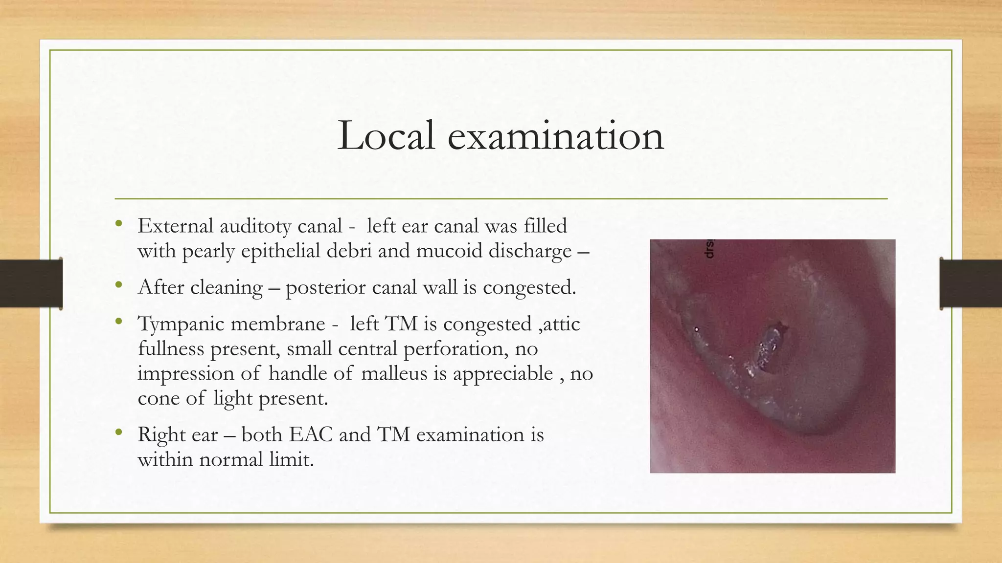
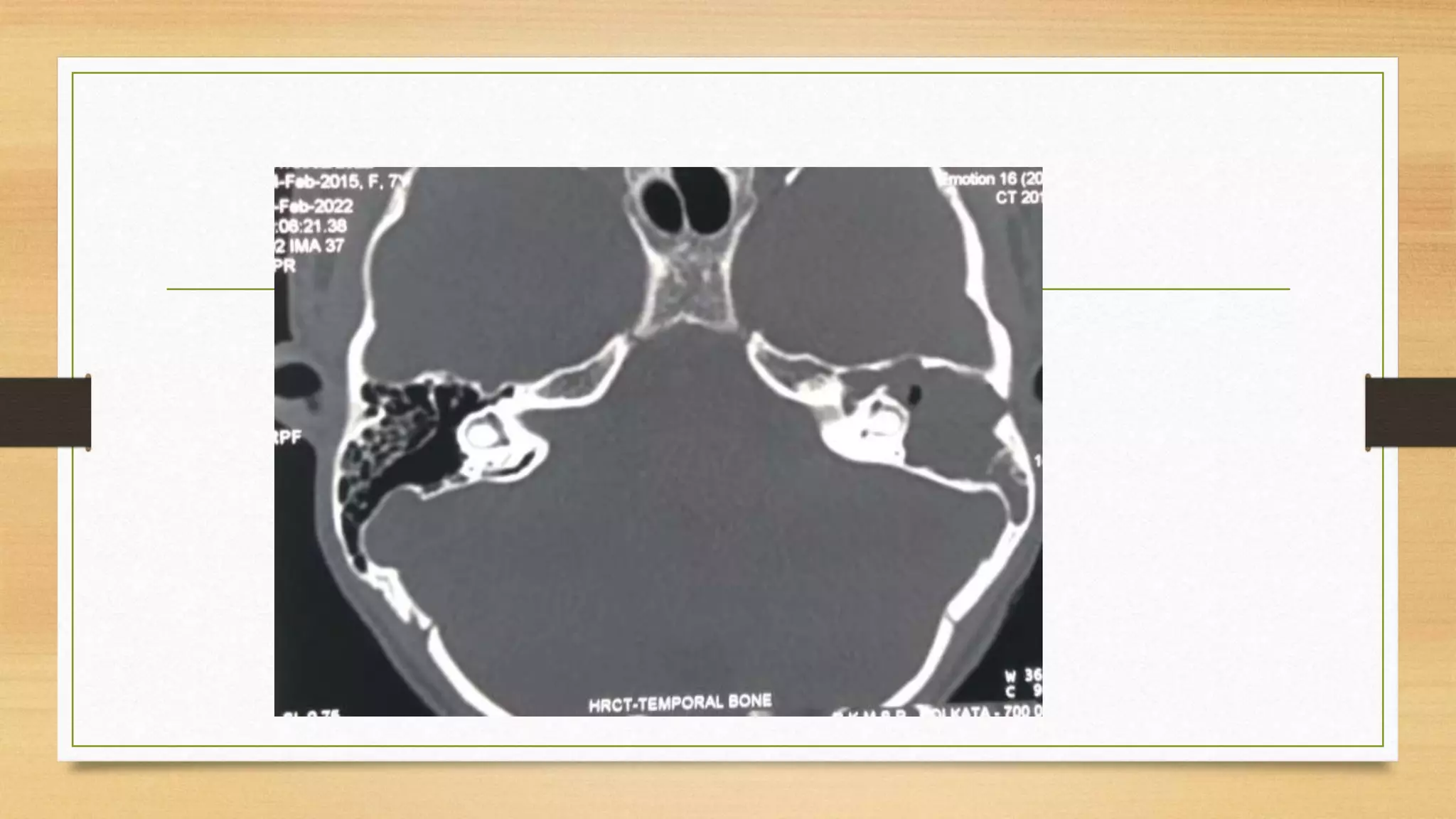
Case Presentation –
Cholesteatoma
Dr Safika Zaman
Dept of ENT and HNS
VIMS- RKMSP
kolkata
Patient details
• Name – S. P
• Age –7 year
• Sex –Female
• Student
• Residence – Kolkata
Chief complaints
• Discharge from left ear from 2 year
• Impaired hearing in the same ear from last 1 year
History of present complaints
• Discharge – from left side , insidious in onset , continuous from last 2 year
• Scanty in amount , mucopurulent , often mixed with blood , foul smelling.
• Decreased hearing – left ear, from last 1 ear , gradually progressive in nature ,
• Persistent , no problem in speech discrimination but difficulty in
understanding soft sound.
History
• No history of ear pain , tinnitus, spinning sensation of head,
• No history of facial muscle weakness, or deviation of face
• No relation of discharge with upper respiratory infection
• No history of severe headache with fever and vomiting
• No history of post auricular abscess or discharging sinus
History
• No previous medical or surgical significant history
• No known food or drug allergen
• Immunised according to NIS
• Fine motor , gross motor , speech and language development is appropriate
for age
• Single child
Treatment history
• In last 2 years patient received multiple course of oral antibiotic and
antibiotic steroid drop , discharge never responded to the therapy and was
persistent .
Personal history
• Sleep , bowel , bladder , appetite – no recent change
General examination
Conscious , oriented and co- operative
Weight – 20 kg
BP – 90 /70
P- 120/min
RR- 18 / min
Temp – Normal
Pallor , cyanosis, clubbing , jaundice – absent
Local examination
• Pinna –both ear - normal in size and shape, no skin changes or lesion
present.
• Local temp of pinna for both ear is normal
• Pre-auricular region – in both ear no swelling or discharge present
• Post auricular region –in both ear no swelling , scar , fistula present .
Local examination
• External auditoty canal - left ear canal was filled
with pearly epithelial debri and mucoid discharge –
• After cleaning – posterior canal wall is congested.
• Tympanic membrane - left TM is congested ,attic
fullness present, small central perforation, no
impression of handle of malleus is appreciable , no
cone of light present.
• Right ear – both EAC and TM examination is
within normal limit.
• Sigalization – no acute onset nystagmus or vertigo
• Examination of nose – within normal limit
• Examination of oral cavity and oropharynx – WNL
• Examination of facial nerve – WNL
Tuning fork test
Right ear Left ear
Rinnes test Positive Negetive
Webers test Towards left ear
Provisional diagnosis
• Active , squamosal chronic otitis media, uncomplicated
Investigations- PTA
Tympanogram
Management
• Canal wall down mastoidectomy with ossiculoplasty and grafting
Thank you

Cholesteatoma Case Presentation .pptx

  • 1.
    Case Presentation – Cholesteatoma DrSafika Zaman Dept of ENT and HNS VIMS- RKMSP kolkata
  • 2.
    Patient details • Name– S. P • Age –7 year • Sex –Female • Student • Residence – Kolkata
  • 3.
    Chief complaints • Dischargefrom left ear from 2 year • Impaired hearing in the same ear from last 1 year
  • 4.
    History of presentcomplaints • Discharge – from left side , insidious in onset , continuous from last 2 year • Scanty in amount , mucopurulent , often mixed with blood , foul smelling. • Decreased hearing – left ear, from last 1 ear , gradually progressive in nature , • Persistent , no problem in speech discrimination but difficulty in understanding soft sound.
  • 5.
    History • No historyof ear pain , tinnitus, spinning sensation of head, • No history of facial muscle weakness, or deviation of face • No relation of discharge with upper respiratory infection • No history of severe headache with fever and vomiting • No history of post auricular abscess or discharging sinus
  • 6.
    History • No previousmedical or surgical significant history • No known food or drug allergen • Immunised according to NIS • Fine motor , gross motor , speech and language development is appropriate for age • Single child
  • 7.
    Treatment history • Inlast 2 years patient received multiple course of oral antibiotic and antibiotic steroid drop , discharge never responded to the therapy and was persistent .
  • 8.
    Personal history • Sleep, bowel , bladder , appetite – no recent change
  • 9.
    General examination Conscious ,oriented and co- operative Weight – 20 kg BP – 90 /70 P- 120/min RR- 18 / min Temp – Normal Pallor , cyanosis, clubbing , jaundice – absent
  • 10.
    Local examination • Pinna–both ear - normal in size and shape, no skin changes or lesion present. • Local temp of pinna for both ear is normal • Pre-auricular region – in both ear no swelling or discharge present • Post auricular region –in both ear no swelling , scar , fistula present .
  • 11.
    Local examination • Externalauditoty canal - left ear canal was filled with pearly epithelial debri and mucoid discharge – • After cleaning – posterior canal wall is congested. • Tympanic membrane - left TM is congested ,attic fullness present, small central perforation, no impression of handle of malleus is appreciable , no cone of light present. • Right ear – both EAC and TM examination is within normal limit.
  • 12.
    • Sigalization –no acute onset nystagmus or vertigo • Examination of nose – within normal limit • Examination of oral cavity and oropharynx – WNL • Examination of facial nerve – WNL
  • 13.
    Tuning fork test Rightear Left ear Rinnes test Positive Negetive Webers test Towards left ear
  • 14.
    Provisional diagnosis • Active, squamosal chronic otitis media, uncomplicated
  • 15.
  • 16.
  • 21.
    Management • Canal walldown mastoidectomy with ossiculoplasty and grafting
  • 22.