Detection of Diabetic Retinopathy using Kirsch Edge Detection and Watershed Transformation Algorithm

Diabetic retinopathy (DR) is a common retinal complication associated with diabetics. A complication of diabetes is that it can also affect various parts of the body. When the small blood vessels have a high level of glucose in the retina, the vision will be blurred and can cause blindness eventually, which is known as diabetic retinopathy. However, if symptoms are identified in the early stage then proper treatment can be provided to prevent blindness. Usually the retinal images obtained from the fundus camera are examined directly and diagnosed. Due to this certain abnormalities due to diabetic retinopathy are not directly visible through the naked eye .Hence by using the image processing techniques these abnormalities can be extracted accurately and required treatments and precautions can be taken. And this also reduces the time for the ophthalmologists to detect the disease and give accurate treatments.
![Divya SN, International Journal of Advance research, Ideas and Innovations in Technology.
© 2015, IJARIIT All Rights Reserved
The blood vessel in human eye is very small and hence more susceptible. The corrosion of blood vessels starts
occurring when the blood sugar levels are increased much above the normal levels for a prolonged period of time.In
DR, the normal vision of a human is hindered and as time passes by, the vision tends to become weaker. Exudates,
micro aneurysms and abnormalities in blood vessels are some features extracted to classify DR. The retina of a human
eye gets damaged when proteins and lipids start leaking from the blood vessels causing exudates. Micro aneurysms are
small areas of balloon like swelling in the retinas tiny blood vessels.
DR can also be detected from abnormality in the structure or extra growth of blood vessels. Early detection
of these features helps the ophthalmologist to detect the DR and also help in preventing blindness. Detection of these
features is done from fundus images. The fundus images are taken from a special type of camera called fundus
cameras. The detection of the mentioned features from the fundus images helps the ophthalmologists to decide on the
severity of the DR and advise the required treatment to the patients.
II. RELATED WORK
Several approaches to diabetic retinopathy detection and classification are found in the literature. Techniques such
as mathematical morphology, neural networks, region growing techniques, fuzzy C-means clustering, matched filters have
been used. A two-dimensional matched filtering technique for Blood vessels detection was proposed to detect the
presence of piecewise linear segments of vessels by rotating 12 different kernel templates at several orientations [1].
Vascular abnormalities were detected by feature extraction and Gabor filter bank outputs at several finer scales were
used to represent energy variations by scale-angle method to classify disease severity as mild, moderate and severe
diabetic retinopathy [2]. Blood vessel detection using top hat transform and watershed transform using mathematical
morphology is described in [3] Gray level variations of the exudates and morphological reconstruction methods were
used in the extraction of exudates [4].
Automatic exudates and cotton-wool spots detection system is developed in [5]. Automated system for
grading the severity of exudates was proposed in [6]. Exudates were detected by top-down segmentation method
followed by local thresholding by combining edge detection techniques and region growing. A polar coordinate
system was used in the grading of severity of exudates. Classification of Diabetic Retinopathy stages as normal, mild,
moderate, severe background retinopathy and as Proliferative by Acharya in [7]. The feature extraction was done
using Higher Order Spectra (HOS). Area and perimeter from the RGB components of the blood vessels were used as
features in grading the severity of retinopathy using a feed forward neural network [8]. Features such as area of
exudates and the area of blood vessels together with texture parameters were input to neural network to grade the
images into normal, Non-Proliferative Retinopathy and Proliferative Retinopathy by Nayak et al. [9].
Wavelet transform based edge detection and Segmentation like simple and global thresholding [10] algorithm was
used and there were some drawbacks in the existing methods like 1. In this method blood vessel detection process in the
retinal images are difficult. 2. In Curved edges, the accuracy of edge localization in wavelet transform is small.
3. Poor Edge detection and Enhancement. In wavelet transform it cannot enhance the curved structure of the retinal
images. 4. Not possible to cluster a fundus Exudates. Radial projection method was proposed in a project to locate the
vessel centerlines of the retina.[11] The previously used algorithms for the four extractions are Curve let transform
and Laplace transform for Blood Vessels, Random forest method for exudates, Hough transform method for Micro
aneurysms and, Bi- histogram equalization and Region growing algorithm for Optic disc.
III. DIABETIC RETINOPATHY
The term retinopathy covers various disorders of the retina, which can affect vision. Retinopathy is usually
due to damage to the tiny blood vessels in the retina. Retinopathy is commonly caused by diabetes, but is sometimes
caused by other diseases such as very high blood pressure. Diabetic retinopathy is a micro vascular complication of
diabetes, causing abnormalities in the retina.](data:image/gif;base64,R0lGODlhAQABAIAAAAAAAP///yH5BAEAAAAALAAAAAABAAEAAAIBRAA7)
Recommended
Recommended
More Related Content
What's hot
What's hot (19)
Viewers also liked
Viewers also liked (18)
Similar to Detection of Diabetic Retinopathy using Kirsch Edge Detection and Watershed Transformation Algorithm
Similar to Detection of Diabetic Retinopathy using Kirsch Edge Detection and Watershed Transformation Algorithm (20)
Recently uploaded
Recently uploaded (20)
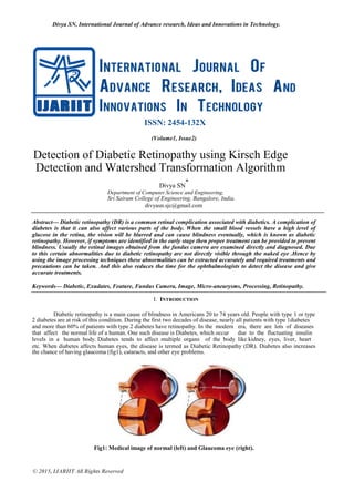
Detection of Diabetic Retinopathy using Kirsch Edge Detection and Watershed Transformation Algorithm
- 1. Divya SN, International Journal of Advance research, Ideas and Innovations in Technology. © 2015, IJARIIT All Rights Reserved ISSN: 2454-132X (Volume1, Issue2) Detection of Diabetic Retinopathy using Kirsch Edge Detection and Watershed Transformation Algorithm Divya SN * Department of Computer Science and Engineering, Sri Sairam College of Engineering, Bangalore, India. divyasn.sjc@gmail.com Abstract— Diabetic retinopathy (DR) is a common retinal complication associated with diabetics. A complication of diabetes is that it can also affect various parts of the body. When the small blood vessels have a high level of glucose in the retina, the vision will be blurred and can cause blindness eventually, which is known as diabetic retinopathy. However, if symptoms are identified in the early stage then proper treatment can be provided to prevent blindness. Usually the retinal images obtained from the fundus camera are examined directly and diagnosed. Due to this certain abnormalities due to diabetic retinopathy are not directly visible through the naked eye .Hence by using the image processing techniques these abnormalities can be extracted accurately and required treatments and precautions can be taken. And this also reduces the time for the ophthalmologists to detect the disease and give accurate treatments. Keywords— Diabetic, Exudates, Feature, Fundus Camera, Image, Micro-aneurysms, Processing, Retinopathy. I. INTRODUCTION Diabetic retinopathy is a main cause of blindness in Americans 20 to 74 years old. People with type 1 or type 2 diabetes are at risk of this condition. During the first two decades of disease, nearly all patients with type 1diabetes and more than 60% of patients with type 2 diabetes have retinopathy. In the modern era, there are lots of diseases that affect the normal life of a human. One such disease is Diabetes, which occur due to the fluctuating insulin levels in a human body. Diabetes tends to affect multiple organs of the body like kidney, eyes, liver, heart etc. When diabetes affects human eyes, the disease is termed as Diabetic Retinopathy (DR). Diabetes also increases the chance of having glaucoma (fig1), cataracts, and other eye problems. Fig1: Medical image of normal (left) and Glaucoma eye (right).
- 2. Divya SN, International Journal of Advance research, Ideas and Innovations in Technology. © 2015, IJARIIT All Rights Reserved The blood vessel in human eye is very small and hence more susceptible. The corrosion of blood vessels starts occurring when the blood sugar levels are increased much above the normal levels for a prolonged period of time.In DR, the normal vision of a human is hindered and as time passes by, the vision tends to become weaker. Exudates, micro aneurysms and abnormalities in blood vessels are some features extracted to classify DR. The retina of a human eye gets damaged when proteins and lipids start leaking from the blood vessels causing exudates. Micro aneurysms are small areas of balloon like swelling in the retinas tiny blood vessels. DR can also be detected from abnormality in the structure or extra growth of blood vessels. Early detection of these features helps the ophthalmologist to detect the DR and also help in preventing blindness. Detection of these features is done from fundus images. The fundus images are taken from a special type of camera called fundus cameras. The detection of the mentioned features from the fundus images helps the ophthalmologists to decide on the severity of the DR and advise the required treatment to the patients. II. RELATED WORK Several approaches to diabetic retinopathy detection and classification are found in the literature. Techniques such as mathematical morphology, neural networks, region growing techniques, fuzzy C-means clustering, matched filters have been used. A two-dimensional matched filtering technique for Blood vessels detection was proposed to detect the presence of piecewise linear segments of vessels by rotating 12 different kernel templates at several orientations [1]. Vascular abnormalities were detected by feature extraction and Gabor filter bank outputs at several finer scales were used to represent energy variations by scale-angle method to classify disease severity as mild, moderate and severe diabetic retinopathy [2]. Blood vessel detection using top hat transform and watershed transform using mathematical morphology is described in [3] Gray level variations of the exudates and morphological reconstruction methods were used in the extraction of exudates [4]. Automatic exudates and cotton-wool spots detection system is developed in [5]. Automated system for grading the severity of exudates was proposed in [6]. Exudates were detected by top-down segmentation method followed by local thresholding by combining edge detection techniques and region growing. A polar coordinate system was used in the grading of severity of exudates. Classification of Diabetic Retinopathy stages as normal, mild, moderate, severe background retinopathy and as Proliferative by Acharya in [7]. The feature extraction was done using Higher Order Spectra (HOS). Area and perimeter from the RGB components of the blood vessels were used as features in grading the severity of retinopathy using a feed forward neural network [8]. Features such as area of exudates and the area of blood vessels together with texture parameters were input to neural network to grade the images into normal, Non-Proliferative Retinopathy and Proliferative Retinopathy by Nayak et al. [9]. Wavelet transform based edge detection and Segmentation like simple and global thresholding [10] algorithm was used and there were some drawbacks in the existing methods like 1. In this method blood vessel detection process in the retinal images are difficult. 2. In Curved edges, the accuracy of edge localization in wavelet transform is small. 3. Poor Edge detection and Enhancement. In wavelet transform it cannot enhance the curved structure of the retinal images. 4. Not possible to cluster a fundus Exudates. Radial projection method was proposed in a project to locate the vessel centerlines of the retina.[11] The previously used algorithms for the four extractions are Curve let transform and Laplace transform for Blood Vessels, Random forest method for exudates, Hough transform method for Micro aneurysms and, Bi- histogram equalization and Region growing algorithm for Optic disc. III. DIABETIC RETINOPATHY The term retinopathy covers various disorders of the retina, which can affect vision. Retinopathy is usually due to damage to the tiny blood vessels in the retina. Retinopathy is commonly caused by diabetes, but is sometimes caused by other diseases such as very high blood pressure. Diabetic retinopathy is a micro vascular complication of diabetes, causing abnormalities in the retina.
- 3. Divya SN, International Journal of Advance research, Ideas and Innovations in Technology. © 2015, IJARIIT All Rights Reserved Fig3: Diabetic Retinopathy Most often, diabetic retinopathy has no symptoms until the damage to your eyes is severe. This is because damage can occur to much of the retina before your vision is affected. Symptoms of diabetic retinopathy include: Blurred vision and slow vision loss over time Floaters Shadows or missing areas of vision Trouble seeing at night Double Vision Eye pain Many people with early diabetic retinopathy have no symptoms before bleeding occurs in the eye. The diabetic retinopathy typically begins as small changes in the retinal capillaries. The smallest detectable abnormalities, micro aneurysms (MA), appear as small red dots in the retina. Due to these damaged capillary walls, the small blood vessels may rupture and cause intra retinal hemorrhages (HA). In the retina, the hemorrhages appear either as small red dots indistinguishable from micro aneurysms or larger round-shaped blots with irregular outline. The diabetic retinopathy also increases the permeability of the capillary walls which results in retinal edema and hard exudates (HE). The hard exudates are lipid formations leaking from the weakened blood vessels and appear yellowish with well-defined borders. If the local capillary circulation and oxygen support fail due to obstructed blood vessels, pale areas with indistinct margins appear in the retina. An extensive lack of oxygen and obstructed capillary in the retina lead to the development of new fragile vessels. These new vessels attempt to grow towards the suffering tissue to supply nutrition and oxygen. However, the new vessels are fragile and tend to grow into the space between the retina and vitreous humour, or directly to the vitreous humour, which can lead to pre-retinal hemorrhage and a sudden loss of vision. IV. PROPOSED METHOD In the proposed system, the automated detection of diabetic retinopathy using feature extraction from digital fundus images is done. This feature extraction is done using MATLAB. The features studied are micro-aneurysms, optic disc, exudates and blood vessels. The block diagram for the proposed system is as shown in the Figure. Fig4: Proposed system
- 4. Divya SN, International Journal of Advance research, Ideas and Innovations in Technology. © 2015, IJARIIT All Rights Reserved Step1: Input Fundus Images Read the input image from Fundus camera. The Fundus camera is more reliable, non-invasive and easy to use compared to traditional ophthalmoscopy. The eye fundus photography results in a better sensitivity rate i.e, a better detection rate of abnormal eye funduses. Step2: Image Pre processing Image Pre processing includes various techniques such as contrast enhancement, gray component, image de- noising etc. Initially we convert the RGB image into gray color image to further process the image. In the RGB images the green channel exhibits the best contrast between the vessels and background while the red and blue ones tends to be more noisy. Since the retinal blood vessels appear darker in gray image, the green channel is used to convert the intensity of the image. Filtering is used to remove the noise which gets added into the fundus image. Here Improved median filtering is used. Fig5: Input image (left) and Image after Improved median filtering (right) The quality of an image can be improved using image enhancement techniques such as Adaptive Histogram equalization. Step3: Feature Extraction A. Blood Vessels Feature Extraction: Kirsch algorithm is used to detect the blood vessels. Kirsch Edge Detection Algorithm: In this algorithm, the Kirsch gradient operator is chosen to extract the contour of the object. The Kirsch edge detection uses eight filters (i.e., eight masks for related eight main directions) that are applied to given image to detect edges. Every pixel has eight outputs. Also, the maximum output of the eight templates is chosen to be the edge magnitude. The direction of edge is defined by the related mask that produces the maximum magnitude. Edge information for a particular pixel is obtained by exploring the brightness of pixels in the neighborhood of that pixel. The algorithm uses a 3x3 table of pixels called a convolution table to store a pixel value. Fig 6: Output of Kirsch edge detection technique B. Exudates Feature Extraction: Exudates are Small yellow white patches with sharp margins and different shapes. These are the accumulations of lipid and protein in the retina. They indicate increased vessel permeability, a connected risk of retinal edema and fluid accumulation in the retina. Fuzzy Clustering algorithm is used for exudates feature extraction.
- 5. Divya SN, International Journal of Advance research, Ideas and Innovations in Technology. © 2015, IJARIIT All Rights Reserved Fuzzy Clustering Algorithm: Fuzzy clustering is an overlapping clustering algorithm, where features with close similarity in an image are grouped into the same cluster. The similarity is defined by the distance of the features vector to the cluster centers. Euclidean distance is used to measure this distance and data will be associated to an appropriate membership value. The output from Fuzzy clustering is a list of cluster centers and n membership-grades for each pixel, where n is a number of desired clusters. Fig 7: Image obtained after Fuzzy clustering algorithm C. Micro Aneurysms Extraction The presence of micro aneurysms is considered as early stage of diabetic retinopathy. Micro aneurysms on the retina appear as small red dots of maximum diameter to be less than the diameter of the major optic veins. Morphological distance based algorithm is used for the detection of micro aneurysms. Morphological Distance Based Algorithm The preprocessing step filters the image, increases the contrast and performs a shading correction in order to balance the non-uniform illumination across the image. The diameter-closing step is a mathematical morphological transformation that fills in all the black dots with diameters smaller than λ. After performing such closing transformation, the grey-scale value of the filled-in dots is higher than in the enhanced preprocessed image, while the vessels and other elements remain virtually unaffected. The black top-hat step uses size and shape criteria to isolate the black components contrasted against the background. The black hat transform is the result of the difference between the images obtained by the diameter closing and preprocessing steps. The automated threshold step identifies all elements in the black top-hat image that are possible μA candidates. Finally a K-nearest neighbors (k -NN) classifier is used for classification. It uses the properties calculated for the candidates to find them as either true μA or false positives based on the learning set in the small database. The classifier acts like a human grader by taking into account features such as size, contrast, circularity, grey-scale level and colour. Then the true micro aneurysms are detected. Fig 8: Image obtained after Morphological Distance Based Algorithm D. Optic Disc Feature Extraction: Optic disc is the brightest part of the retina and it is in oval in shape and for diabetic retinopathy affected person the oval structure is irregular in structure. Watershed Transformation algorithm is used for optic disc feature extraction. Watershed Transformation
- 6. Divya SN, International Journal of Advance research, Ideas and Innovations in Technology. © 2015, IJARIIT All Rights Reserved Watershed transformation is a segmentation technique for gray-scale images. This algorithm is a powerful segmentation tool whenever the minima of the image represent the objects of interest and the maxima are the separation boundaries between objects. Due to this fact, the input image of this method is usually a gradient image. If the gradient image is considered as input image, the watershed transformation produces a segmentation which can be viewed as a set of closed contours of segmented regions. Fig 9: Image obtained after watershed transformation III. RESULTS The results obtained and the severities of the disease are tabulated at the end of this paper. Depending on the severity, there are three categories such as mild, moderate and severe stage. A treatment can also be based on the severity. Certain known treatments are Vitrectomy, Scatter laser treatment, Focal laser treatment and Laser photocoagulation. IV. CONCLUSION In this paper, the techniques for early detection of Diabetic Retinopathy are given. The abnormalities that cannot be seen via the naked eye can be detected accurately. Finally the different algorithms used, which follows the International council of Ophthalmology’s criteria for the assessment of the severity of the disease, helps in establishing the severity of the disease so that the patient can accordingly be referred to the specialist and hence treated accordingly. Treatments such as Vitrectomy, Scatter Laser Treatment, Focal Laser Treatment, and Laser Photocoagulation can be taken. Image analysis tools can be used for automated detection of these various features. REFERENCES [1] Vinita Automated Diabetic Retinopathy Severity Classification using Support Vector Machine [2] Vallabha, D., Dorairaj, R., Namuduri, K., and Thompson, H., Automated detection and classification of vascular abnormalities in diabetic retinopathy, Proceedings of 13th IEEE Signals, Systems and Computers 2:1625-1629, 2004. [3] Nidhal Khdhair El Abbadi, Enas Hamood Al Saadi., Blood vessels extraction using mathematical morphology, Journal of Computer Science 9 (10): 1389-1395, 2013,ISSN: 1549-3636. [4] Walter T, Massin P, Erginay A, Ordonez R, Jeulin C, Klein J-C "Automatic Detection of Micro aneurysms in Color Fundus Images Medical Image analysis"2007. [5] Niemeijer, M., van Ginneken, B., Russell, R. S., Suttorp-Schulten,S. A. M., and Abramoff, D. M., Automated detection and differentiation of drusen, exudates, and cotton-wool spots in digital color fundus photographs for diabetic retinopathy diagnosis. Invest. Ophthalmol. Vis. Sci. 48(5):2260–2267, 2007. [6] Hussain F. Jaafar, Asoke K. Nandi and Waleed Al-Nuaimy,A utomated detection and grading of hard exudates from retinal fundus images, 9th European Signal Processing Conference (EUSIPCO 2011), Barcelona, Spain, August 29 - September 2, 2011. [7] Acharya, U. R., Chua, K. C., Ng, E. Y. K., Wei, W., and Chee, C. Application of higher order spectra for the identification of diabetes retinopathy stages. J. Med. Systems, 2008, 32(6), 48 1-488. [8] Wong, L. Y., Acharya, U. R., Venkatesh, Y. V., Chee, C., Lim, C. M., and Ng, E. Y. K., Identification of different stages of diabetic retinopathy using retinal optical images. Information Sciences 178(1):106–121, 2008. [9] Manual micro aneurysms detection support with size and shape based detection by petra varsanyi ,zsolt fegyvari ,szaboles sergyan , zoltan vammosy, 2014 IEEE paper pg no: 361 to 365. [10] Retinal blood vessels segmentation using the radial projection and Supervised classification by quinmu peng,xinge you,long zhou,yiuming cheung, 2010 international conference on pattern recognition pg no: 1489 to 1492.
- 7. Divya SN, International Journal of Advance research, Ideas and Innovations in Technology. © 2015, IJARIIT All Rights Reserved Results and Disease Severity Input ima ge Blood vessel Exudates Microaneurysms Optic disc Severity Mild moderate severe moderate severe About Author: Divya.S.N is currently pursuing her Bachelor of Engineering in Computer Science and Engineering at Sri Sairam College of Engineering, VTU University, India. Her research interests include Image Processing, Programming languages, Web Designing and Development, Animations and Graphics.