Automatic Blood Vessels Segmentation of Retinal Images
•Download as PPTX, PDF•
9 likes•4,961 views

a review on blood vessels segmentation methods. with one proposed method

Recommended
Recommended
More Related Content
What's hot
What's hot (20)
Viewers also liked
Viewers also liked (20)
Similar to Automatic Blood Vessels Segmentation of Retinal Images
Similar to Automatic Blood Vessels Segmentation of Retinal Images (20)
Recently uploaded
Recently uploaded (20)
Automatic Blood Vessels Segmentation of Retinal Images
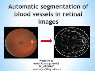
- 1. Presented by Harish Rajula -p14ec009 SV_NIT-SURAT harish.rajula91@gmail.com
- 2. Introduction Need of Automatic Segmentation in retinal images Segmentation Methods Literature Survey Proposed System Simulation Results Conclusion
- 3. In clinical ophthalmology colour retinal images acquired from digital fundus camera(low power microscope with attached camera). These images are widely used for detection and diagnosis of diseases like diabetic retinopathy, hyper-tension and various vascular disorders. Retinal images provide information about the blood supply system of the retina.
- 4. Screening pro-gram of eye results in large number of retinal images needed to be examined by ophthalmologists. Manual diagnosis is usually performed by analysing the images from a patient, as not all images show signs of diabetic retinopathy. It increases the time and decreases the efficiency of ophthalmologists. Therefore, an automatic segmentation of the vasculature could save workload of the ophthalmologists and may assist in characterizing the detected lesions and to identify false positives. In Automatic retinal vessel segmentation, the registration of retinal images of the same patient taken over period of time. The registered images are useful in monitoring the progression of disease and to observe the effect of treatment.
- 5. 1. Local Segmentation It deals with segmenting sub images which are small windows on whole image 2. Global Segmentation It is concerned with segmenting a whole image.this further divided into some categories. i. Region Approach ii. Threshold based approach iii. Edge Approach iv. Clustering Approach v. Matching
- 6. Threshold based segmentation. Histogram thresholding and slicing techniques are used to segment the image. They may be applied directly to an image, but can also be combined with pre- and post-processing techniques. Region based segmentation. Where an edge based technique may attempt to find the object boundaries and then locate the object itself by filling them in, a region based technique takes the opposite approach, by (e.g.) starting in the middle of an object and then “growing” outward until it meets the object boundaries. Edge based segmentation. With this technique, detected edges in an image are assumed to represent object boundaries, and used to identify these objects. Matching. When we know what an object we wish to identify in an image (approximately) looks like, we can use this knowledge to locate the object in an image. This approach to segmentation is called matching.
- 7. In This literature they used graph based approach for blood vessel boundary delineation. The widths of the retinal blood vessels are measured and its edges are segmented. The graph is constructed based on the vessels weight. The REVIEW database was used in this work. This paper has some deficiencies, such as the crossing points and branching points are currently not treated individually, and consequently the blood vessel detection points are not clearly indicated[1].
- 8. In this they proposed line-shape concavity measuring model to remove dark lesions which have an intensity structure different from the line- shaped vessels in a retina. This method achieved 95.67% of an average accuracy for the blood vessel detection with respect to ground truth images in DRIVE database. While provided 95.56 % of an average accuracy for the blood vessel detection with respect to ground truth images in STARE database. In this they presented multi-scale feature extraction and region growing algorithm for retinal blood vessels segmentation. This implementation allowed a faster processing of these images and was based on a data partitioning.
- 9. In this they proposed a pixel feature based method that additionally analysed the vessels as elongated structures. The edge-based methods can be further classified into window- based and tracking-based methods. Window-based method estimates a match at each pixel against the pixel‘s surrounding window. In order to trace the vessels, the tracking approach makes use of local image properties from an initial point.
- 10. The proposed system consists of three stages-first is pre- processing of retinal image to separate the green channel and second stage is retinal image enhancement and third stage is blood vessel segmentation using morphological operations and SVM Classifier. The proposed system for blood vessel segmentation is illustrated in Fig. 1.
- 11. Initially the retinal image is enhanced using adaptive histogram equalization technique in order to enhance the blood vessels. Then, the retinal fundus image is divided in to three primary components such as Red Channel (R), Green Channel (G) and Blue Channel (B). The green channel is high sensitive to the blood vessels. Hence this channel is considered for the detection and segmentation of retinal blood vessels from the retinal image.
- 13. The morphological operations are applied on the pre-processed green channel. Morphological operation processes the pre processed image with structuring element. The retinal blood vessels are detected by applying dilation and erosion process to a pre-processed image. The morphological opening and closing operation are applied to an image based on multi structure elements to enhance the vessel edges. Morphological opening and closing operation is performed by using dilation and erosion. The morphologically processed opened image and morphologically processed closed images are absolutely subtracted to detect the blood vessels from retina fundus image. The combination of dilation and erosion operations is performed on image with different structuring element of radius . Then, an absolute difference mapping image is formed by absolute subtraction of retinal image from the morphologically processed sub-band image.
- 17. The blood vessel detection and segmentation is an important for diabetic retinopathy diagnosis at earlier stage. The morphological and SVM classifier is proposed in this paper to detect and segment the blood vessels from the retinal image . The local binary pattern and GLCM features are extracted from the morphologically processed image and used as blood vessels features. The proposed method detected blood vessels with an average sensitivity of 78%, average specificity of 97.99% and an average accuracy of 99.6% in the retinal fundus images.