The document presents a study on a system for detecting diabetic retinopathy from retinal images using image-processing techniques. The proposed algorithm incorporates contrast limited adaptive histogram equalization and classification through support vector machine and k-nearest neighbors classifiers, aiming to enhance early detection of the disease. It emphasizes the importance of automated screening for efficient diagnosis to prevent vision loss in diabetic patients.
![International Journal of Trend in Scientific Research and Development (IJTSRD) @ www.ijtsrd.com eISSN: 2456-6470
@ IJTSRD | Unique Paper ID – IJTSRD38353 | Volume – 5 | Issue – 2 | January-February 2021 Page 195
disease is the level of blood sugar. Due to high level of sugar
in blood, blood vessels in retina are heavily damaged.
Various lesions like microaneurysms, hemorrhages, hard
exudates, blood vessels are explained as: Microaneurysms
are out-pouching oftheretinal capillaries,appearingassmall
red dots on the fundus image. They appear as small bulges
developed from weak blood vessels. They are the earliest
clinical sign of DR. Leakage of oily formations from the poor
end blood vessels are known as exudates. HardExudatesare
well-circumscribed, shiny white or cream deposits located
within retina. They indicate accumulation of fluid in the
retina and considered sight threateningifappearcloseto the
macula center. They are generally seen together with
Microaneurysms. At every different retinopathy stage their
shape and size varies; as it starts emerging, the DR is termed
as moderate non-proliferative DR [36]. When retinopathy
increases with time, blood vessels in retina get blocked by
micro infarcts known as soft exudates. Presence of these
three abnormalities is termed as severe non-proliferative
diabetic retinopathy. Hemorrhages may take various shape
and sizes depending on their location within retina. Most
common DR hemorrhages are dot hemorrhages.
III. RELATED WORKS
[4], presents an approach for localizing different lesionsand
features in retinal fundus image. The authors proposed a
constraint in detecting optic disk. Blood vessels, exudates,
microaneurysms and hemorrhages were detected using
different morphological operations which success rate of
97.1% for disk localization.
[23], presents SVM classifier for the classification of the
disease. They proposed a methodology for detecting optic
disc, blood vessels and exudates and then feature extraction
is carried out followed by classification. This yielded an
average accuracy of 94.17%. The problem in the proposed
method of exudate classification was less that was mainly
due to false intensity computation features.
V. Ramya et al [24] proposed a method for the recognition of
DR using SVM classifier. The focus of this paper is on
distinguishing patients with PDR and NPDR. The proposed
methodology begins by pre-processing techniques: median
sifting and histogram equalization followed by feature
extraction. Based on the extracted featureslevelsofDR were
classified using SVM.Theproposedcalculationaccomplished
overall recognition rate of 84% based on hemorrhages.
An approach for early detection of DR from fundus images
presented in [16]. Startingwithpre-processingofrawretinal
fundus images using extraction of green channel, histogram
equalization, image enhancement and resizing techniques.
For evaluation of the results, is done by consideringthearea,
mean value and standard deviation for the extracted
features. Detection of DR is done using machine learning
techniques. From the results obtained, they showed that
exudate area was the best feature out of blood vessel and all
other features for DR detection, which finally concludesthat
exudate is one of the major feature responsible for diabetic
retinopathy.
Sahana Shetty et al [25] have presented an approach for
detecting Diabetic Retinopathy using SVM. The author’s
study began by pre-processing, then eliminating optic disk,
and separating vascular tissue of damaged area oftheretina.
Mathematical morphology methods were carried out to
detect dark lesions then followed by feature extraction.
Extracted features were classified using SVM. Performance
of SVM showed better results than using image processing
with AUC above 90%.
Ahmad Z. F, Muhammad F. et al [26] and S Deva Kumar and
Gnanneswara Rao Nitta et al [32] have taken up GLCM as
method for feature extractionandSVMforclassification.[32]
Includes CLAHE, Kirsch’s operator for detecting blood
vessels, in [26, 32] featuresobtainedbyGLCM.SVMclassifier
is used to classify DR [26, 32]. Comparing [26, 32], accuracy
[32] was shown better compared to [26].
A method of an automatic detection and classification of DR
system is been proposed in [30, 33]. For detection [30] uses,
Circular Hough Transform and image processing while
morphological operations are used in [33].Textural features
and HOG, SURF features are extracted [30, 33]. The authors
use Support Vector Machine in order to classify the retinal
fundus image as normal, NPDR and PDR.
[2, 5] used ANN for the classification of the disease. [2] used
the classifier for classifying the image as normal and
abnormal and [5] for classifying the imageasmoderate,mild
and severe stages. However, one of the methods from [5]
fails because of the non-detection of soft exudates that
occurred in the optic disk because of its removal.
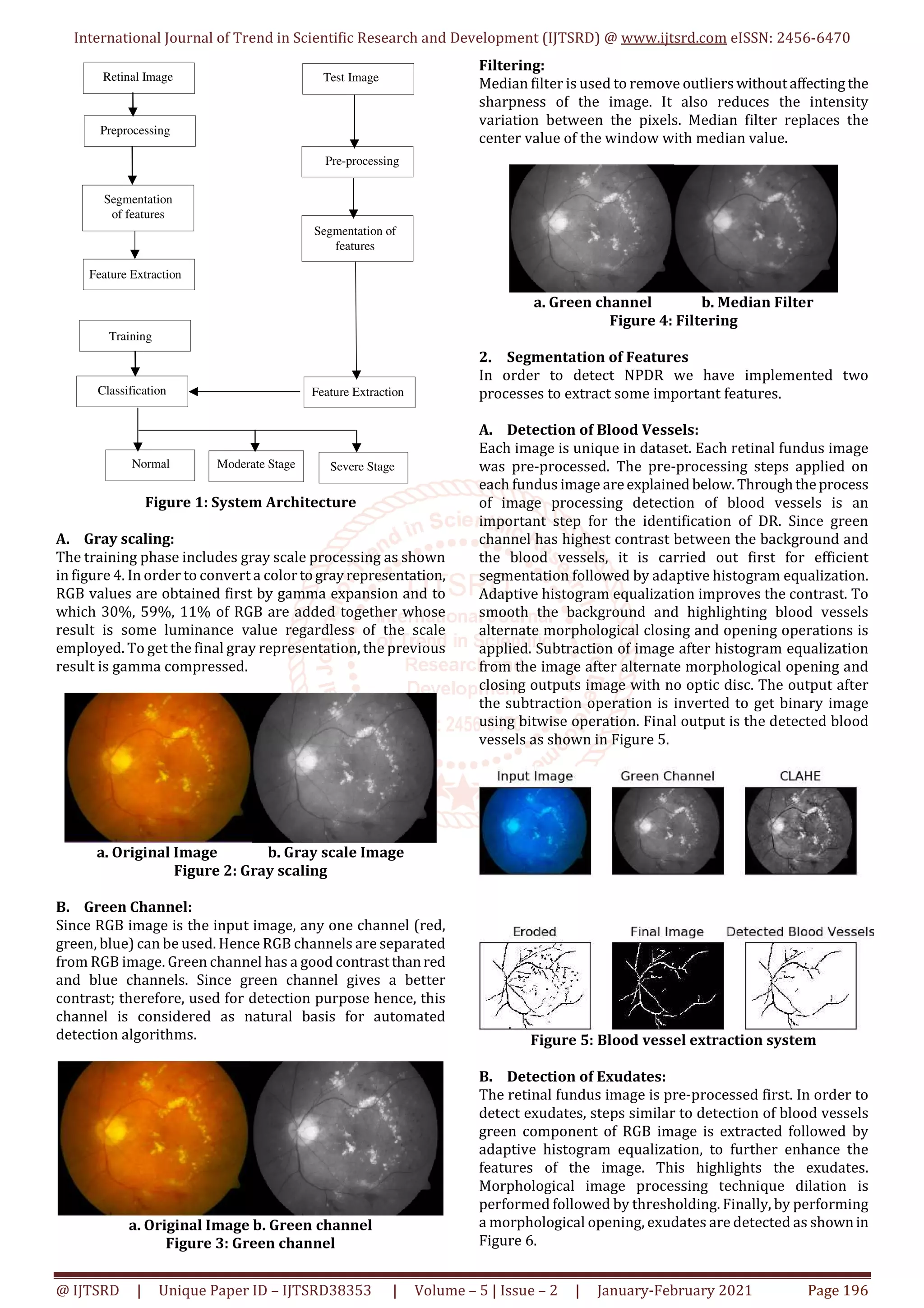
IV. METHODOLOGY
In this section, we discuss the proposed methodology of the
general Diabetic Retinopathy Detection System. The
proposed system comprises of two phases:
Training Phase
Testing Phase
The Training Phase comprises of Pre-processing,
Segmentation of retinal features, Feature Extraction, Storing
features in the database and Training. Testing Phase
comprises of loading the test image, Pre-processing,
Segmentation of Retinal features, Feature extraction and
Classification as normal, moderate or severe, done using
SVM and k-NN classifiers.
A. Training Phase
This step deals with training the system for diabetic
retinopathy recognition. SVM and k-NN will be used in this
case. Each image is unique in dataset. Each retinal fundus
image was pre-processed. The pre-processing steps applied
on each fundus image are explained below.
1. Pre-Processing
Image pre-processing is the initial step in DR detection
system. Pre-processing should be applied before feature
extraction. Preprocessing steps applied on the image are:-](https://image.slidesharecdn.com/36diabeticretinopathydetectionsystemfromretinalimages-210324055752/75/Diabetic-Retinopathy-Detection-System-from-Retinal-Images-2-2048.jpg)
![International Journal of Trend in Scientific Research and Development (IJTSRD) @ www.ijtsrd.com eISSN: 2456-6470
@ IJTSRD | Unique Paper ID – IJTSRD38353 | Volume – 5 | Issue – 2 | January-February 2021 Page 199
evaluated on different measures. As the existingsystemsare
quiet slow in the operation, a real time implementing
screening system will provide better performance.
ACKNOWLEDGMENT
I express my deep sense of gratitude towards my Project
guide Prof. Amit Patil who from the very onset has taken
keen interest in the study and has skillfullyledmetoexecute
each step involved in this undertaking. In a very special way
I am thankful and also grateful to my parents, brothers and
friends for their immense trust and persistent support in all
my endeavors.
REFERENCES
[1] C. Sinthanayothin, V. Konbunkait, S.
Phoojaruenchanachai, A. Singalavanija. “Automated
Screening system for diabetic retinopathy.”
Proceedings of 3rd International Symposium Conf.
Image and Signal Processing and Analysis, vol. 2, pp.
915-920, October 2003.
[2] E. M. Shahin, T. E. Taha, W. Al-Nuaimy, S. El Rabaie, O.
F. Zharan, F. E. A. El-Samie, “Automated Detection of
Diabetic Retinopathy in Blurred Digital Fundus
Images”, 8th International Computer Engineering
Conference (ICENCO), Cairo, 2012, pp. 20-25.
[3] Y. Kumaran, C. M. Patil, “A Breif Review of the
Detection of Diabetic Retinopathy in Human Eyes
Using Pre-Processing & Segmentation Techniques. ”,
International Journal of Recent Technology and
Engieering, vol. 7, pp. 310-320, December 2018.
[4] S. Ravishankar, A. Jain and A. Mittal, “Automated
Feature Extraction for Early Detection of Diabetic
Retinopathy infundusimages,”2009IEEEConference
on Computer Vision and Pattern Recognition, Miami,
FL, pp. 210-217, June 2009.
[5] B. Sumathy, S. Poomachandra, “Automated DR and
prediction of various related diseases of retinal
fundus images”, Artificial Intelligent Techniques for
Bio Medical Signal Processing, January 2018.
[6] S. Sayed, V. Inamdar, S. Kapre, “Detection of Diabetic
Retinopathy using Image Processing and Machine
Learning”, International Journal of Innovative
Research in Science Engineering and Technology,vol.
6, Issue 1, January 2007.
[7] C. Sinthanayothin, J. F. Boyce, T. Williamson, H. L.
Cook, E. Mensah, S. Lal, D. Usher. “Automated
Detection of Diabetic Retinopathy on Digital fundus
images. ” Diabetic Medicine, vol. 19, no. 2, pp. 105-
112, February 2002.
[8] Li, Chutatape. “A model-based approach for
automated feature extractioninfundusimages”,Proc.
9th IEEE International Conference on Computer
Vision, vol. 1, pp. 394, Nov. 2003.
[9] A. Osareh, “Automated Identification of Diabetic
Retinal Exudates and the Optic Disc”, PhD thesis,
University of Bristol, January 2004.
[10] X. Zhang, O. Chutatape, “Top-down and bottom-up
strategies in lesion detection of background diabetic
retinopathy”, IEEE Computer Society Conference on
Computer Vision and Pattern Recognition (CVPR’05),
vol. 2, pp. 422-428, July 2005.
[11] X. Zhang, O. Chutatape,“Detectionandclassificationof
bright lesions in color fundus images”, 2004
International Conference of Image Processing, ICIP
‘04, vol. 1, pp. 139-142, Nov. 2004.
[12] H. Wang, W. Hsu, G. K. Goh, L. Lee, “An effective
approach to detect lesions in color retinal images”,
Proceedings IEEE Conference Computer Vision and
Pattern Recognition, vol. 2, pp. 181-186, Feb. 2000.
[13] A. Sopharak, K. T. New, Y. A. Moe, M. N. Dailey, B.
Uyyanonvara, “Automatic exudate detection with a
naïve bayes classifier.” International Conference of
Embedded Systems and Intelligent Technology
(ICESIT), pp. 139-142, Feb. 2008.
[14] A. Gupta, R. Chhikara, “Diabetic Retinopathy: Present
and Past”, Procedia Computer Science, International
Conference on Computational Intelligence and Data
Science (ICCIDS), ELSEVEIR, vol. 132, pp. 1432-1440,
January 2018.
[15] K. Narasimhan, V. C. Neha, K. Vijayarekha,
“Hypertensive retinopathy diagnosis from fundus
images by estimation of AVR”, International
Conference on modelingoptimizationandcomputing,
Published by Elesevier Ltd. , Procedia Engineering,
vol. 38, pp. 980-993, December 2012.
[16] S. S. Dilip, S. Nair, K. Pooja, “Diabetic Retinal Fundus
Images: Preprocessing and Feature Extraction for
early Detection of Diabetic Retinopathy”, Biomedical
and Pharmacology Journal, vol. 10, no. 2, pp. 615-26,
June 2017.
[17] M. Foracchia, E. Grisan, A. Ruggeri, “DetectionofOptic
Disc in Retinal Images by means of a Geometrical
Model of Vessel Structure”, IEEE Transactions on
Medical Imaging, vol. 23, no. 10, pp. 1189-95, October
2004.
[18] K. A. Vermeer, F. M. Vos, H. Lemij, A. M. Vossepoel. “A
model based method for retinal blood vessel
detection”, Computers in Biology and Medicine,
ELSEVIER, vol. 34, no. 3, pp. 209-219, May 2004.
[19] S. Chaudhari, C. Shankar, N. Katz, M. Nelson, M.
Goldbaum, “Detection of Blood Vessels in Retinal
Images using two dimensional Matched filters”, IEEE
Transactions on Medical Imaging,vol.8,no.3,pp.263-
269, September 1989.
[20] H. Li, C. Opas, “Fundus Image Features Extraction”,
Proceedings of the 22nd Annual International
Conference of the IEEE, Conf. EngineeringinMedicine
and Biology Society, vol. 4, pp. 3071-3073, February
2000.
[21] C. I. Sanchez, R. Hornero, M. I. Lopez, J. Poza, “Retinal
Image Analysis to detect and quantify lesions
associated with diabetic retinopathy”, Proceedingsof
the 26th Annual International Conference of the IEEE
EMBS, San Francisco, CA, USA, pp. 1-5, September
2004.
[22] H. Li, O. Chutatape, “Automated Feature Extraction in
Color Retinal Images by a Model Based Approach”,
IEEE Transactions onBiomedical Engineering,vol.51,
no. 2, pp. 246-254, February 2004.](https://image.slidesharecdn.com/36diabeticretinopathydetectionsystemfromretinalimages-210324055752/75/Diabetic-Retinopathy-Detection-System-from-Retinal-Images-6-2048.jpg)
![International Journal of Trend in Scientific Research and Development (IJTSRD) @ www.ijtsrd.com eISSN: 2456-6470
@ IJTSRD | Unique Paper ID – IJTSRD38353 | Volume – 5 | Issue – 2 | January-February 2021 Page 200
[23] N. B. Prakash, G. R. Hemalakshmi, M. Stella,
“Automated grading of DR stages in fundus images
using SVM classifier”, Journal of Chemical and
Pharmaceutical Research, vol. 8, no. 1 pp. 537-541,
Jan. 2016.
[24] V. Ramya, “SVM Based Detection for Diabetic
Retinopathy”, International Journal of Research and
Scientific Innovation (IJRSI), vol. 5, issue 1, January
2018.
[25] S. Sahana, B. K. Kaveri, A. R. Jayantkumar, “Detection
of Diabetic Retinopathy using Support Vector
Machine (SVM)”, International Journal of Emerging
Technology in Computer Science and Electronics
(IJETCSE), vol. 23, issue 6, October 2016.
[26] A. Z. Foeady, F. Muhammad. , D. C. R. Novitasari, A. H.
Asyhar, M. Firmansjah, “AutomatedDiagnosisSystem
of Diabetic Retinopathy using GLCM MethodandSVM
Classifier”, 5th International Conference on Electrical
Engineering, Computer Science and Informatics
(EECSI), pp. 154-160, October 2018.
[27] A. Taj, K. Kumari, “Detection of Exudates in Retinal
Images using Support Vector Machine”, International
Research Journal of Engineering and Technology
(IRJET), vol. 04, Oct. 2017.
[28] M. Bhagyashri, R. Nitin, “Automatic Detection of
Diabetic Retinopathy using Morphological Operation
and Machine Learning”, International Journal of
Engineering and Technology, vol. 3, no. 5 May 2016.
[29] K. Malathi, R. Nedunchelian, “A recursive support
vector machine (RSVM) algorithm to detect and
classify Diabetic Retinopathy in fundus retinal
images”, Biomedical Research, Computational Life
Sciences and Smarter Technological Advancement,
Jan. 2018.
[30] A. Biran, P. B. Sobhe, A. Almazroe, A. Laxshminarayan,
K. Raahemifar, “Automatic Detection and
Classification of Diabetic Retinopathy using Retinal
Fundus Images”, International Journal of Computer
and Information Engineering, vol. 10, no. 7, pp. 1308-
1311, 2016.
[31] V. Enrique, G. Andres, C. Ricardo, P. Colegio,
“Automated Detection of Diabetic Retinopathy using
SVM”, IEEE XXIV International Conference on
Electronics, Electrical Engineering and Computing
(INTERCON), Cusco, vol. 1, pp. 1-4, August 2017.
[32] S. K. Deva, G. R. Nitta, “Early Detection of Diabetic
Retinopathy in Fundus Images using GLCMandSVM”,
International Journal of Recent Technology and
Engineering (IJRTE), vol. 7, February 2019.
[33] S. Hemavathi, Dr. S. Padmapriya, “Detection of
Diabetic RetinopathyonRetinal ImagesusingSupport
Vector Machine”, SSRG International Journal of
Computer Science and Engineering (SSRG-IJCSE)
ICMR, pp. 5-8 March 2019.
[34] V. A. Aswale, J. A. Shaikh, “Detection of
Microaneurysms in fundus retinal images using SVM
Classifier”, International Journal of Engineering
Development and Research, vol. 5, 2017.
[35] P. N. Sharath, R. U. Deepak, S. Anuja, V.
Sahasranamam, K. R. Rajesh, “Automated Detection
System of Diabetic Retinopathy using field fundus
photography”, 6th International Conference on
Advances in Computingand Communications,6-8,pp.
486-494, September 2016.
[36] Ramanjit Sihota, Radhika Tandon, “Parson’s Diseases
of the Eye” Elsevier Health Sciences India, 22nd
Edition, 15 July 2015.
[37] Aurelien Geron, “Hands-On Machine Learning with
Scikit-Learn and Tensorflow Concepts, Tools and
Techniques to build intelligent systems”, O’reilly
Media, Inc. 1005 Gravenstein Highway North,
Sebastopol, CA, 1st Edition, March 2017.](https://image.slidesharecdn.com/36diabeticretinopathydetectionsystemfromretinalimages-210324055752/75/Diabetic-Retinopathy-Detection-System-from-Retinal-Images-7-2048.jpg)