Downloaded 354 times
![RETINAL IMAGES ENTROPY CONTRAST HOMOGENITY
6.652 [0.3241 0.3396] [0.8751 0.8754]
6.8912 [0.1504 0.1527] [0.9308 0.9344]
6.1124 [0.1268 0.1470] [0.9377 0.9324]
TEXTURE ANALYSIS RESULTS](https://image.slidesharecdn.com/finalppt-170316081234/75/Diabetic-Retinopathy-Analysis-using-Fundus-Image-30-2048.jpg)
![RETINAL IMAGES ENTROPY CONTRAST HOMOGENITY
6.5160 [0.5008 0.4678] [0.8519 0.8600]
7.228 [0.3114 0.2509] [0.8853 0.8988]
6.409 [0.4226 0.5190] [0.8487 0.8466]](https://image.slidesharecdn.com/finalppt-170316081234/75/Diabetic-Retinopathy-Analysis-using-Fundus-Image-31-2048.jpg)


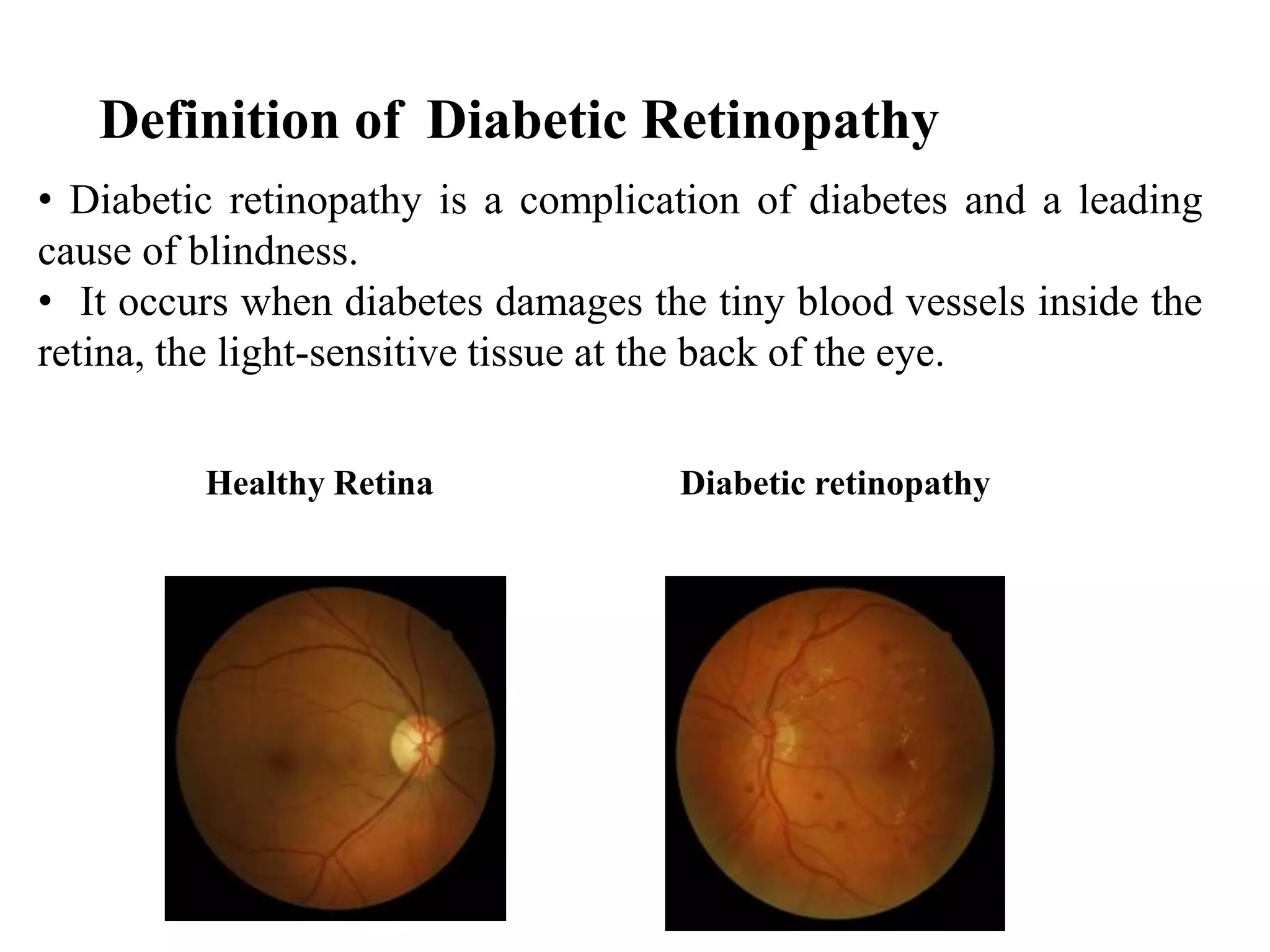
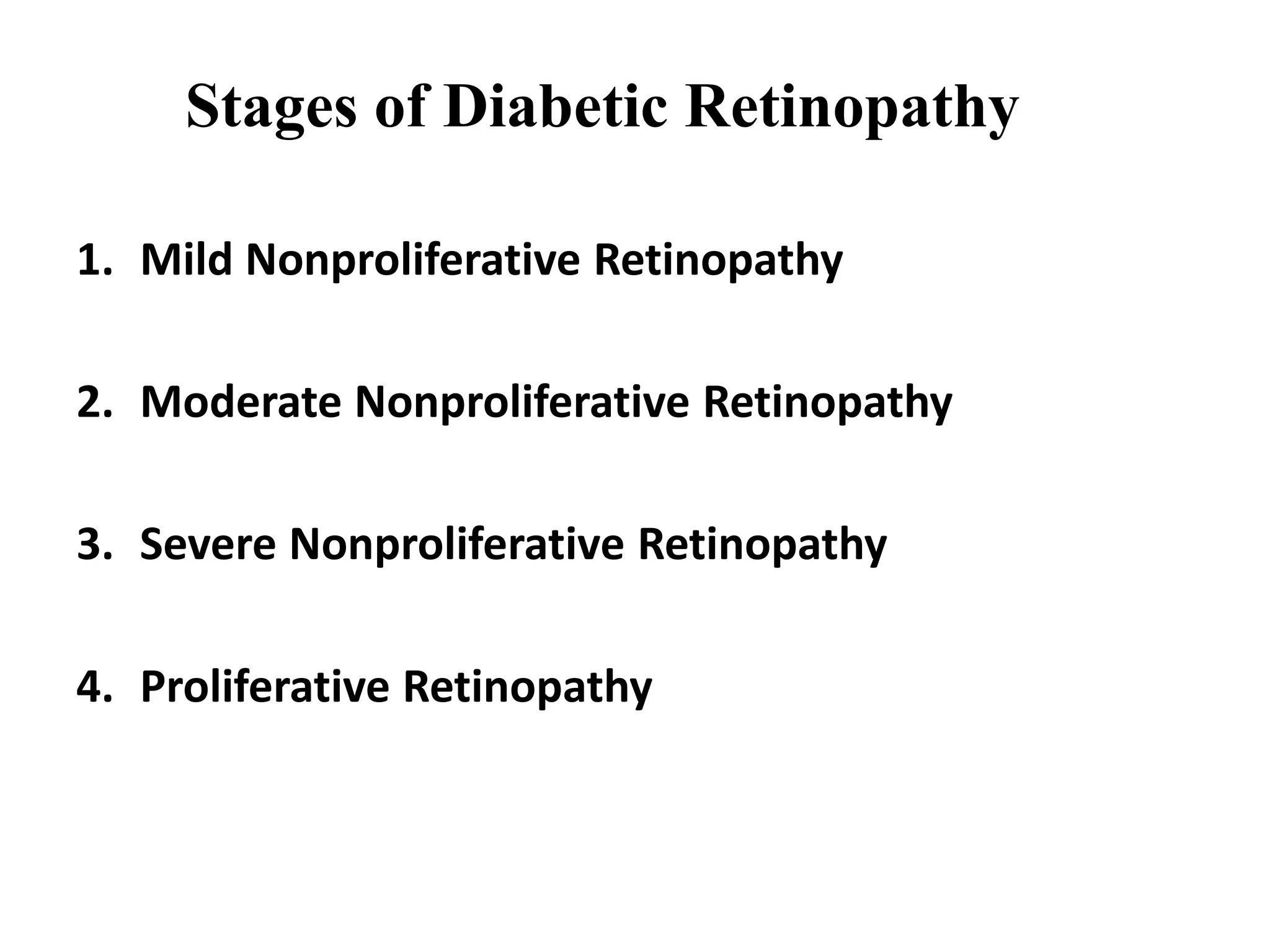
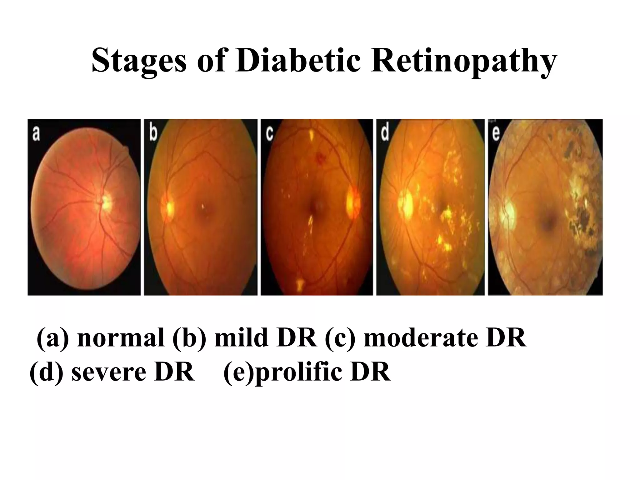
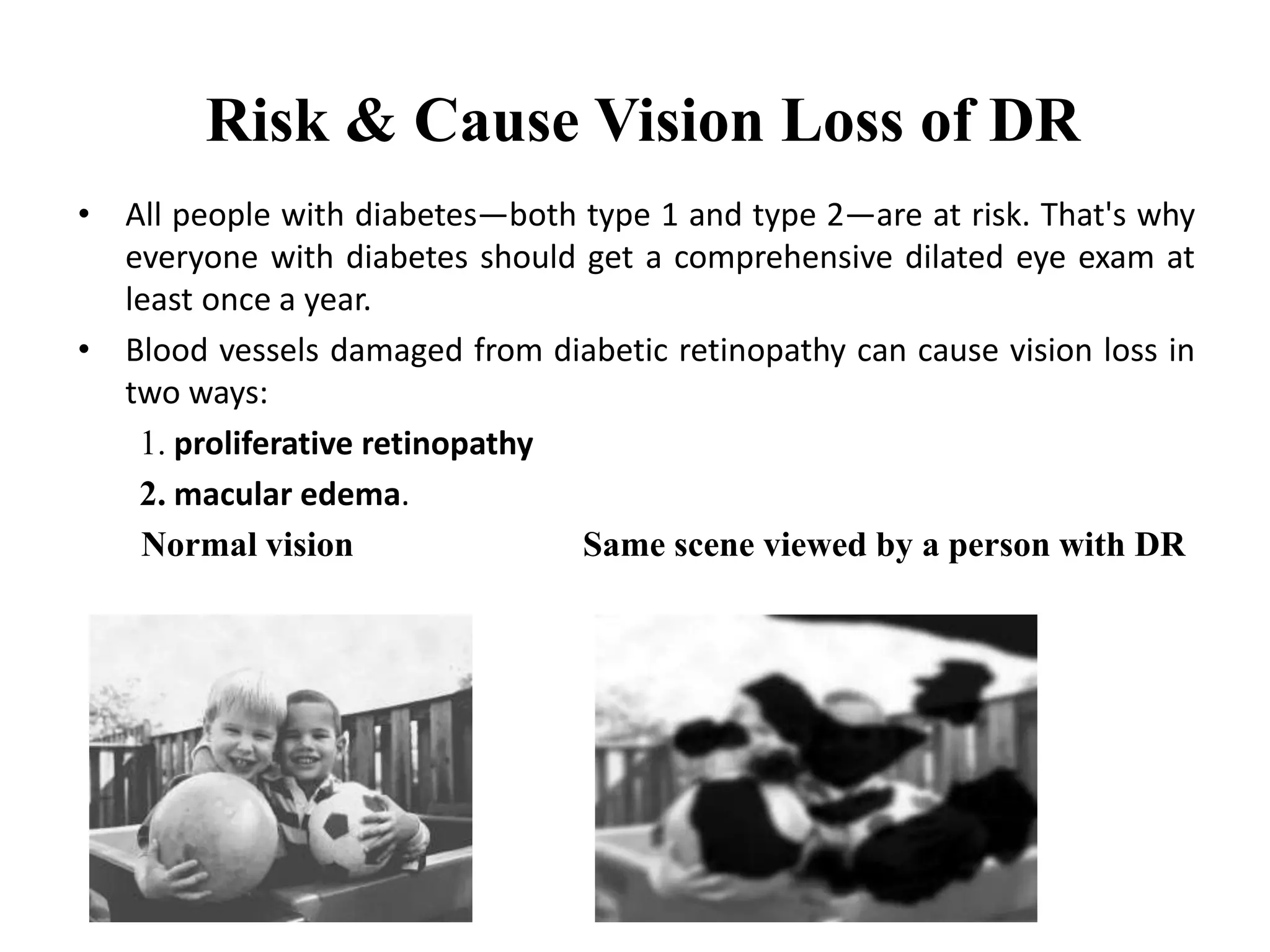
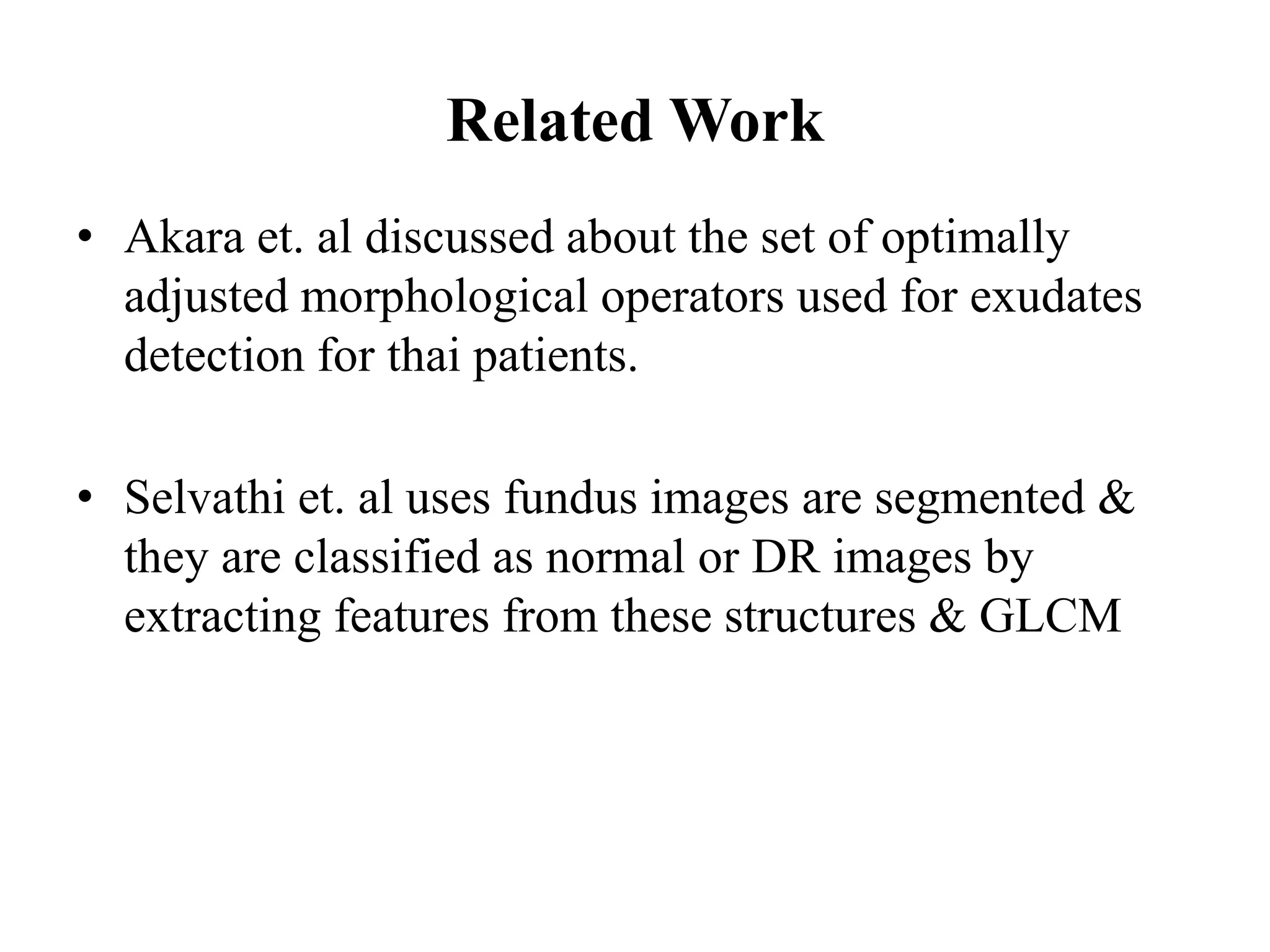
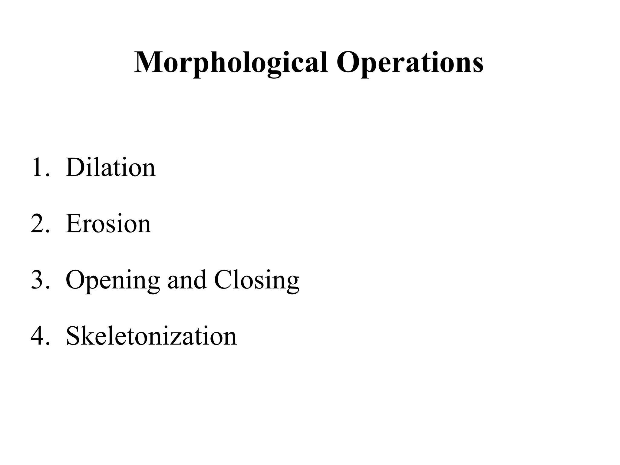
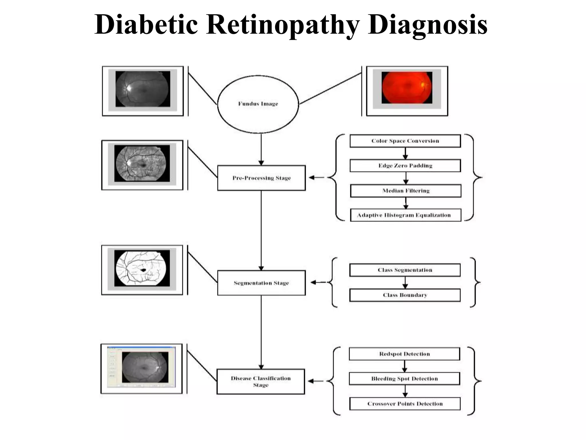
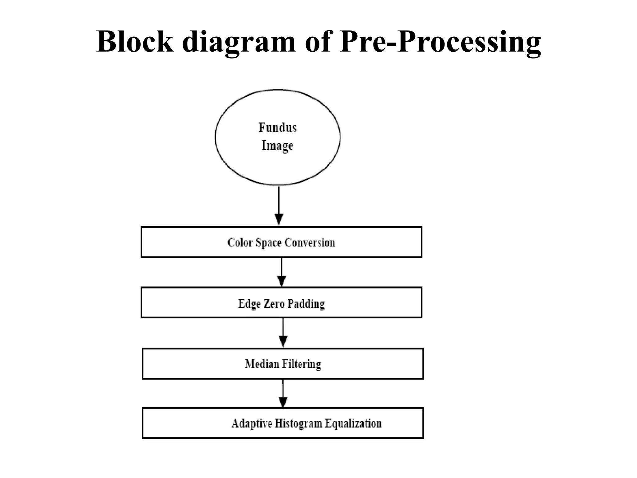
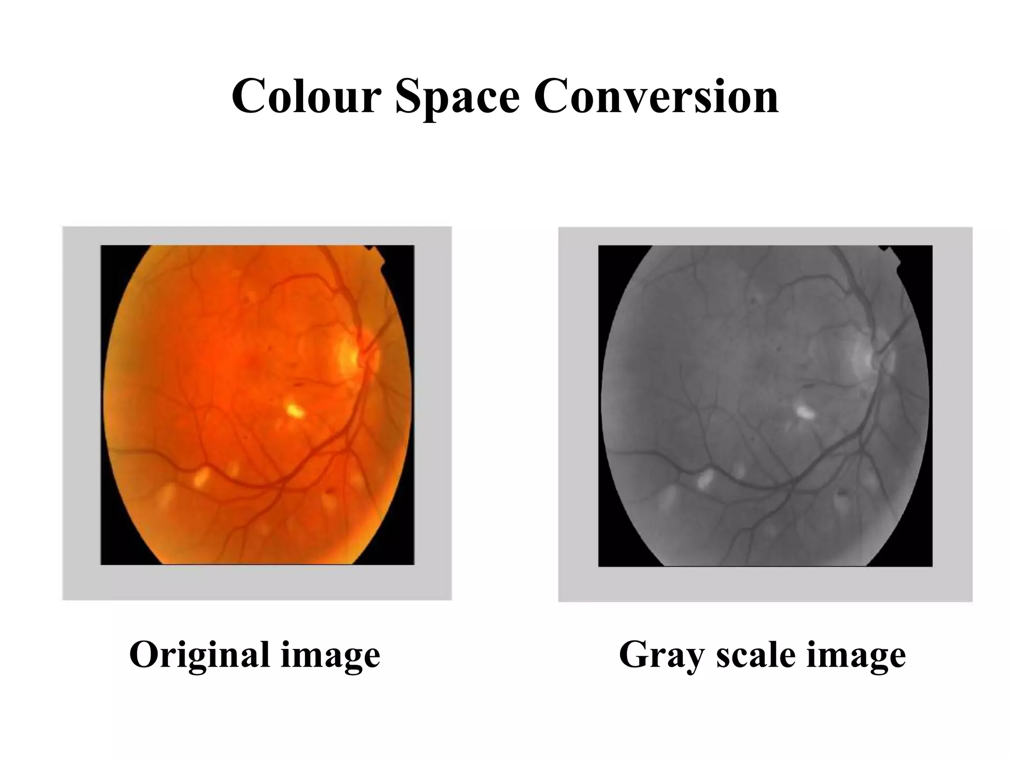
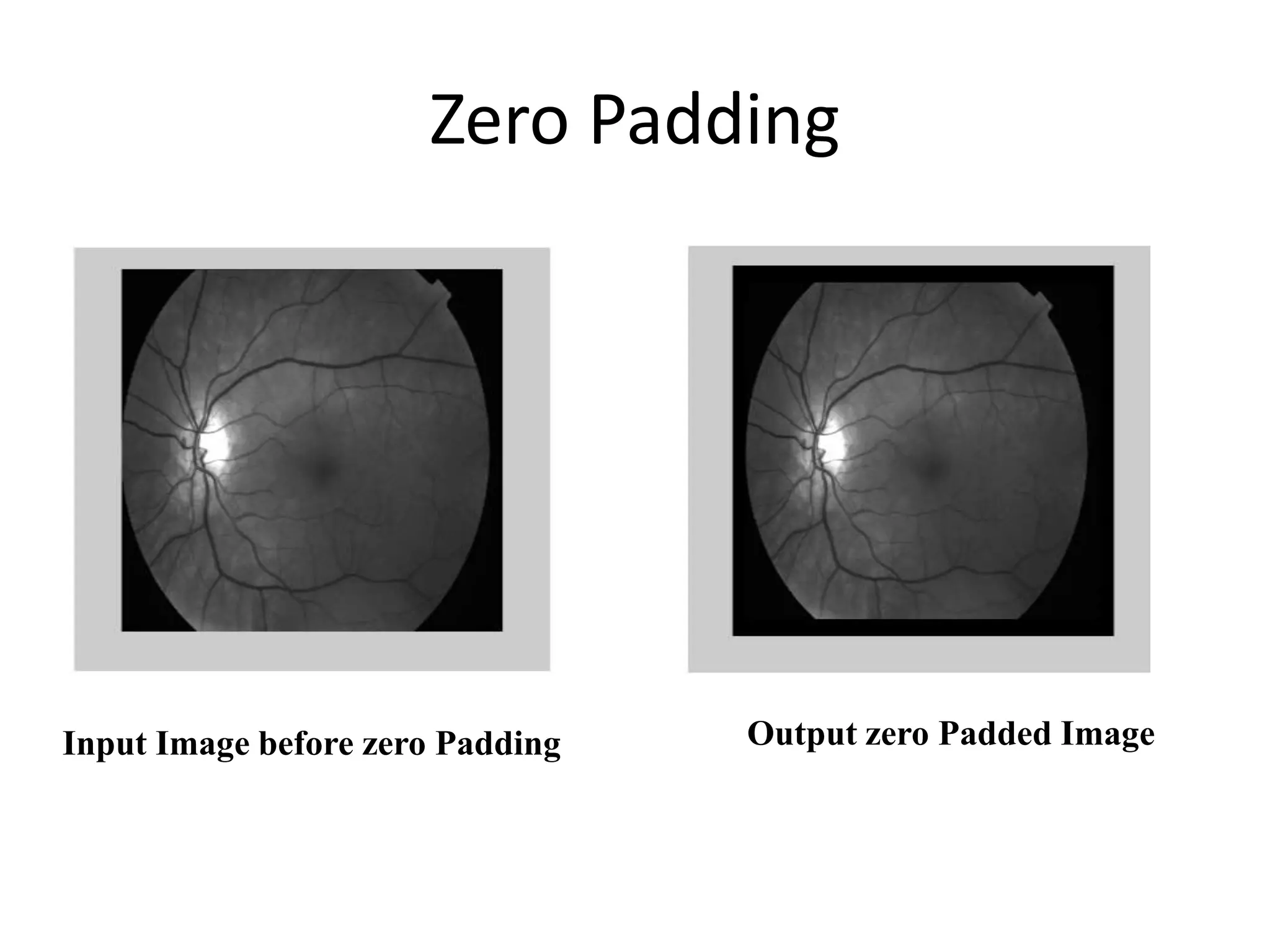
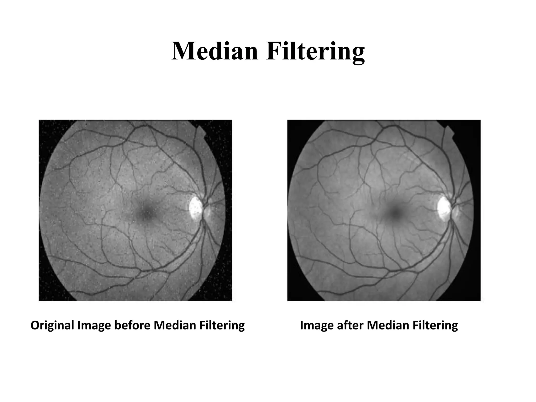
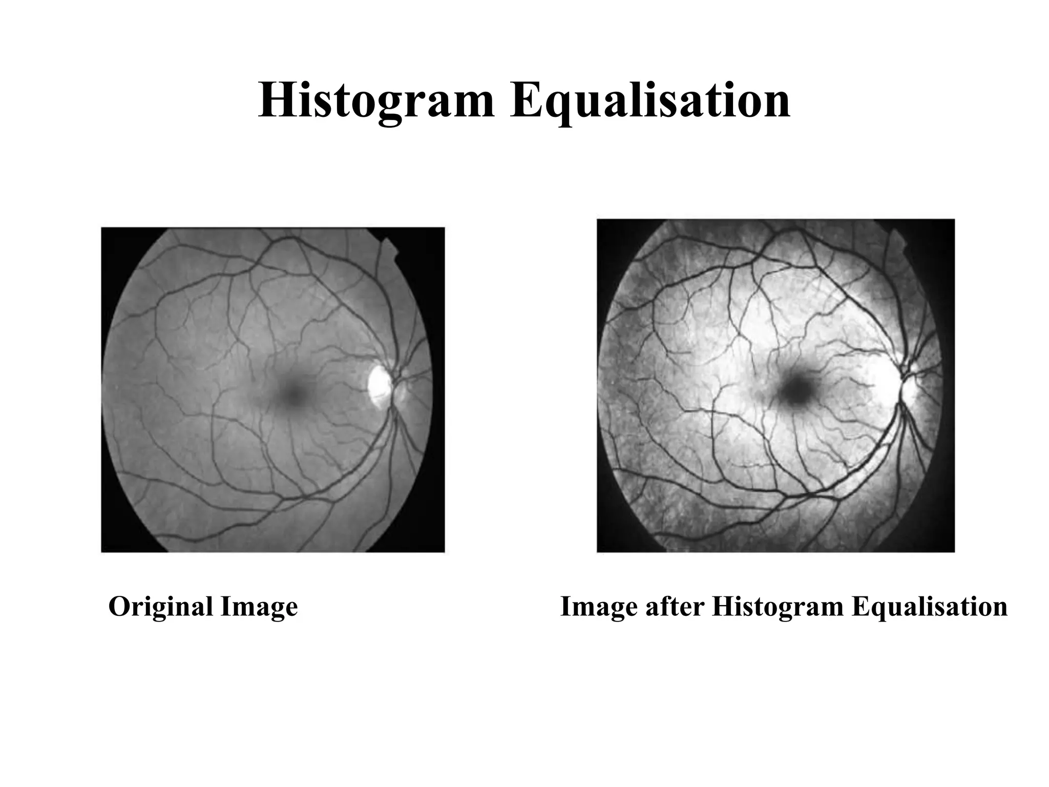
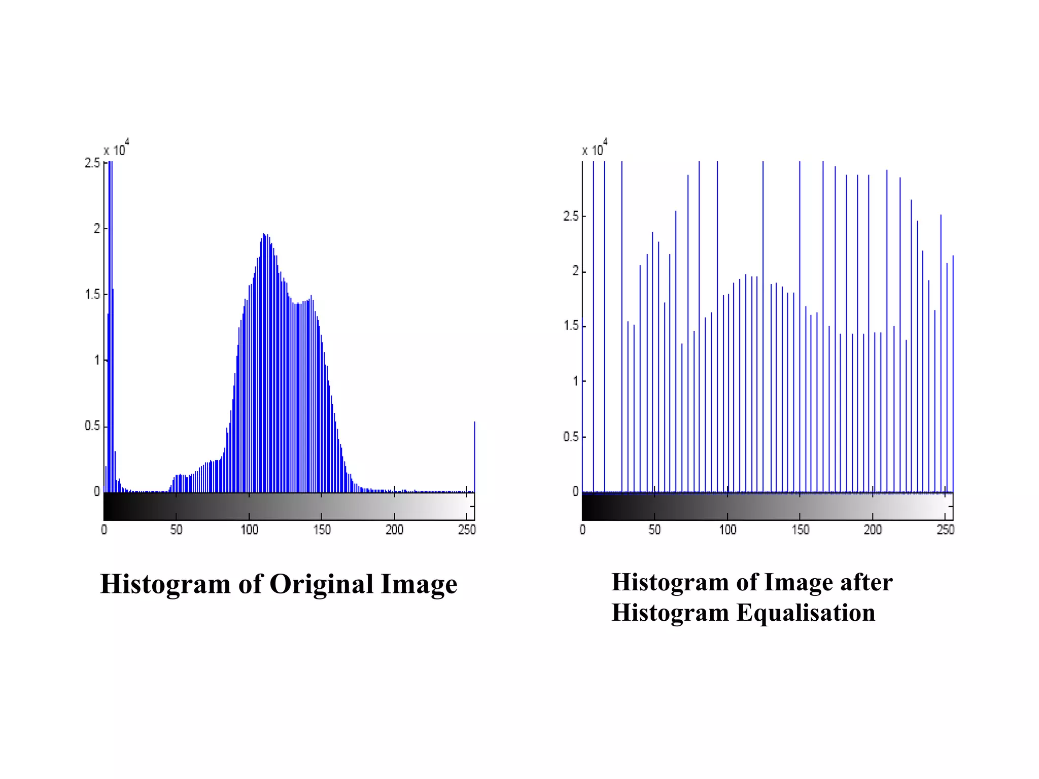
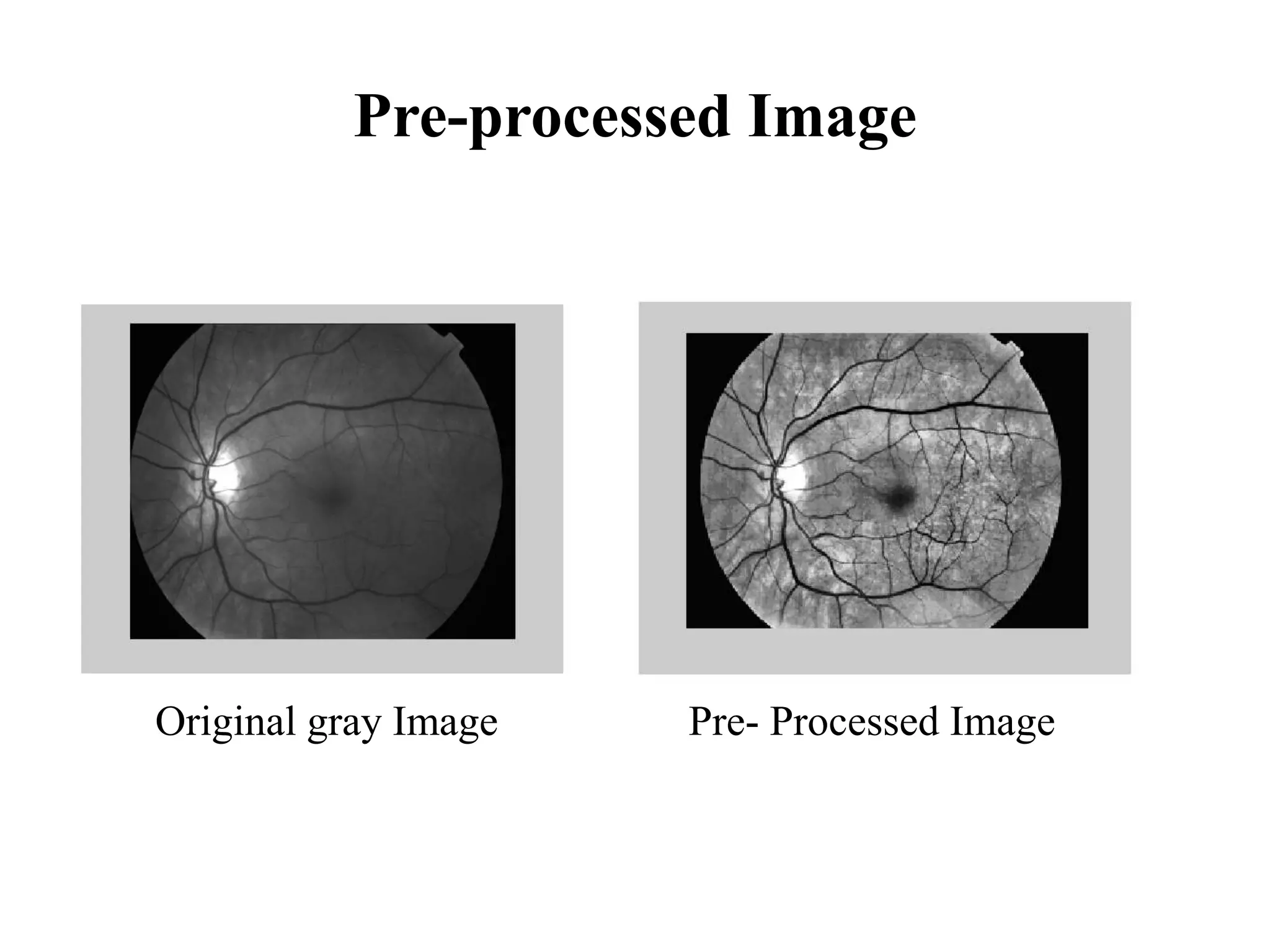
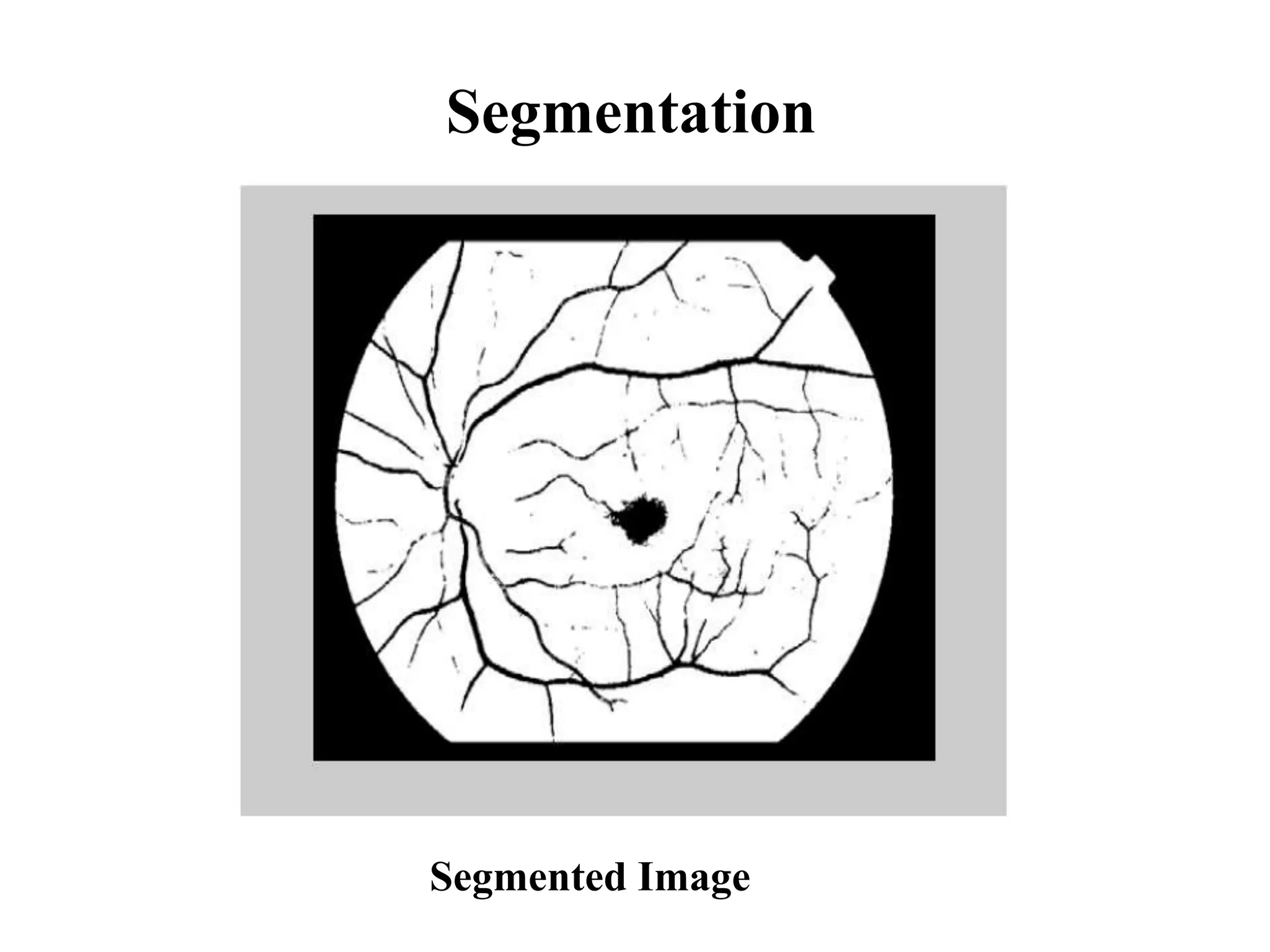
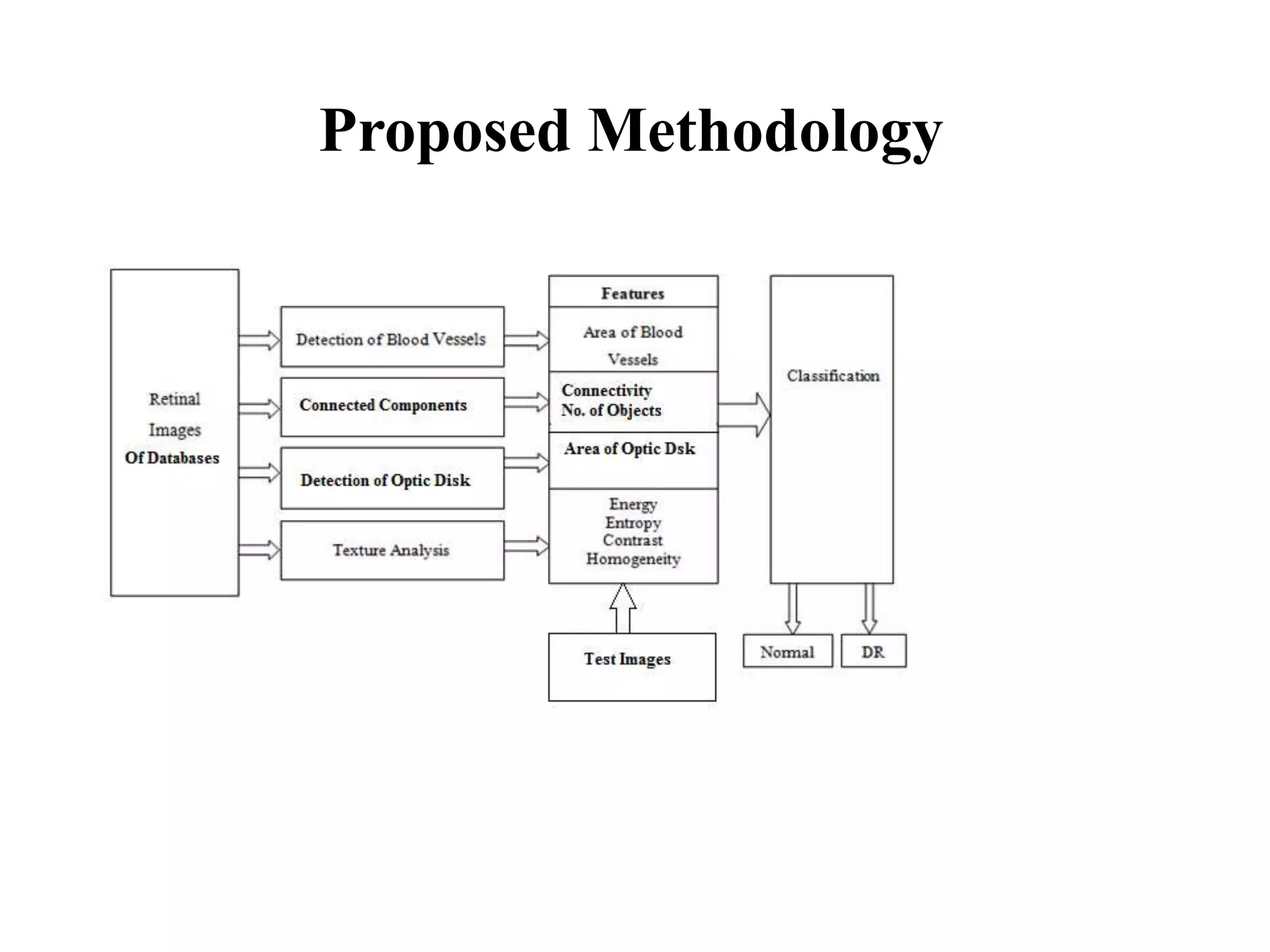
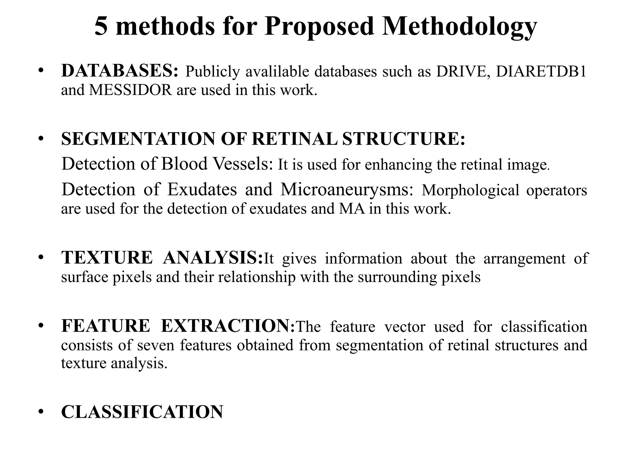
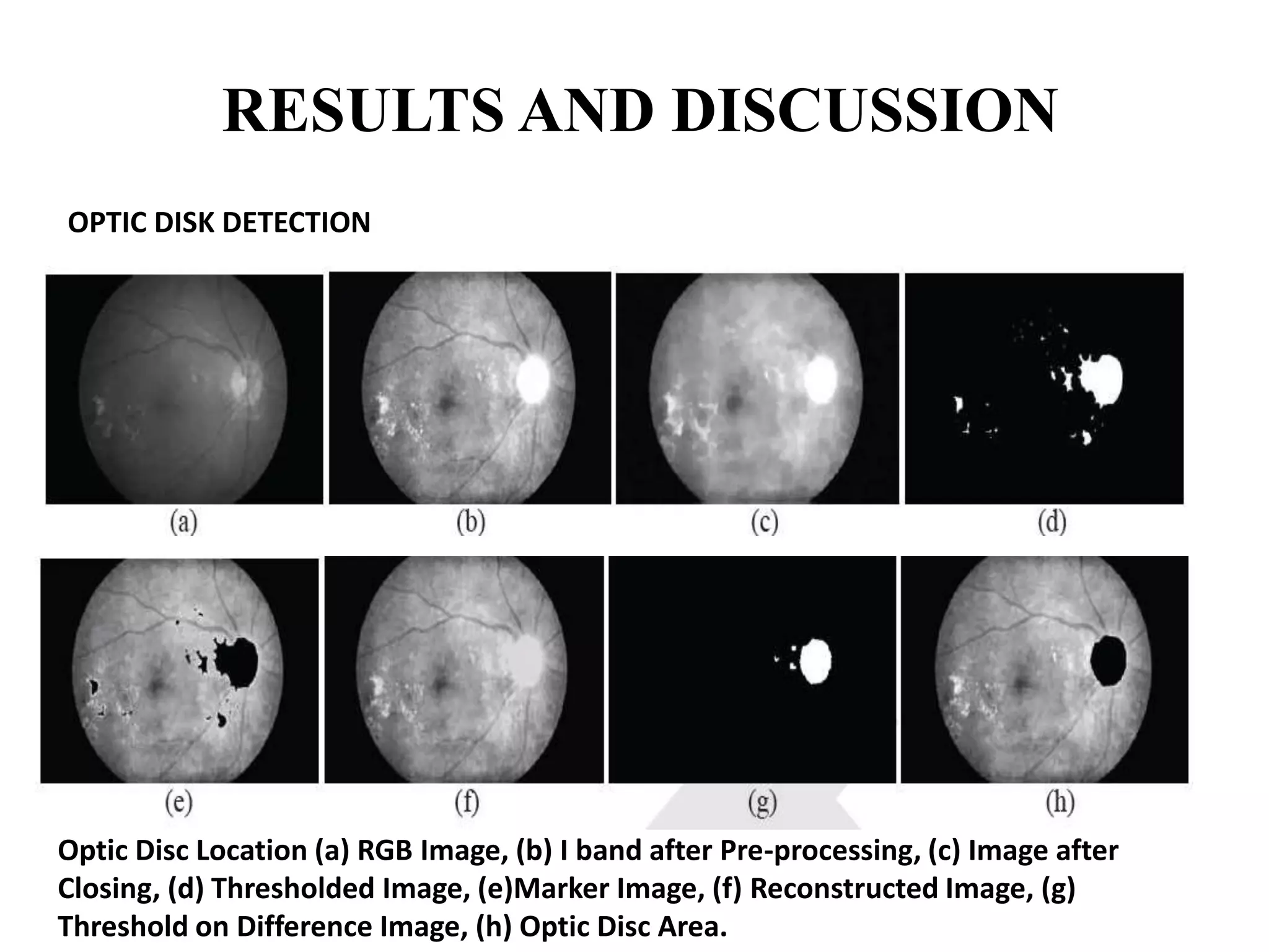
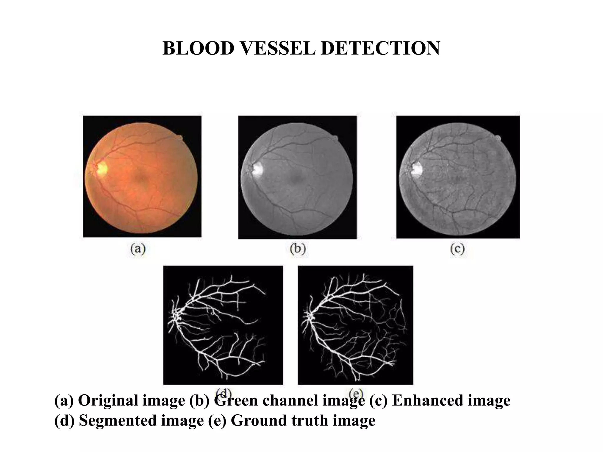
This document provides an overview of diabetic retinopathy diagnosis through the analysis of retinal images. It discusses the aims of identifying patients with different stages of diabetic retinopathy. The stages of diabetic retinopathy and associated symptoms are defined. Pre-processing steps like color conversion, filtering and segmentation are described. A proposed methodology includes blood vessel and lesion detection through morphological operations, texture analysis, feature extraction and classification. Results of optic disc detection, blood vessel segmentation and texture analysis are shown. The conclusion discusses developing more accurate detection techniques and extracting smaller blood vessels to aid in diagnosis.


![[IJET-V1I4P17] Authors: Fahimuddin. Shaik, Dr.Anil Kumar Sharma, Dr.Syed.Must...](https://cdn.slidesharecdn.com/ss_thumbnails/ijet-v1i4p17-151213125610-thumbnail.jpg?width=640&height=640&fit=bounds)