Download to read offline
![IOSR Journal of VLSI and Signal Processing (IOSR-JVSP)
Volume 5, Issue 1, Ver. II (Jan - Feb. 2015), PP 37-40
e-ISSN: 2319 – 4200, p-ISSN No. : 2319 – 4197
www.iosrjournals.org
DOI: 10.9790/4200-05123740 ww.iosrjournals.org 37 | Page
An Automated Systems for the Detection of Macular Ischemia
based-Diabetic Retinopathy
Komal Lende1
, Prof. W. V. Patil2
1,2, (Department of Electronics Engineering,G.H. Raisoni College of Engineering, Nagpur(M.S),India)
Abstract: The proposed methodology in this paper marks out application for automatic detection of eye
diseases called Macular Ischemia using image processing techniques. In semi urban and rural areas large
percentages of people suffer from various eye diseases. For diagnoses of various eye diseases, Image processing
technique is used. . Diseases occur in Macula from retinal images have a huge type of textures, shapes and at
times they are difficult to be recognised and identified by doctors. Thus we are trying to optimize and develop
such system which is based on smart image recognition/classification algorithms. This proposed system
provides accuracy, uniformity and speed in performance and a high credence coefficient in results interpreting.
Keywords: Macular Ischemia, diagnosis, textures, consistence
Keywords: consistence, diagnosis ,Macular Ischemia, optimize, textures.
I. Introduction
The Macula is a delicate and hypersensitive area responsible for high acuity vision, color vision and
central vision in the central part of the human eye retina. Macular Ischemia is a process where small blood
vessels close causing lack of blood supply to the macula. Macular diseases have a large variety of shapes,
textures and sometimes they are difficult to be determined and recognized by expert doctors. Many researchers
are working in the domain of image processing for early detection of diabetic retinopathy based Macular
Ischemia. The earliest forms of diabetic retinopathy are recognized by distortion and leaking of the blood
vessels in retina. Early detection of DR based Macular Ischemia is very important because it enables timely
treatment that can ease the burden of the disease on the patients by a sufficient quality of vision and prevention
severe loss and blindness. Our proposed technique gives information on retinal blood vessel morphology which
is adjustable for normal to expected blood vessel diameters which can detect minute and tiny blood vessel
abnormalities that distinguish the blood vessel and hence support for timely detection of Macular Ischemia [1]-
[5].
II. Literature Review
In [6] author present an approach using neural classification method and segmentation to separate out
arteries and veins. Using two-dimensional matched filters Blood vessels are segmented, which derived from
Gaussian functions, first the enhancement of retinal images had done using Homomorfic filter. The Match filter
is used to detect blood vessels. Here for each segment, feature vectors is used which is based on vessel profile
extraction. Blood vessel was cropped a Vessel Profile Based Feature Vectors had extracted then used for neural
classifier. The obtained features introduced as the vector input of a Multi-Layer Perceptron (MLP) for
classification of artery/vein[6].
According to [7] a novel technique is introduced to detect true vessels from retinal images. The
methodology proposed by author is a post processing step to vessel segmentation in images. The trouble
introduced in finding the optimal vessel forest from a graph with constraints on the vessel tree. All vessel trees
are consider while finding ideal forest; hence, the commonly approach is a severely aware of wrong vessels
coupling.
In [9] to detect the retinal disorder, author proposes one candidate extraction and two preprocessing
method in this literature. Based on various features like standard deviation, area, mean, entropy etc.
classification of various retinal abnormality is done. For effective screening of retinal abnormalities Adaptive
NeuroFuzzy Inference System (ANFIS) is an efficient tool is used which classify the retinal images as normal,
mild, severe depending on their severity.
From literature [10], a graph formulation was implemented with Dijkstra’s shortest path algorithm for
detection of central vein. Likewise, In [11] author introduced Dijkstra’s algorithm for determine vessels and
calculated their proposed method on a group of 15retinal images. Although, this method is incorrect for vessel
detection due to selecting proper vessel segments to meet at a branches or crossover needs information from
another neighboring vessel in retina. In paper[12] author proposed expert rules to solved vessels crossover. At](https://image.slidesharecdn.com/f05123740-151126060959-lva1-app6892/75/An-Automated-Systems-for-the-Detection-of-Macular-Ischemia-based-Diabetic-Retinopathy-1-2048.jpg)
![An automated systems for the detection of Macular Ischemia based-Diabetic Retinopathy
DOI: 10.9790/4200-05123740 ww.iosrjournals.org 38 | Page
these crossovers, rarely lineup segments to provide a vascular network. Although, they are failed determined
complete vessels.
In literature [13] a new technique of blood-vessel identification in human retinal images, using Quad
trees of edges and post-filtration of edges depends on the regional circulative hierarchical decomposition using a
fixed difference operator. The blood vessels presents as blackening of blurred edges and focal, indicated by an
determine intensity slope/gradient, which is distributed in rejection of false alarms to a huge intensity. This
technique gives information of blood vessel morphology in retina that progress to diameter of normal expected
blood vessel, which detect the neat and rare(fine) blood vessel abnormalities that identified blood vessels and
thus benefit for detection of Diabetic Retinopathy.
In [14] author proposed vessel enhancement approach for evolution microneurysms filter.
Microaneurysms filter detects microaneurysms and hemorrhages present in retinal images. These classification
output helped to integrate different types of diagnosis with the help of various ophthalmologist.
III. Proposed Work
In this methodology, the proposed system is focusing on increasing sensitivity, specificity and the
accuracy for the detection of any type of retinal disease present in the patient. The system can be used for
automatic screening of Macular Ischemia with an additive capability of grading the retinal image on basis of
abnormalities present in it and implementing it by using computer aided system to calculate the computational
processing time[5].
Fig 1: Proposed System Design
Retinal images acquired from database or retinal investigation camera, then two retinal images from
database are compare simultaneously for comparing parameters of two images and arrange them according to
severity.
The given retinal dataset images are converted into gray image by separate any one of RED, GREEN,
and BLUE. Because the retinal abnormalities have better visualization in the gray scale when compared to
others. Vessels are segmented in gray image using Morphological method. Normally retinal image green
channel give the better performance result.
Fig 2: GUI of Grey Image conversion](https://image.slidesharecdn.com/f05123740-151126060959-lva1-app6892/75/An-Automated-Systems-for-the-Detection-of-Macular-Ischemia-based-Diabetic-Retinopathy-2-2048.jpg)
![An automated systems for the detection of Macular Ischemia based-Diabetic Retinopathy
DOI: 10.9790/4200-05123740 ww.iosrjournals.org 40 | Page
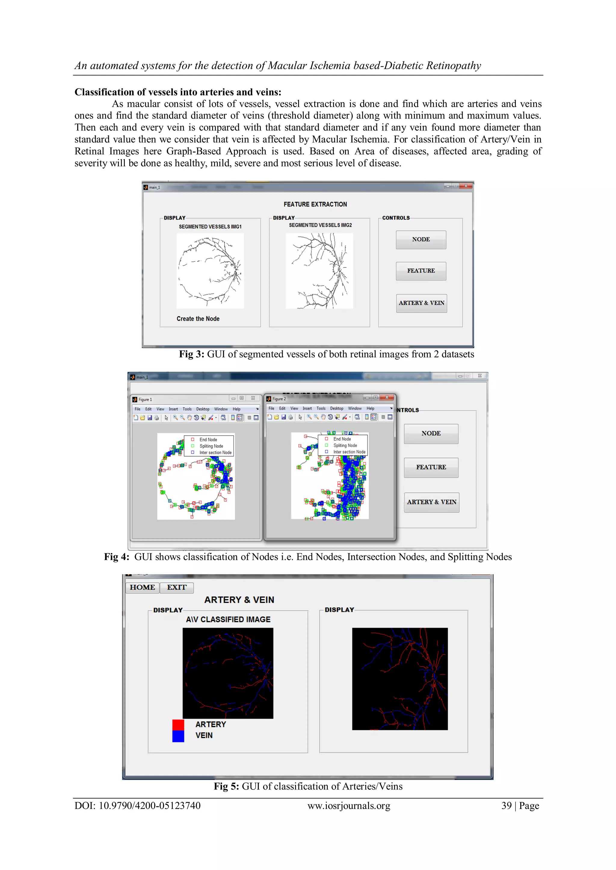
Classification of various stages of abnormality:
Using SVM algorithm, grading of severity is done and retinal abnormalities are classify as Normal,
Moderate and Severe class depending on their severity and save each images in separate database accordingly.
Images of different stages of diabetic retinopathy, which is collected from DRIVE database.
Figure 6: Images of Different Stages of DR based Mi (a) Normal Stage (b) Mild Stage (c) Severe Stage
IV. Conclusion
Design the ensemble based system using various preprocessing and feature extraction methods. Also
finds the severity of the Ischemia diseases extracted from the retinal images and detect the Macular Ischemia
caused by diabetes from the retinal images database by applying appropriate methodology to obtain high
accuracy, sensitivity and specificity.
References
[1]. ManjulaSri Rayudu, Vaibhav Jain, MM.Rao Kunda,,”Review of Image Processing Techniques for Automatic Detection of Eye
Diseases”, ©2012 IEEE
[2]. M. Usman Akram, Sundus Mujtaba, Anam Tariq, “Automated Drusen Segmentation in Fundus Images For Diagnosing Age related
Macular Degeneration”, ©2013 IEEE
[3]. M. Luculescu1
, “Macular Diseases Recognition Using Neural Networks”, The First National Conference of Medical Engineering –
COPTOMIM 2006, Braşov, 2006, pp. 155-158.
[4]. M. Luculescu, Researches on Biological Human Visual Structures Concerning the Diagnosis of Macular Diseases, PhD Thesis,
2007.
[5]. M. C. Luculescu1, S. Lache1 “Using Artificial Neural Networks in a ComputerAided Diagnosis System for Macular
Diseases”,@2008IEEE.
[6]. Jihene Malek, Rached Tourki, “Blood Vessels Extraction and Classification into Arteries and Veins in Retinal Images”,10th
International Multi-Conference on Systems,Signals & Devices (SSD),Hammamet, Tunisia, March 18-21, 2013.
[7]. Qiangfeng Peter Lau, Mong Li Lee, Wynne Hsu, Tien Yin Wong, “Simultaneously Identifying All True Vessels From Segmented
Retinal Images”, IEEE TRANSACTIONS ON BIOMEDICAL ENGINEERING, VOL. 60, NO. 7, JULY 2013
[8]. Maria Hameed, Muhammad Sharif, Syed Waqas Haider, Muhammad Iqbal, “Framework for Comparison of Classifiers for Medical
Image Segmentation with Transform and Moment based features”, Research Journal of Recent Sciences Vol. 2(6), 1-10, June
(2013)
[9]. T.Yamuna, S.Maheswari, “Detection of Abnormalities in Retinal Images”, 2013 IEEE International Conference on Emerging
Trends in Computing, Communication and Nanotechnology (ICECCN 2013)
[10]. H. Azegrouz, E. Trucco, “Max-min central vein detection in the retinal fundus images,” in Proc. IEEE Int. Conf. Image Process.,
Oct. 2006,pp. 1925–1928.
[11]. V. S. Joshi, M. K. Garvin, J. M. Reinhardt, M. D. Abramoff, “Automated method for identification and analysis of vascular tree
structures in retinal vessel network,” inProc. SPIEConf.Med. Imag., 2011, vol. 7963,no. 1, pp. 1–11.
[12]. B. Al-Diri, A. Hunter,D. Steel, M.Habib, “Automated analysis of the retinal vascular network connectivity,” Comput. Med. Imag.
Graph., vol. 34,no. 6, pp. 462–470, 2010.
[13]. Sumeet Dua, Naveen Kandiraju, Hilary W. Thompson, “Design and Implementation of a Unique Blood-vessel Detection Algorithm
towards Early Diagnosis of the Diabetic Retinopathy”, Proceedings of the International Conference on Information Technology:
Coding and Computing(ITCC’05)
[14]. R. Vidyasari1 , I. Sovani2, and T.L.R. Mengko3 ,H. Zakaria4 “Vessel Enhancement Algorithm in Digital Retinal Fundus
Microaneurysms Filter for Nonproliferative Diabetic Retinopathy Classification” International Conference on Communication,
Information Technology and Biomedical Engineering,2011.](https://image.slidesharecdn.com/f05123740-151126060959-lva1-app6892/75/An-Automated-Systems-for-the-Detection-of-Macular-Ischemia-based-Diabetic-Retinopathy-4-2048.jpg)

The document discusses an automated system for detecting macular ischemia in diabetic retinopathy using image processing techniques. It highlights the importance of early detection and classification of retinal abnormalities for timely treatment, employing algorithms for blood vessel segmentation and grading of disease severity. The proposed methodology aims to enhance sensitivity, specificity, and accuracy while processing retinal images for better diagnostics.