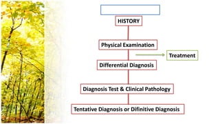
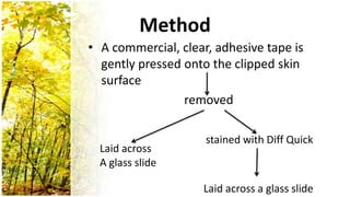
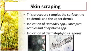
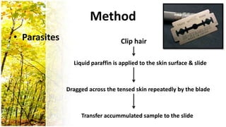
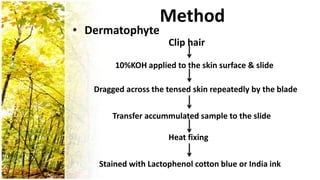
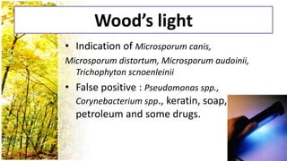
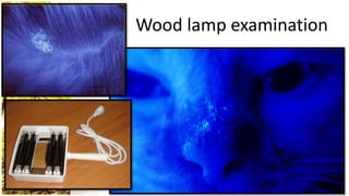
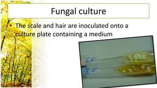
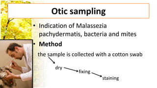
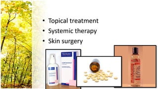
Dermatological History and Examination
        Natapol Pumipuntu,DVM
Contents
•
•
    - History taking
    - Physical Examination
    - Differential Diagnosis
    - Diagnosis Test & Clinical pathology
    - Tentative Diagnosis & Definitive Diagnosis
•
•   Parasitic diseases
•   Bacterial skin diseases
•   Viral, Protozoal and Rickettsial skin disease
•   Fungal skin diseases
•   Hypersensitivities
•   Autoimmune dermatoses
•   Endocrine dermatoses
•   Nutritional dermatoses
•   Neoplastic dermatoses
•   Psychogenic dermatoses
•   Congenital and hereditary dermatoses
•   Miscellaneous dermatoses
Parasitic diseases
•   Demodex canis
•   Sarcoptes scabiei
•   Otodectes cynotis
•   Cheyletiella spp.
•   Tick
•   Flea
•   Fly
Demodex canis
Sarcoptic mange
Otodectes cynotis
Bacterial skin diseases
• Staphylococcus intermedius
    coagulase-positive & Gram-positive coccus
Bacterial skin diseases cont
• Surface infections
    - pyotraumatimatic dermatitis
   Hotspot
  - fold pyoderma
Bacterial skin diseases cont
• Superficial infections
    - impetigo
  - Superficial folliculitis
Bacterial skin diseases cont
• Deep infections
    - Deep folliculitis
  - Deep pyoderma
  - cellulitis
  - mycobacterial infections
Viral, protozoal and
     Ricketsettsial skin diseases
• Viral
  : canine distemper virus
    canine viral papillomatosis etc.
• Protozoal
  : canine leishmaniasis etc.
• Bacteria
  : Ehrlichia canis etc.
Fungal skin diseases
• Fungi
  - Microsporum canis
  - Microsporum gypseam
  - Trichophyton mentagrophytes
• Yeast
  - Malassezia patchydermatis
Hypersensitivites
•   Allergic contact dermatitis
•   Inhalant allergy
•   Adverse reaction to food
•   Drug eruption
    etc.
Autoimmune dermatoses
•   Lupus erythematosus
•   Pemphigus foliaceus
•   Pemphigoid syndrome
•   Alopecia areata
•   Discoid lupus erythematosus
•   Pemphigus vulgaris
Endocrine dermatoses
 •   Hypothyroidisim
 •   Hyperadrenocorticism
 •   Gonadal hormones
 •   Growth hormone
       etc.
Nutritional dermatoses
 •   Protein/calorie deficiency
 •   Essential fatty acid deficiency
 •   Vitamin A deficiency
 •   Vitamin E deficiency
 •   Zinc deficiency
Neoplastic dermatoses
 • Tumors of epithelial origin
   : Papilloma, Basal cell tumor,
     Sebaceous gland tumor,
     Squamous cell carcinoma etc.
Neoplastic dermatoses cont
 • Tumors of mesenchymal origin
   : Fibroma, Fibrosarcoma, Lipoma,
     Liposacoma, Mast cell tumor etc.
Neoplastic dermatoses cont
 • Tumors of lymphohistiocytic origin
   : Histiocytosis, cutaneous lymphoma,
     Transmissible venereal tumor etc.
Psychogenic dermatoses
• Lick dermatitis
Miscellaneous dermatoses
•   Congenital dermatoses
•   Hereditary dermatoses
•   Keratinization disorder
•   Metabolic dermatitis
HISTORY


           Physical Examination
                                      Treatment
          Differential Diagnosis


   Diagnosis Test & Clinical Pathology


Tentative Diagnosis or Difinitive Diagnosis
History Taking
•
•

    CHIEF COMPLAINT

•
•   /
•

•           …
•

•
•               /
    /
•       /
•
•   /

•               …
        /
•           …
History
•   Breed
•   Age
•   Sex
•   Environment
•   Diet
•   Health status
•   History of disease
•   Response to previous treatments
Physical Examination
•
Physical Examination
•
      - Bilaterally symmetric lesion
    - Asymmetric lesion
Physical Examination
•
      - Single
    - Linear
    - Annular
LINEAR LESION
                    ANNULAR LESION


  ARCIFORM LESION
Physical Examination
•
      - Primary Lesions
    - Secondary Lesions
<<Primary Lesions>>
• Macules & Patches
  - Discoloration of the skin
  - macules  less than 1 cm in diameter
  - patches  larger than 1 cm in diameter
***The change is colour may result from pigment
   loss or excess, erythema or haemorrhage.
Papules & Plaques
• A circumscribed, solid elevation of the skin
• Papules  less than 1 cm in diameter often
  pink and red
• Plaques  larger than 1 cm in diameter
Pustules
• Circumscribed elevations of the superficial
  layers of the epidemis
• Bacterial infection
• The infiltrate will contain neutrophils,
  bacteria, debris and perhaps a few free
  keratinocytes
PUSTULE


**The most important cause is
Staphylococcus intermedius
Wheals
• More or less well-defined
  elevations of the skin due to edema
• Round, oval or plaque-like
• Disappears within minutes or hours
**angioedema


        *Localised mast cell
        degranulation within
        the skin
Vesicles & Bullae
• A well-demarcated elevation of the
  superficial layers of the skin
• Vesicles  less than 1 cm in diameter
• Bullae  lager than 1 cm in diameter
Nodules
• Circumscribed elevations of the
  skin that are larger than the 1 cm in
  diameter deeper skin layers
Tumors
• Masses of neoplastic origin
  whether benign or malignant
• May extend into the deeper
  subcutaneous tissue
Cysts
• Cavities within the skin that are
  lined by epithelium
<<Secondary Lesions>>
• Comedones
  - dilated hair follicle which contains
    a pigmented impaction of lipid
    and keratinaceous debris
  “BLACK HEAD”
Scale
• Associated with the shedding of large rafts
  of keratinocytes accumulation of loose
  cornified fragment of the epidermis
Crust
• Composed of dried exudate mixtd
  with debris on the skin surface
   serum/blood/pus
Exocoriation
• Areas of epidermal damage
   caused by scratching, biting or
  rubbing
Ulceration
• The integrity of the basal layer
   basement membrane is
  destroyed
• The dermis is involved and healing
  is often by scar formation
Lichenification
• A thickening of the epidermis with an
  accompanying hyperpigmentation in
  most instances elephant skin like
Hyperpigmentation
• Usually a consequence of melanin
  excess
          Hypopigmentation
• Loss of pigment
Scar
• Occurs when the basal layers of the
  epidermis have been breached and
  the underlying dermis is damaged
&


Problem list ?
Problem list?
•   Pruritus
•   Focal / generalized/symmetrical alopecia
•   Crust
•   Hyperpigmentation
•   Scale
•   Nodule
•   Patch
•   Plaque
Diagnostic Test
• Trichogram
Identification of fur mites, adherent egg
  cases, fractured ends to the hair shaft
** Trichograms are not a reliable method of diagnosing, or ruling
   out, ectoparasite infections
Method
• Remove a small tuft of hair with fingers
  or forceps

• Laid onto trasparent adhesive tape

• Laid onto a glass slide

• Microscopic examination
Adhesive tape stripping
• Allows direct sampling of the coat or skin
  surface&hair
• Observes squamous morphology, ectoparasites and
  micro-organisms
Method
• A commercial, clear, adhesive tape is
  gently pressed onto the clipped skin
  surface
                 removed

                     stained with Diff Quick
  Laid across
  A glass slide

                    Laid across a glass slide
Skin scraping
• This procedure samples the surface, the
  epidermis and the upper dermis
• Indication of Demodex spp., Sarcoptes
  scabiei and Cheyletiella spp.
• Indication of dermatophytosis spores
Method
• Parasites                        Clip hair

                Liquid paraffin is applied to the skin surface & slide


              Dragged across the tensed skin repeatedly by the blade


                   Transfer accummulated sample to the slide
Method
• Dermatophyte
                      Clip hair

        10%KOH applied to the skin surface & slide

   Dragged across the tensed skin repeatedly by the blade

       Transfer accummulated sample to the slide

                      Heat fixing

    Stained with Lactophenol cotton blue or India ink
Wood’s light
• Indication of Microsporum canis,
Microsporum distortum, Microsporum audoinii,
  Trichophyton scnoenleinii
• False positive : Pseudomonas spp.,
  Corynebacterium spp., keratin, soap,
  petroleum and some drugs.
Wood lamp examination
Fungal culture
• The scale and hair are inoculated onto a
  culture plate containing a medium
Otic sampling
• Indication of Malassezia
  pachydermatis, bacteria and mites
• Method
 the sample is collected with a cotton swab

        dry
                 fixing
                          staining
Impression cytology
• Used to sample cells from the surface of
  erosions and ulcers or from the orifices of
  discharging fistulae
• Medthod
  Pressing a cleaned glass slide against the lesion

                        dry

              Staining with Diff Quick
Aspiration cytology
• Used to sample cells from within lymph
  nodes, nodules and neplasms
• Method       Needle aspiration

                      smear

                        dry

       Staining with Diff Quick or special stain
Bacteria Culture
• Indication of aerobic bacteria
Method
the sample is collected with a cotton swab /
  aspiration
Intradermal skin testing
 • Demonstrate the presence of allergen-
   specific IgG on the mast cells at the site of
   the intradermal injection
 • Method
Punch biopsy
• Obtain samples for histopathological
  examination
• Method        Select site

                 Local anesthesia

          Push biopsy punch through the skin

       Remove sample & placed into 10% formaline
Treatment
•
    -
    -
    -
• Topical treatment
• Systemic therapy
• Skin surgery
THANK YOU FOR YOUR ATTENTION
                         -

Dermatological history and examination

  • 1.
    Dermatological History andExamination Natapol Pumipuntu,DVM
  • 2.
    Contents • • - History taking - Physical Examination - Differential Diagnosis - Diagnosis Test & Clinical pathology - Tentative Diagnosis & Definitive Diagnosis •
  • 3.
    Parasitic diseases • Bacterial skin diseases • Viral, Protozoal and Rickettsial skin disease • Fungal skin diseases • Hypersensitivities • Autoimmune dermatoses
  • 4.
    Endocrine dermatoses • Nutritional dermatoses • Neoplastic dermatoses • Psychogenic dermatoses • Congenital and hereditary dermatoses • Miscellaneous dermatoses
  • 5.
    Parasitic diseases • Demodex canis • Sarcoptes scabiei • Otodectes cynotis • Cheyletiella spp. • Tick • Flea • Fly
  • 6.
  • 7.
  • 8.
  • 9.
    Bacterial skin diseases •Staphylococcus intermedius coagulase-positive & Gram-positive coccus
  • 10.
    Bacterial skin diseasescont • Surface infections - pyotraumatimatic dermatitis Hotspot - fold pyoderma
  • 11.
    Bacterial skin diseasescont • Superficial infections - impetigo - Superficial folliculitis
  • 12.
    Bacterial skin diseasescont • Deep infections - Deep folliculitis - Deep pyoderma - cellulitis - mycobacterial infections
  • 13.
    Viral, protozoal and Ricketsettsial skin diseases • Viral : canine distemper virus canine viral papillomatosis etc. • Protozoal : canine leishmaniasis etc. • Bacteria : Ehrlichia canis etc.
  • 14.
    Fungal skin diseases •Fungi - Microsporum canis - Microsporum gypseam - Trichophyton mentagrophytes • Yeast - Malassezia patchydermatis
  • 15.
    Hypersensitivites • Allergic contact dermatitis • Inhalant allergy • Adverse reaction to food • Drug eruption etc.
  • 16.
    Autoimmune dermatoses • Lupus erythematosus • Pemphigus foliaceus • Pemphigoid syndrome • Alopecia areata • Discoid lupus erythematosus • Pemphigus vulgaris
  • 17.
    Endocrine dermatoses • Hypothyroidisim • Hyperadrenocorticism • Gonadal hormones • Growth hormone etc.
  • 18.
    Nutritional dermatoses • Protein/calorie deficiency • Essential fatty acid deficiency • Vitamin A deficiency • Vitamin E deficiency • Zinc deficiency
  • 19.
    Neoplastic dermatoses •Tumors of epithelial origin : Papilloma, Basal cell tumor, Sebaceous gland tumor, Squamous cell carcinoma etc.
  • 20.
    Neoplastic dermatoses cont • Tumors of mesenchymal origin : Fibroma, Fibrosarcoma, Lipoma, Liposacoma, Mast cell tumor etc.
  • 21.
    Neoplastic dermatoses cont • Tumors of lymphohistiocytic origin : Histiocytosis, cutaneous lymphoma, Transmissible venereal tumor etc.
  • 22.
  • 23.
    Miscellaneous dermatoses • Congenital dermatoses • Hereditary dermatoses • Keratinization disorder • Metabolic dermatitis
  • 24.
    HISTORY Physical Examination Treatment Differential Diagnosis Diagnosis Test & Clinical Pathology Tentative Diagnosis or Difinitive Diagnosis
  • 25.
  • 26.
    • • CHIEF COMPLAINT •
  • 27.
    / • • … • • • / / • /
  • 28.
    • • / • … / • …
  • 29.
    History • Breed • Age • Sex • Environment • Diet • Health status • History of disease • Response to previous treatments
  • 30.
  • 31.
  • 32.
    Physical Examination • - Bilaterally symmetric lesion - Asymmetric lesion
  • 33.
    Physical Examination • - Single - Linear - Annular
  • 34.
    LINEAR LESION ANNULAR LESION ARCIFORM LESION
  • 35.
    Physical Examination • - Primary Lesions - Secondary Lesions
  • 36.
    <<Primary Lesions>> • Macules& Patches - Discoloration of the skin - macules  less than 1 cm in diameter - patches  larger than 1 cm in diameter
  • 37.
    ***The change iscolour may result from pigment loss or excess, erythema or haemorrhage.
  • 38.
    Papules & Plaques •A circumscribed, solid elevation of the skin • Papules  less than 1 cm in diameter often pink and red • Plaques  larger than 1 cm in diameter
  • 40.
    Pustules • Circumscribed elevationsof the superficial layers of the epidemis • Bacterial infection • The infiltrate will contain neutrophils, bacteria, debris and perhaps a few free keratinocytes
  • 41.
    PUSTULE **The most importantcause is Staphylococcus intermedius
  • 42.
    Wheals • More orless well-defined elevations of the skin due to edema • Round, oval or plaque-like • Disappears within minutes or hours **angioedema *Localised mast cell degranulation within the skin
  • 43.
    Vesicles & Bullae •A well-demarcated elevation of the superficial layers of the skin • Vesicles  less than 1 cm in diameter • Bullae  lager than 1 cm in diameter
  • 45.
    Nodules • Circumscribed elevationsof the skin that are larger than the 1 cm in diameter deeper skin layers
  • 46.
    Tumors • Masses ofneoplastic origin whether benign or malignant • May extend into the deeper subcutaneous tissue
  • 47.
    Cysts • Cavities withinthe skin that are lined by epithelium
  • 48.
    <<Secondary Lesions>> • Comedones - dilated hair follicle which contains a pigmented impaction of lipid and keratinaceous debris “BLACK HEAD”
  • 49.
    Scale • Associated withthe shedding of large rafts of keratinocytes accumulation of loose cornified fragment of the epidermis
  • 50.
    Crust • Composed ofdried exudate mixtd with debris on the skin surface serum/blood/pus
  • 51.
    Exocoriation • Areas ofepidermal damage caused by scratching, biting or rubbing
  • 52.
    Ulceration • The integrityof the basal layer basement membrane is destroyed • The dermis is involved and healing is often by scar formation
  • 53.
    Lichenification • A thickeningof the epidermis with an accompanying hyperpigmentation in most instances elephant skin like
  • 54.
    Hyperpigmentation • Usually aconsequence of melanin excess Hypopigmentation • Loss of pigment
  • 55.
    Scar • Occurs whenthe basal layers of the epidermis have been breached and the underlying dermis is damaged
  • 56.
  • 57.
    Problem list? • Pruritus • Focal / generalized/symmetrical alopecia • Crust • Hyperpigmentation • Scale • Nodule • Patch • Plaque
  • 58.
    Diagnostic Test • Trichogram Identificationof fur mites, adherent egg cases, fractured ends to the hair shaft ** Trichograms are not a reliable method of diagnosing, or ruling out, ectoparasite infections
  • 59.
    Method • Remove asmall tuft of hair with fingers or forceps • Laid onto trasparent adhesive tape • Laid onto a glass slide • Microscopic examination
  • 60.
    Adhesive tape stripping •Allows direct sampling of the coat or skin surface&hair • Observes squamous morphology, ectoparasites and micro-organisms
  • 61.
    Method • A commercial,clear, adhesive tape is gently pressed onto the clipped skin surface removed stained with Diff Quick Laid across A glass slide Laid across a glass slide
  • 62.
    Skin scraping • Thisprocedure samples the surface, the epidermis and the upper dermis • Indication of Demodex spp., Sarcoptes scabiei and Cheyletiella spp. • Indication of dermatophytosis spores
  • 63.
    Method • Parasites Clip hair Liquid paraffin is applied to the skin surface & slide Dragged across the tensed skin repeatedly by the blade Transfer accummulated sample to the slide
  • 64.
    Method • Dermatophyte Clip hair 10%KOH applied to the skin surface & slide Dragged across the tensed skin repeatedly by the blade Transfer accummulated sample to the slide Heat fixing Stained with Lactophenol cotton blue or India ink
  • 65.
    Wood’s light • Indicationof Microsporum canis, Microsporum distortum, Microsporum audoinii, Trichophyton scnoenleinii • False positive : Pseudomonas spp., Corynebacterium spp., keratin, soap, petroleum and some drugs.
  • 66.
  • 67.
    Fungal culture • Thescale and hair are inoculated onto a culture plate containing a medium
  • 68.
    Otic sampling • Indicationof Malassezia pachydermatis, bacteria and mites • Method the sample is collected with a cotton swab dry fixing staining
  • 69.
    Impression cytology • Usedto sample cells from the surface of erosions and ulcers or from the orifices of discharging fistulae • Medthod Pressing a cleaned glass slide against the lesion dry Staining with Diff Quick
  • 70.
    Aspiration cytology • Usedto sample cells from within lymph nodes, nodules and neplasms • Method Needle aspiration smear dry Staining with Diff Quick or special stain
  • 71.
    Bacteria Culture • Indicationof aerobic bacteria Method the sample is collected with a cotton swab / aspiration
  • 72.
    Intradermal skin testing • Demonstrate the presence of allergen- specific IgG on the mast cells at the site of the intradermal injection • Method
  • 73.
    Punch biopsy • Obtainsamples for histopathological examination • Method Select site Local anesthesia Push biopsy punch through the skin Remove sample & placed into 10% formaline
  • 74.
  • 75.
    • Topical treatment •Systemic therapy • Skin surgery
  • 76.
    THANK YOU FORYOUR ATTENTION -