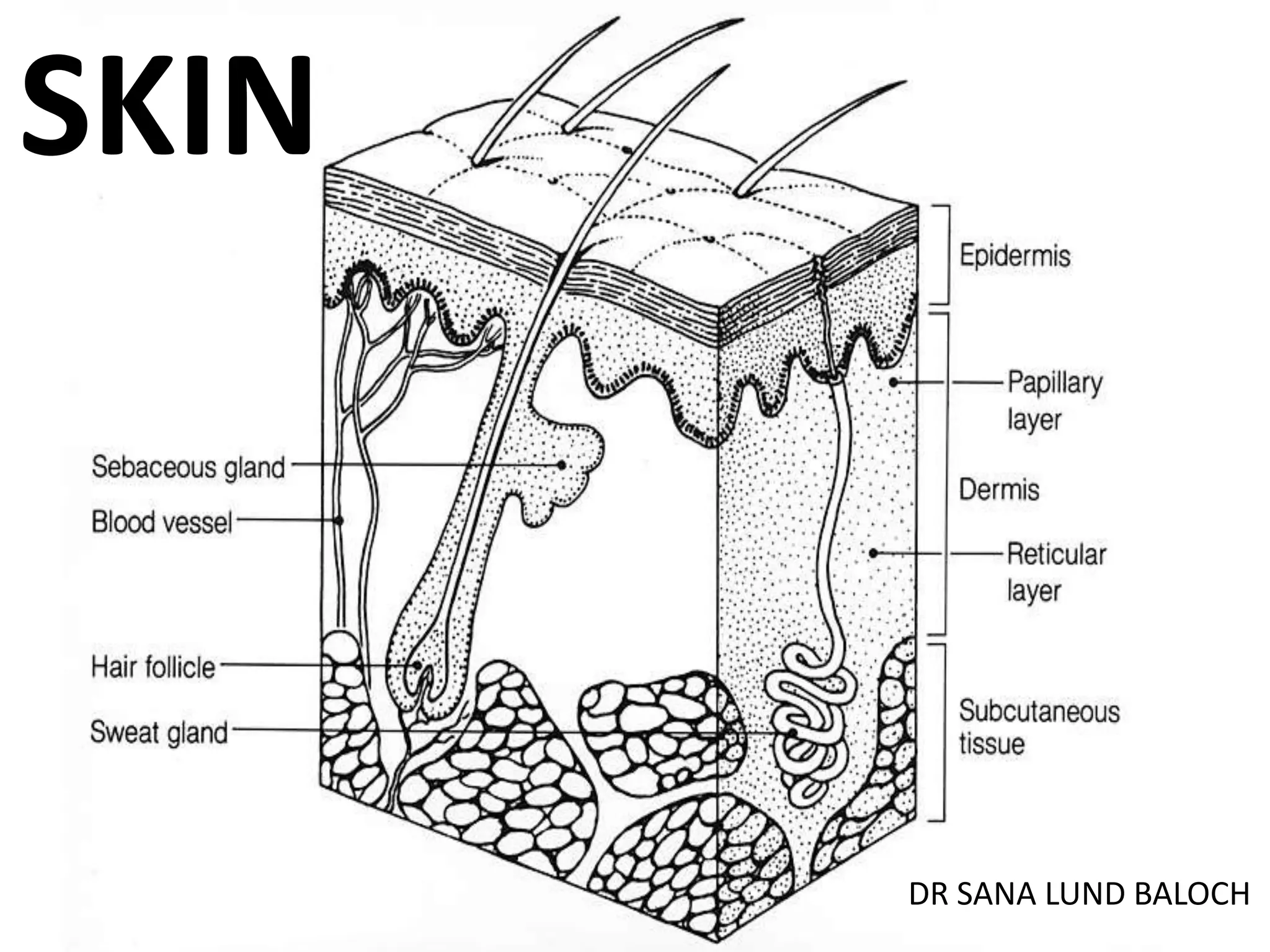
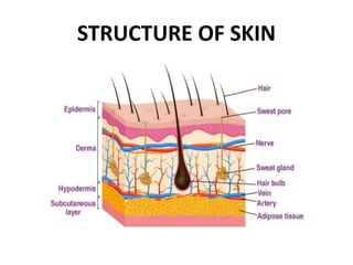
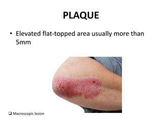
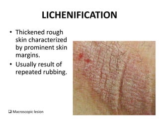
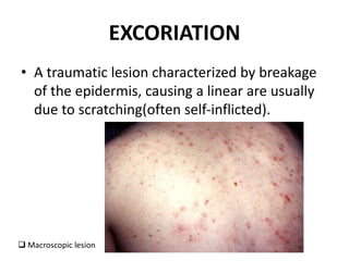
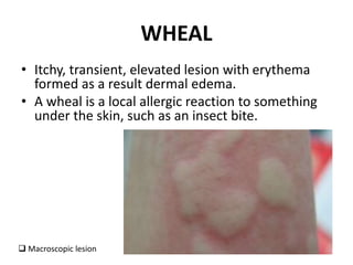
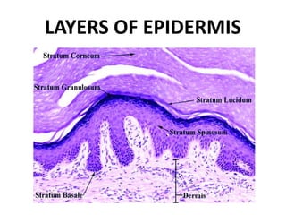
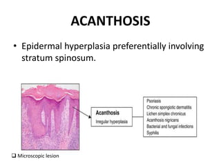
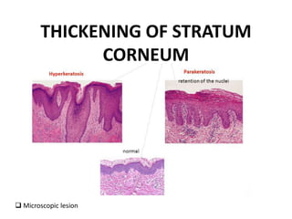
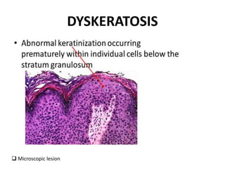
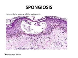
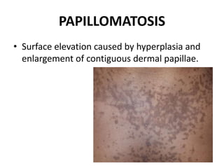
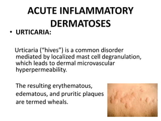
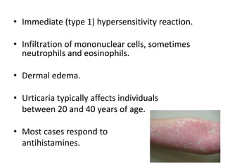
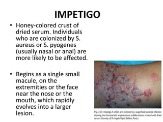
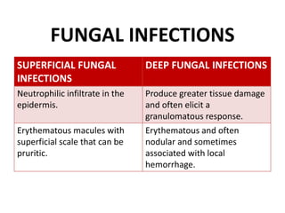
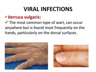
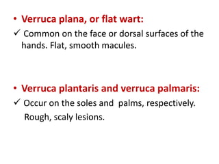
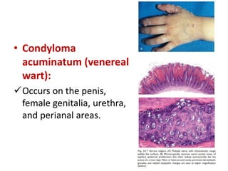
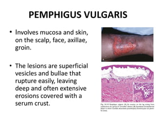
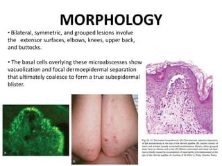
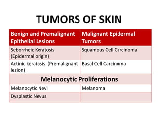
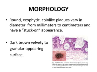
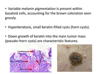
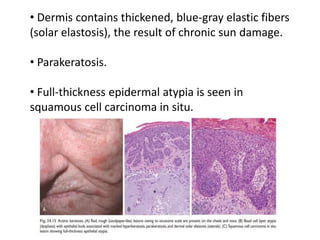
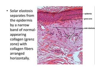
2. INTRODUCTION
• Cutaneous membrane covering external
surface of body.
• Sensory organ the largest organ in the body
covering a total area of about 1.8 square
meters.
• Skin, when stretched out is 2 square meters.
3. • Skin accounts for around 15% of your body
weight.
• Integumentary system composed of skin, hair,
nail and glands.
5. APPENDAGES OF SKIN
• Hair follicles and hair
• Sweat and sebaceous glands
• Nails
6. FUNCTIONS OF SKIN
• Skin controls heat regulation by responding to
changes in temperature with vasoconstriction
and vasodilatation.
• Helps to maintain homeostasis through fluid
and electrolyte balance.
• Sebum and sweat are secreted by skin and
lubricates skin surface.
• Endogenous synthesis of vit: D, occurs in
epidermis.
10. FLAT LESSION
• MACULE:
Flat circumscribed area distinguished from
surrounding skin by coloration (hyperpigmented
skin patch). Macules are 5mm in diameter or less.
Macroscopic lesion
11. PUSTULE
• Discrete, pus filled raised area.
• Is not always infected (e.g. sterile pustule)
Macroscopic lesion
14. EXCORIATION
• A traumatic lesion characterized by breakage
of the epidermis, causing a linear are usually
due to scratching(often self-inflicted).
Macroscopic lesion
15. WHEAL
• Itchy, transient, elevated lesion with erythema
formed as a result dermal edema.
• A wheal is a local allergic reaction to something
under the skin, such as an insect bite.
Macroscopic lesion
22. ACUTE INFLAMMATORY
DERMATOSES
• URTICARIA:
Urticaria (“hives”) is a common disorder
mediated by localized mast cell degranulation,
which leads to dermal microvascular
hyperpermeability.
The resulting erythematous,
edematous, and pruritic plaques
are termed wheals.
23. • Immediate (type 1) hypersensitivity reaction.
• Infiltration of mononuclear cells, sometimes
neutrophils and eosinophils.
• Dermal edema.
• Urticaria typically affects individuals
between 20 and 40 years of age.
• Most cases respond to
antihistamines.
24. ACUTE ECZEMATOUS
DERMATITIS
• Erythematous papules, with overlying vesicles,
which ooze and become crusted.
• Pruritus is characteristic.
• With persistence, these lesions become
raised, scaling plaques.
25. CLINICAL SUBTYPES OF ACUTE
ECZEMATOUS DERMATITIS
• Allergic contact dermatitis—exposure to
an allergen (Delayed hypersensitivy reactions).
• Atopic dermatitis—allergen exposure,
defects in keratinocyte barrier function,
defined as skin with increased permeability to
substances to which it is exposed, such as
potential antigens
26. • Drug-related eczematous dermatitis—
hypersensitivity reaction to a drug.
• Photoeczematous dermatitis—appears as
an abnormal reaction to UV or visible light.
• Primary irritant dermatitis—results from
exposure to substances that chemically,
physically, or mechanically damage the skin
27. MORPHOLOGY
• Skin involvement in contact dermatitis is limited to
sites of direct contact with the triggering agent.
• Spongiosis, or epidermal edema, characteristic of
acute eczematous dermatitis—hence the synonym
spongiotic dermatitis.
• Edema of dermal papillae, and mast cell
degranulation.
• Eosinophils may be present.
28. CLINICAL FEATURES
• Pruritic, edematous, oozing
plaques, vesicles and bullae.
• With persistent antigen
exposure, lesions may
become scaly
(hyperkeratotic) as the
epidermis thickens
(acanthosis).
29. ERYTHEMA MULTIFORME
• Erythema multiforme is characterized by
epithelial injury mediated by skin-homing CD8+
cytotoxic T lymphocytes.
• Uncommon, usually self-limiting disorder.
• Hypersensitivity response to certain infections
(herpes simplex, mycoplasma, and some fungi)
and drugs (sulfonamides, penicillin, anti-
malarials).
30. MORPHOLOGY
• Macules, papules, vesicles, and bullae (hence
the term multiforme).
• Well-developed lesions have a characteristic
“targetoid” appearance.
• Lymphocytic infiltrate with dermal edema.
• Basal epidermal necrosis, toxic epidermal
necrolysis.
31. CLINICAL FEATURES
• Associated with infection (most often
herpesvirus) are less severe.
• Erythema multiforme caused by
medications may progress to Stevens-
Johnson syndrome or toxic epidermal
necrolysis.
• Life-threatening, as they may cause
sloughing of epidermis, fluid loss and
infections complications similar to
those seen in burn-injured patients.
32. CHRONIC INFLAMMATORY
DERMATOSES
• Persistent skin conditions.
• Months to years, although they may begin
with an acute stage.
• Skin roughened as a result of excessive or
abnormal scale formation and shedding
(desquamation).
33. PSORIASIS
• Psoriasis is a common chronic inflammatory
dermatosis.
• Associated with an increased risk for heart
attack, stroke and arthritis.
34. PATHOGENESIS
• Pathogenesis Psoriasis is a T cell-mediated
inflammatory disease, autoimmune in origin,
although the antigens (self antigen and
enviromental antigens) are not well described.
• Both genetic and environmental factors
contribute to the risk.
35. MORPHOLOGY
• The typical lesion is a well-demarcated, pink to
salmon– colored plaque covered by loosely
adherent silver-white scale.
• Acanthosis.
• The pattern of this downward growth has been
likened to “test tubes in a rack”.
• Loss of the stratum granulosum and extensive
parakeratosis.
36. • Vessels bleed readily when the scale is removed,
giving rise to multiple punctate bleeding points,
pin point bleeding (Auspitz sign).
• Parakeratotic stratum corneum.
37. CLINICAL FEATURES
• Skin of the elbows, knees, scalp, lumbosacral
areas, intergluteal cleft, glans penis, and vulva.
• Nail changes on the fingers and toes.
38. LICHEN PLANUS
• “Pruritic, purple, polygonal, planar papules,
and plaques” are the tongue-twisting Ps.
• The lesions result from a CD8+ T cell–
mediated cytotoxic response against antigens
in the basal cell layer and the dermoepidermal
junction that are produced by viral infection or
drug exposure.
39. MORPHOLOGY
• Pruritic, flat-topped papules highlighted by white dots
or lines termed Wickham striae.
• Sawtoothing,
Zigzag angulated dermoepidermal junction.
• Civatte bodies,
Anucleated necrotic basal cells
• Epidermal hyperplasia and
hyperkeratosis.
41. CLINICAL FEATURES
• Middle-aged adults.
• Cutaneous lesions are multiple, usually
symmetrically distributed, on the extremities,
around the wrists and elbows.
• 70% of cases also involve the oral mucosa.
• The cutaneous lesions of lichen planus usually
resolve spontaneously within 1 to 2 years, but the
oral lesions may persist and interfere with food
intake.
42. LICHEN SIMPLEX CHRONICUS
• It manifests as roughening of the skin.
• It is a response to local repetitive trauma, usually
from rubbing or scratching.
• Nodular forms exist that are referred to as
prurigo nodularis.
• The pathogenesis is not understood, but the
trauma induces epithelial hyperplasia and
eventual dermal scarring.
44. INFECTIOUS DERMATOSES
• BACTERIAL
Superficial
Deep
• FUNGAL
Superficial: Tinea or Candida spp, (stratum corneum,
hair, and nails)
Deep (dermis)
Systemic, arising through hematogenous spread,in an
immunocompromised patient(Aspergillus)
• VIRAL
45. BACTERIAL SUPERFICIAL
INFECTION (IMPETIGO)
• Common bacterial infections of the skin,
affecting children.
• Caused by Staphylococcus aureus, less
commonly, Streptococcus pyogenes.
• Direct contact with a source.
• Accumulation of neutrophils beneath the
stratum corneum often produces a subcorneal
pustule.
• Demonstrated by Gram stain.
46. IMPETIGO
• Honey-colored crust of
dried serum. Individuals
who are colonized by S.
aureus or S. pyogenes
(usually nasal or anal) are
more likely to be affected.
• Begins as a single small
macule, on the
extremities or the face
near the nose or the
mouth, which rapidly
evolves into a larger
lesion.
47. FUNGAL INFECTIONS
SUPERFICIAL FUNGAL
INFECTIONS
DEEP FUNGAL INFECTIONS
Neutrophilic infiltrate in the
epidermis.
Produce greater tissue damage
and often elicit a
granulomatous response.
Erythematous macules with
superficial scale that can be
pruritic.
Erythematous and often
nodular and sometimes
associated with local
hemorrhage.
48. VERRUCAE (Warts)
• Proliferative lesions of squamous epithelial
cells caused by human papillomavirus (HPV).
• Common in children and adolescents, may
encounter in any age group.
• Self-limited.
49. VIRAL INFECTIONS
• Verruca vulgaris:
The most common type of wart, can occur
anywhere but is found most frequently on the
hands, particularly on the dorsal surfaces.
50. • Verruca plana, or flat wart:
Common on the face or dorsal surfaces of the
hands. Flat, smooth macules.
• Verruca plantaris and verruca palmaris:
Occur on the soles and palms, respectively.
Rough, scaly lesions.
52. BLISTERING (BULLOUS)
DISORDERS
• Pemphigus:
Uncommon autoimmune blistering disorder, loss
of normal intercellular attachments (desmogleins,
proteins) within the epidermis and the squamous
mucosal epithelium.
• There are three major variants:
Pemphigus vulgaris (Most common type)
Pemphigus foliaceus
Paraneoplastic pemphigus
53. PEMPHIGUS VULGARIS
• Involves mucosa and skin,
on the scalp, face, axillae,
groin.
• The lesions are superficial
vesicles and bullae that
rupture easily, leaving
deep and often extensive
erosions covered with a
serum crust.
55. PEMPHIGUS FOLIACEUS
• A rare, milder form of
pemphigus, results in bullae
mainly confined to the skin,
with only infrequent
involvement of mucous
membranes.
• Blisters are superficial, limited
zones of erythema and
crusting of ruptured blisters.
56. PATHOGENESIS OF VULGARIS AND
FOLIACEUS
• Autoimmune diseases caused by antibody-
mediated (type II) hypersensitivity reactions.
• The pathogenic antibodies are IgG
autoantibodies that bind to intercellular
proteins (desmoglein types 1 and 3) found in
the skin and mucous membranes.
58. CLINICAL FEATURES OF
PEMPHIGUS
• Older adults, often in women than in men.
• Lesions are painful.
• Oropharyngeal involvement.
• Treatment is immunosuppressive therapy.
59. BULLOUS
PEMPHIGOID
• Blistering in bullous
pemphigoid is triggered by
the linear deposition of
autoreactive IgG antibodies
and complement in the
epidermal basement
membrane.
60. DERMATITIS HERPETIFORMIS
• Autoimmune blistering disorder associated
with gluten sensitivity characterized by
extremely pruritic grouped vesicles and
papules.
• Predominantly affects males. 80% of cases are
associated with celiac disease.
• Like celiac disease, dermatitis herpetiformis
responds to a gluten-free diet.
61. PATHOGENESIS
• Genetically predisposed individuals develop
IgA antibodies to dietary gluten (derived from
the wheat protein gliadin).
• IgA autoantibodies that cross-react with tissue
transglutaminases, including epidermal
transglutaminase expressed by keratinocytes.
62. MORPHOLOGY
• Bilateral, symmetric, and grouped lesions involve
the extensor surfaces, elbows, knees, upper back,
and buttocks.
• The basal cells overlying these microabscesses show
vacuolization and focal dermoepidermal separation
that ultimately coalesce to form a true subepidermal
blister.
64. SEBORRHEIC KERATOSIS
• Common pigmented epidermal tumors.
• Frequently in middle-age or older individuals.
• Arise spontaneously and are numerous on the
trunk, extremities, head, and neck sites of
involvement.
• Caused by the benign proliferation of immature
keratinocytes.
65. MORPHOLOGY
• Round, exophytic, coinlike plaques vary in
diameter from millimeters to centimeters and
have a “stuck-on” appearance.
• Dark brown velvety to
granular-appearing
surface.
66. • Variable melanin pigmentation is present within
basaloid cells, accounting for the brown coloration seen
grossly.
• Hyperkeratosis, small keratin-filled cysts (horn cysts).
• Down growth of keratin into the main tumor mass
(pseudo–horn cysts) are characteristic features.
67. ACTINIC KERATOSIS
• Premalignant lesion caused by UV-induced DNA
damage associated with mutations in TP53.
• Chronic exposure to sunlight, associated with
hyperkeratosis, they are called actinic (sun-
related) keratoses.
• Rate of progression to squamous cell carcinoma
is less 0.1% to 2.6% per year.
• Most regress or remain stable.
68. MORPHOLOGY
• Usually are less than 1 cm in diameter, tan brown
or red, and rough (sandpaper-like) to the touch.
• Microscopically, lower portions of the epidermis
show cytologic atypia, associated with
hyperplasia of basal cells.
• Atrophy and diffuse thinning of the epidermal
surface.
69. • Dermis contains thickened, blue-gray elastic fibers
(solar elastosis), the result of chronic sun damage.
• Parakeratosis.
• Full-thickness epidermal atypia is seen in
squamous cell carcinoma in situ.
70. • Solar elastosis
separates from
the epidermis
by a narrow
band of normal-
appearing
collagen (grenz
zone) with
collagen fibers
arranged
horizontally.
71. CLINICAL FEATURES
• Common in fair-skinned individuals.
• Increase in incidence with age and sun exposure.
• Sunexposed areas (face, arms, dorsum of the
hands).
• Cryotherapy (superficial freezing) or topical
agents is effective and safe.
72. MALIGNANT SKIN TUMORS
• Three important types of skin cancer:
• Basal Cell Carcinoma ( BCC): Most common,
locally aggressive
• Squamous Cell Carcinoma (SCC): Second most
common, Aggressive
• MELANOMA: Most LETHAL!
73. MALIGNANT EPIDERMAL
TUMORS
Squamous Cell Carcinoma:
• Common tumor that typically arises on sun-
exposed sites in older adults.
• Higher incidence in men than in women.
• Predisposing factors include industrial
carcinogens (tars and oils), chronic non healing
ulcers, old burn scars, ingestion of arsenicals, and
ionizing radiation.
74. PATHOGENESIS
• Mainly caused by UV light exposure, which
leads to widespread DNA damage.
• TP53 mutations are common.
• Immunosuppression, (organ transplant
recipients) is associated with an increased
incidence of cutaneous squamous cell
carcinomas likely to be associated with HPV
infection.
75. MORPHOLOGY
• Sharply defined, red, scaling plaques;
association with prior actinic keratoses.
• Microscopically, Highly atypical cells at all
levels of the epidermis.
• Invasive squamous cell carcinomas are
nodular, often scaly lesions that may undergo
ulceration.
76. CLINICAL FEATURES
• Invasive squamous cell carcinomas of the skin
often are discovered while small and
resectable.
• Less than 1% will have metastasized to
regional lymph nodes at diagnosis.
• Tumors arising from actinic keratoses may be
locally aggressive but generally metastasize
only after long periods of time.
77. • While those arising in burn
scars, ulcers, and non–sun-
exposed skin often behave
more aggressively.
• SCC arising at internal sites
(oropharynx, lung, esophagus)
are invasive and aggressive,
because (unlike in the skin)
early lesions go unrecognized.
78. BASAL CELL CARCINOMA
• Basal cell carcinoma is a common 90%, slow-
growing, locally invasive cancer that rarely
metastasizes.
• It tends to occur at sites subject to chronic sun
exposure and in lightly pigmented individuals.
• The molecular hallmark of basal cell carcinoma is
loss-of function mutations in PTCH1, a tumor
suppressor gene.
79. • “Majority of the BCC’s occur in head and neck
region. The most common location would be
above the line which is drawn from the angle
of mouth to the pinna of the ear”.
• Referred to as “rodent ulcer.”
• Gross: Nodulo- ulcerative is the most common
morphological type. The lesion is typically an
elevated lesion with ulcer surrounded by
rolled out edges.
80.
81. MORPHOLOGY
• Pearly papules, prominent, dilated subepidermal
blood vessels (telangiectasia) .
• Tumor resembles that of the basal layer of
epidermis. Arranged in the form of nests, which
are seen arising from epidermis and extending
into the dermis.
• These cells have scanty cytoplasm, elongated
hyperchromatic nuclei. At the periphery of these
nests, the cells are radially arranged with long
axis parallel to each other. This is referred to as
“ palisading arrangement”
82. • These nests are separated from the
surrounding stroma by cleft like space. These
cleft like spaces were earlier thought to be an
artefact, but are now said to be as a result
of peritumoral mucin deposition.
• Presence of scattered areas myxoid stroma (
appear bluish in H&E Stained sections) along
with varying amounts of chronic inflammatory
cell infiltrates comprised of lymphocytes and
plasma cells.
86. CLINICAL FEATURES
• Flat, firm, pale area that is small, raised, pink or red,
translucent, shiny, and waxy, and the area may bleed
following minor injury.
• Tumors usually are cured by local excision, but
approximately 40% of patients develop another basal
cell carcinoma within 5 years.
• Advanced lesions may ulcerate, and extensive local
invasion of bone or facial sinuses may occur if the
lesions are neglected.
• Metastasis is exceedingly rare.
88. MELANOCYTIC PROLIFERATION
MELANOCYTIC NEVI DYSPLASTIC NEVUS
Benign congenital or acquired
neoplasm of melanocytes
May be sporadic or familial
Pathogenesis: Benign
neoplasms caused by
mutations in BRAF(Proto-
oncogene) or RAS.
RAS or BRAF mutations
Tan-to-brown, uniformly
pigmented, small papules (5
mm or less across) with well-
defined, rounded borders
Larger than most acquired nevi
(often more than 5 mm across)
and may number in the
hundreds
91. DYSPLASTIC NEVUS
• Flat macules to slightly raised plaques, with a
“pebbly” surface.
• Variable pigmentation (variegation) and irregular
borders.
• Microscopically, are mostly compound nevi.
• Associated with prolonged sun exposure,
especially in people with light skin.
92. CLINICAL FEATURES
• Familial dysplastic nevus syndrome is strongly
associated with melanoma.
• In sporadic cases, only individuals with 10 or
more dysplastic nevi appear to be at an increased
risk for melanoma.
• Cytologic atypia with irregular, often angulated,
nuclear contours and hyperchromasia is observed
95. MELANOMA
• Melanoma is less common but much more
deadly than basal or squamous cell carcinoma.
• Caused by UV light–induced DNA damage.
• The incidence is highest in sun-exposed skin
and in geographic locales such as Australia,
where sun exposure is high and much of the
population is fair-skinned.
97. PATHOGENESIS
• Key phases of melanoma development are
marked by radial and vertical growth.
• 25-50% of people with hereditary melanoma
have a mutation in the CDKN2A tumour
suppressor gene, which leads to uncontrolled
melanocyte proliferation.
• 10% of all melanomas are considered familial.
98. MORPHOLOGY
• Striking variations in pigmentation, including
shades of black, brown, red, dark blue, and
gray.
• Irregular borders.
• Pagetoid melanocytosis refers to the presence
of solitary and small groups of melanocytes in
the superficial layers of the epidermis
(hallmark of melanoma).
99. MELANOMA
CAUSES OF MELANOMA
Unprotected UV radiation exposure
History of childhood tanning and sunburn
Having a lot of moles (naevi) – more than 50 on the body and more
than 10 above the elbows on the arms
Increased numbers of unusual moles (dysplastic naevi)
Immunosuppressed individuals
Family history, Fair skin
100. • Breslow thickness is the measurement of the
depth of the melanoma from the surface of
your skin down through to the deepest point
of the tumour.
101. • Individual melanoma cells usually are
considerably larger than nevus cells.
• They have large nuclei with irregular
contours, prominent “cherry red”
eosinophilic nucleoli.
102. CLINICAL FEATURES
• Lesions arise in the skin, also may occur in the
oral and anogenital mucosal surfaces, the
esophagus and the eye.
• Melanoma of the skin usually is
asymptomatic, although pruritus may be an
early manifestation.
103. • The most important clinical sign is a change in the color
or size of a pigmented lesion. The main clinical warning
signs are as follows:
1. Rapid enlargement of a preexisting nevus
2. Itching or pain in a lesion
3. Development of a new pigmented lesion during adult
life
4. Irregularity of the borders of a pigmented lesion
5. Variegation of color within a pigmented lesion
104. DIAGNOSIS
PHYSICAL EXAMINATION
• A - Asymmetry, irregular
• B - Border (uneven or scalloped edges)
• C - Color (differing shades and colour patches)
• D - Diameter (usually over 6mm)
• E - Evolving (changing and growing).
• Biopsy and lymph node palpation