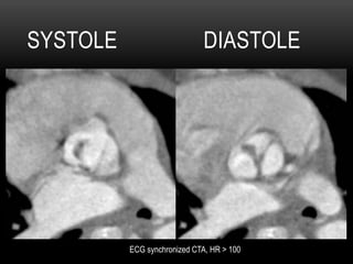
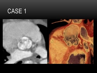
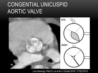
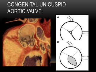
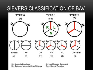
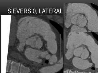
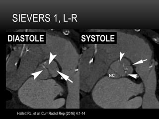
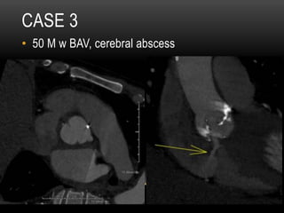
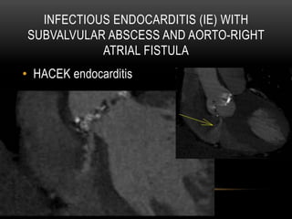
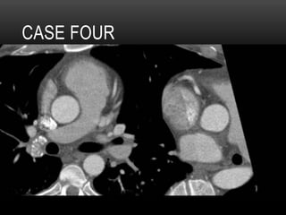
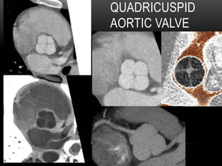
Richard L. Hallett, MD
Chief, Cardiovascular Imaging
Northwest Radiology Network – Indianapolis, IN
Adjunct Assistant Professor of Radiology
Stanford University Hospital and Clinics
Stanford, CA
NASCI 2016 Baltimore, MD October 15, 2016
DISCLOSURES
•  None
HANDOUT:
http://stanford.edu/~hallett
Choose folder: NASCI 2016
CASE 1
•  5 yo male with murmur, FTT
SYSTOLE DIASTOLE
ECG synchronized CTA, HR > 100
CASE 1
CONGENTIAL UNICUSPID
AORTIC VALVE
SYS
DIAST
Line drawings: Petit CJ, et al Am J Cardiol 2016; 117(6) 972-9.
CONGENITAL UNICUSPID
AORTIC VALVE
CASE 1 QUESTION:
WHICH OF THE FOLLOWING IS TRUE
REGARDING UNICUSPID AORTIC VALVE?
A. Most cases present in adulthood
B. UAV have better results from balloon
valvuloplasty than bicuspid aortic valve (BAV)
C. UAV shares some overlap with BAV patients,
including risk of aortopathy and coarctation
D. Have larger valve opening area than BAV
CASE 1 QUESTION: ANSWER
WHICH OF THE FOLLOWING IS TRUE
REGARDING UNICUSPID AORTIC VALVE?
A. Most cases present in adulthood
B. UAV have better results from balloon
valvuloplasty than bicuspid aortic valve (BAV)
C. UAV shares some overlap with BAV patients,
including risk of aortopathy and coarctation
D. Have larger valve opening area than BAV
Mookadam F, et al. J Heart Valve Dis 2010, 19(6)678-83
CONGENITAL “UNICUSPID”
AORTIC VALVE
•  Unicuspid unicommisural (one lateral commisural
attachment, slit-like orifice)
•  Unicuspid acomissural (no lateral attachments,
pinhole orifice, neonates)
•  Mean age of diagnosis 14 months
•  #1 presentation: CHF
•  #1 lesion: isolated AS (37%)
Singh S, et al. Texas Heart J. 2015; 42: 273-276
Mookadam F, et al. J Heart Valve Dis 2010; 19(6)678-83
CONGENITAL “UNICUSPID”
AORTIC VALVE
•  Associated anomalies:
•  Coarctation (37%)
•  VSD (12%)
•  PDA (5%)
•  Aortic Aneurysm (5%)
•  Coronary artery anomalies
Singh S, et al. Texas Heart J. 2015; 42: 273-276
Mookadam F, et al. J Heart Valve Dis 2010; 19(6)678-83
CONGENITAL “UNICUSPID”
AORTIC VALVE
•  Unicuspid (or functionally unicuspid) valves1,2:
•  Have lower associated LVEF than BAV
•  Lower opening area (AVOA)
•  Greater fusion length
•  Higher incidence of leaflet tears and resultant
aortic insufficiency after balloon v-plasty
1.  Gao, K, et al. J Invasive Cardiol. 2016; 28(9):381-8.
2.  Petit CJ, et al Am J Cardiol 2016; 117(6) 972-9.
CASE 2
BICUSPID AORTIC VALVE
SAM QUESTION:
Bicuspid aortic valve (BAV) is best characterized as:
A. An isolated defect causing aortic valve
abnormalities
B. A systemic disorder with aortic tissue showing
decreased levels of matrix metalloproteinases
C. A systemic disorder that shares some
histopathologic findings with Marfan syndrome
D. A systemic disorder with sporadic heritance
SAM QUESTION: ANSWER
Bicuspid aortic valve (BAV) is best characterized as:
A. An isolated defect causing aortic valve
abnormalities
B. A systemic disorder with aortic tissue showing
decreased levels of matrix metalloproteinases
C. A systemic disorder that shares some
histopathologic findings with Marfan syndrome
D. A systemic disorder with sporadic heritance
Hanneman K, et al. Pre- and Postoperative Imaging of the Aortic Root.
Radiographics. 2016;36:19–37.
BICUSPID AORTIC VALVE
•  1-2% of the population
•  Decreased Fibrillin-1 levels (M)
•  Increased matrix metalloproteinases (M)
•  NOTCH1 and ACTA2 genes
•  Some inherited - autosomal dominant
BICUSPID AORTIC VALVE
•  Aortic Dilatation / Aneurysms
•  Aortic Dissection
•  Aortic Coarctation
•  Family members of BAV patients can show
aortic dilatation even though they have
trileaflet valves
BAV: CLASSIFICATION
•  Sievers and Schmidtke – 2007
•  Classification based on 304 surgical BAV
specimens
•  Raphe: conjoint or fused portions of two
adjacent leaflets that become a malformed
commissure
SIEVERS CLASSIFICATION OF BAV
SIEVERS 0, LATERAL
SIEVERS 1, L-R
Hallett RL, et al. Curr Radiol Rep (2016) 4:1-14
DIASTOLE SYSTOLE
CASE 3
•  50 M w hx BAV, cerebral abscess, new heart
failure
CASE 3
•  50 M w BAV, cerebral abscess
INFECTIOUS ENDOCARDITIS (IE) WITH
SUBVALVULAR ABSCESS AND AORTO-RIGHT
ATRIAL FISTULA
•  HACEK endocarditis
CASE 3 QUESTION
Regarding HACEK infectious endocarditis (IE), which
is true?
A. HACEK are gram positive bacteria that are part
of normal skin flora
B. HACEK organisms cause a majority of infectious
endocarditis (IE) cases
C. HACEK IE patients tend to be younger than
other IE patients
D. Outcomes are poorer than other IE patients
CASE 3 QUESTION - ANSWER
Regarding HACEK infectious endocarditis (IE), which
is true?
A. HACEK are gram positive bacteria that are part
of normal skin flora
B. HACEK organisms cause a majority of infectious
endocarditis (IE) cases
C. HACEK IE patients tend to be younger than
other IE patients
D. Outcomes are poorer than other IE patients
Chambers ST et al. HACEK Infective Endocarditis: Characteristics and Outcomes from a Large,
Multi-National Cohort
Abbate A, editor. PLoS ONE. 2013;8:e63181.
HACEK ENDOCARDITIS
• Haemophilus spp.
• Aggregatibacter spp.
• Cardiobacterium hominus
• Eikenella corodens
• Kingella spp.
HACEK ENDOCARDITIS
•  Heterogenous group of gram negative fastidious
bacteria
•  Frequently colonize oropharyngeal
•  Cause < 5% of IE cases
•  Better outcomes
•  11% 1-year mortaility (vs 39%)
Chambers ST et al. HACEK Infective Endocarditis: Characteristics and Outcomes from a Large, Multi-National Cohort
PLoS ONE. 2013;8:e63181.
AORTO-RIGHT ATRIAL FISTULA1,2
•  Congenital
•  Aneurysm / Dissection
•  Infective endocarditis
•  Aorto-cavernous fistulae in only 1-2% IE pts
•  Staphlyococcus: worse outcomes
John ES, et al. Journal of Cardiothoracic Surgery 2014, 9:124
Nadgji GRJ, et al. Heart 2005 91: 7-10.
CASE 4
CASE 4
•  32 yr old male
•  Atypical CP, equivocal stress echo
•  Cath: “No vessel coming off R sinus”, concern
for anomalous coronary artery
CASE FOUR
QUADRICUSPID
AORTIC VALVE
QUESTION
The most common complication of Quadricuspid
Aortic Valve (QAV) is:
A. Valvular aortic stenosis
B. Aortic regurgitation
C. Atrial fibrillation
D. Left ventricular hypertrophy
Douglas, H., Moore, M., & Purvis, J. (2012). Comprehensive assessment of a quadricuspid
aortic valve and coronary arteries by multidetector cardiac CT. Heart, 98(24), 1838-1838.
QUESTION: ANSWER
The most common complication of Quadricuspid
Aortic Valve (QAV) is:
A. Valvular aortic stenosis
B. Aortic regurgitation
C. Atrial fibrillation
D. Left ventricular hypertrophy
Douglas, H., Moore, M., & Purvis, J. Heart, 2012: 98(24), 1838-1838.
QUADRICUSPID AORTIC VALVE
(QAV)
•  Rare, 1/6000 aortic valve surgery patients1
•  M = F, avg. age at Dx ~ 50
•  Classification by size of cusps2
•  Most common: 3 same size + 1 smaller cusp
(type B)
•  Echo: “X”-shaped SAX view
•  CT/MR: confirmatory; perform planimetry and/or
flow measurement3
1.  Douglas, H., Moore, M., & Purvis, J. (2012) Heart 2012; 98(24), 1838-1838.
2.  Hurwitz L, et al. Am J Cardiol 1973; 31(5) 623-626.
3.  Khan SK, Tamin SS, Araoz PA. J Comput Assist Tomogr. 35 (5): 637-41.
CLASSIFICATION OF QAV1
A 4 equal sized cusps
B 3 equal + 1 smaller (most common)
C 2 equal + 2 equal smaller
D 1 large + 2 intermediate + 1 smaller
E 3 equal + 1 larger
F 2 equal large + 2 smaller unequal sizes
G 4 unequal sized cusps
1.  Hurwitz L, et al. Am J Cardiol 1973; 31(5) 623-626.
QUADRICUSPID AORTIC VALVE
(QAV)
•  Usually isolated, but can be associated with:
•  Single or Anomalous Coronary Arteries
•  Displacement of coronary ostia (from addl
cusp)
•  HCM / Subaortic Stenosis
•  PDA, VSD
•  Endocarditis
1. Jagganath AD, et al. Echocardiography 2011; 28(9), 1035-1040.
2. Zhu J, et al. J Cardiothor Surg 2013; 8(1) 87.
3. Tutarel O. J. Heart Valve Dis. 2004;13 (4): 534-7.
QUADRICUSPID AORTIC VALVE
(QAV)
•  Complications:
•  Aortic Insufficiency (#1)
• Up to 75% at time of Dx
•  LVH
•  Conduction problems (BBB)
•  TX: Reconstruction and/or Surgical valve
replacement
1. Jagganath AD, et al. Echocardiography 2011; 28(9), 1035-1040.
2. Zhu J, et al. J Cardiothor Surg 2013; 8(1) 87.
3. Tutarel O. J. Heart Valve Dis. 2004;13 (4): 534-7.
CONCLUSION
•  Whether 1, 2, 3, or
4 cusps, aortic
valve pathology
can be well
assessed by ECG
synchronized CTA
THANKS FOR YOUR ATTENTION!!
HANDOUT:
http://stanford.edu/~hallett
Choose folder: NASCI 2016
xraydoc97@yahoo.com
hallett@stanford.edu

Aortic Valve Cases

  • 1.
    Richard L. Hallett,MD Chief, Cardiovascular Imaging Northwest Radiology Network – Indianapolis, IN Adjunct Assistant Professor of Radiology Stanford University Hospital and Clinics Stanford, CA NASCI 2016 Baltimore, MD October 15, 2016
  • 2.
  • 3.
    CASE 1 •  5yo male with murmur, FTT
  • 4.
  • 5.
  • 6.
    CONGENTIAL UNICUSPID AORTIC VALVE SYS DIAST Linedrawings: Petit CJ, et al Am J Cardiol 2016; 117(6) 972-9.
  • 7.
  • 8.
    CASE 1 QUESTION: WHICHOF THE FOLLOWING IS TRUE REGARDING UNICUSPID AORTIC VALVE? A. Most cases present in adulthood B. UAV have better results from balloon valvuloplasty than bicuspid aortic valve (BAV) C. UAV shares some overlap with BAV patients, including risk of aortopathy and coarctation D. Have larger valve opening area than BAV
  • 9.
    CASE 1 QUESTION:ANSWER WHICH OF THE FOLLOWING IS TRUE REGARDING UNICUSPID AORTIC VALVE? A. Most cases present in adulthood B. UAV have better results from balloon valvuloplasty than bicuspid aortic valve (BAV) C. UAV shares some overlap with BAV patients, including risk of aortopathy and coarctation D. Have larger valve opening area than BAV Mookadam F, et al. J Heart Valve Dis 2010, 19(6)678-83
  • 10.
    CONGENITAL “UNICUSPID” AORTIC VALVE • Unicuspid unicommisural (one lateral commisural attachment, slit-like orifice) •  Unicuspid acomissural (no lateral attachments, pinhole orifice, neonates) •  Mean age of diagnosis 14 months •  #1 presentation: CHF •  #1 lesion: isolated AS (37%) Singh S, et al. Texas Heart J. 2015; 42: 273-276 Mookadam F, et al. J Heart Valve Dis 2010; 19(6)678-83
  • 11.
    CONGENITAL “UNICUSPID” AORTIC VALVE • Associated anomalies: •  Coarctation (37%) •  VSD (12%) •  PDA (5%) •  Aortic Aneurysm (5%) •  Coronary artery anomalies Singh S, et al. Texas Heart J. 2015; 42: 273-276 Mookadam F, et al. J Heart Valve Dis 2010; 19(6)678-83
  • 12.
    CONGENITAL “UNICUSPID” AORTIC VALVE • Unicuspid (or functionally unicuspid) valves1,2: •  Have lower associated LVEF than BAV •  Lower opening area (AVOA) •  Greater fusion length •  Higher incidence of leaflet tears and resultant aortic insufficiency after balloon v-plasty 1.  Gao, K, et al. J Invasive Cardiol. 2016; 28(9):381-8. 2.  Petit CJ, et al Am J Cardiol 2016; 117(6) 972-9.
  • 13.
  • 15.
  • 16.
    SAM QUESTION: Bicuspid aorticvalve (BAV) is best characterized as: A. An isolated defect causing aortic valve abnormalities B. A systemic disorder with aortic tissue showing decreased levels of matrix metalloproteinases C. A systemic disorder that shares some histopathologic findings with Marfan syndrome D. A systemic disorder with sporadic heritance
  • 17.
    SAM QUESTION: ANSWER Bicuspidaortic valve (BAV) is best characterized as: A. An isolated defect causing aortic valve abnormalities B. A systemic disorder with aortic tissue showing decreased levels of matrix metalloproteinases C. A systemic disorder that shares some histopathologic findings with Marfan syndrome D. A systemic disorder with sporadic heritance Hanneman K, et al. Pre- and Postoperative Imaging of the Aortic Root. Radiographics. 2016;36:19–37.
  • 18.
    BICUSPID AORTIC VALVE • 1-2% of the population •  Decreased Fibrillin-1 levels (M) •  Increased matrix metalloproteinases (M) •  NOTCH1 and ACTA2 genes •  Some inherited - autosomal dominant
  • 19.
    BICUSPID AORTIC VALVE • Aortic Dilatation / Aneurysms •  Aortic Dissection •  Aortic Coarctation •  Family members of BAV patients can show aortic dilatation even though they have trileaflet valves
  • 20.
    BAV: CLASSIFICATION •  Sieversand Schmidtke – 2007 •  Classification based on 304 surgical BAV specimens •  Raphe: conjoint or fused portions of two adjacent leaflets that become a malformed commissure
  • 21.
  • 22.
  • 23.
    SIEVERS 1, L-R HallettRL, et al. Curr Radiol Rep (2016) 4:1-14 DIASTOLE SYSTOLE
  • 24.
    CASE 3 •  50M w hx BAV, cerebral abscess, new heart failure
  • 25.
    CASE 3 •  50M w BAV, cerebral abscess
  • 26.
    INFECTIOUS ENDOCARDITIS (IE)WITH SUBVALVULAR ABSCESS AND AORTO-RIGHT ATRIAL FISTULA •  HACEK endocarditis
  • 27.
    CASE 3 QUESTION RegardingHACEK infectious endocarditis (IE), which is true? A. HACEK are gram positive bacteria that are part of normal skin flora B. HACEK organisms cause a majority of infectious endocarditis (IE) cases C. HACEK IE patients tend to be younger than other IE patients D. Outcomes are poorer than other IE patients
  • 28.
    CASE 3 QUESTION- ANSWER Regarding HACEK infectious endocarditis (IE), which is true? A. HACEK are gram positive bacteria that are part of normal skin flora B. HACEK organisms cause a majority of infectious endocarditis (IE) cases C. HACEK IE patients tend to be younger than other IE patients D. Outcomes are poorer than other IE patients Chambers ST et al. HACEK Infective Endocarditis: Characteristics and Outcomes from a Large, Multi-National Cohort Abbate A, editor. PLoS ONE. 2013;8:e63181.
  • 29.
    HACEK ENDOCARDITIS • Haemophilus spp. • Aggregatibacterspp. • Cardiobacterium hominus • Eikenella corodens • Kingella spp.
  • 30.
    HACEK ENDOCARDITIS •  Heterogenousgroup of gram negative fastidious bacteria •  Frequently colonize oropharyngeal •  Cause < 5% of IE cases •  Better outcomes •  11% 1-year mortaility (vs 39%) Chambers ST et al. HACEK Infective Endocarditis: Characteristics and Outcomes from a Large, Multi-National Cohort PLoS ONE. 2013;8:e63181.
  • 31.
    AORTO-RIGHT ATRIAL FISTULA1,2 • Congenital •  Aneurysm / Dissection •  Infective endocarditis •  Aorto-cavernous fistulae in only 1-2% IE pts •  Staphlyococcus: worse outcomes John ES, et al. Journal of Cardiothoracic Surgery 2014, 9:124 Nadgji GRJ, et al. Heart 2005 91: 7-10.
  • 32.
  • 33.
    CASE 4 •  32yr old male •  Atypical CP, equivocal stress echo •  Cath: “No vessel coming off R sinus”, concern for anomalous coronary artery
  • 34.
  • 35.
  • 36.
    QUESTION The most commoncomplication of Quadricuspid Aortic Valve (QAV) is: A. Valvular aortic stenosis B. Aortic regurgitation C. Atrial fibrillation D. Left ventricular hypertrophy Douglas, H., Moore, M., & Purvis, J. (2012). Comprehensive assessment of a quadricuspid aortic valve and coronary arteries by multidetector cardiac CT. Heart, 98(24), 1838-1838.
  • 37.
    QUESTION: ANSWER The mostcommon complication of Quadricuspid Aortic Valve (QAV) is: A. Valvular aortic stenosis B. Aortic regurgitation C. Atrial fibrillation D. Left ventricular hypertrophy Douglas, H., Moore, M., & Purvis, J. Heart, 2012: 98(24), 1838-1838.
  • 38.
    QUADRICUSPID AORTIC VALVE (QAV) • Rare, 1/6000 aortic valve surgery patients1 •  M = F, avg. age at Dx ~ 50 •  Classification by size of cusps2 •  Most common: 3 same size + 1 smaller cusp (type B) •  Echo: “X”-shaped SAX view •  CT/MR: confirmatory; perform planimetry and/or flow measurement3 1.  Douglas, H., Moore, M., & Purvis, J. (2012) Heart 2012; 98(24), 1838-1838. 2.  Hurwitz L, et al. Am J Cardiol 1973; 31(5) 623-626. 3.  Khan SK, Tamin SS, Araoz PA. J Comput Assist Tomogr. 35 (5): 637-41.
  • 39.
    CLASSIFICATION OF QAV1 A4 equal sized cusps B 3 equal + 1 smaller (most common) C 2 equal + 2 equal smaller D 1 large + 2 intermediate + 1 smaller E 3 equal + 1 larger F 2 equal large + 2 smaller unequal sizes G 4 unequal sized cusps 1.  Hurwitz L, et al. Am J Cardiol 1973; 31(5) 623-626.
  • 40.
    QUADRICUSPID AORTIC VALVE (QAV) • Usually isolated, but can be associated with: •  Single or Anomalous Coronary Arteries •  Displacement of coronary ostia (from addl cusp) •  HCM / Subaortic Stenosis •  PDA, VSD •  Endocarditis 1. Jagganath AD, et al. Echocardiography 2011; 28(9), 1035-1040. 2. Zhu J, et al. J Cardiothor Surg 2013; 8(1) 87. 3. Tutarel O. J. Heart Valve Dis. 2004;13 (4): 534-7.
  • 41.
    QUADRICUSPID AORTIC VALVE (QAV) • Complications: •  Aortic Insufficiency (#1) • Up to 75% at time of Dx •  LVH •  Conduction problems (BBB) •  TX: Reconstruction and/or Surgical valve replacement 1. Jagganath AD, et al. Echocardiography 2011; 28(9), 1035-1040. 2. Zhu J, et al. J Cardiothor Surg 2013; 8(1) 87. 3. Tutarel O. J. Heart Valve Dis. 2004;13 (4): 534-7.
  • 42.
    CONCLUSION •  Whether 1,2, 3, or 4 cusps, aortic valve pathology can be well assessed by ECG synchronized CTA
  • 43.
    THANKS FOR YOURATTENTION!! HANDOUT: http://stanford.edu/~hallett Choose folder: NASCI 2016 xraydoc97@yahoo.com hallett@stanford.edu