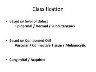
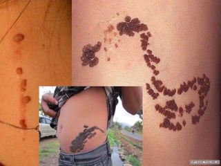
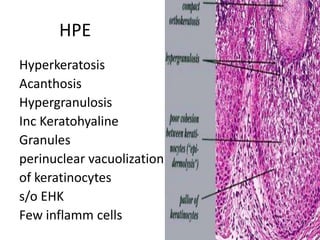
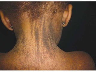
This document discusses epidermal naevi, which are congenital developmental defects or birthmarks of the skin and mucosa. It describes different types of epidermal naevi classified based on the level of defect (epidermal, dermal, subcutaneous) and component cell (vascular, connective tissue, melanocytic). Verrucous epidermal naevi, also known as verrucous nevus or nevus verrucosus, are discussed in detail. They are keratinocyte hamartomas that can be either epidermolytic or non-epidermolytic types, with the latter having greater malignant potential and possible associations with extracutaneous abnormalities. Clinical features
Pigmentation disorders of skin dermatology revision notesTONY SCARIA
dermatology revision notes for neet pg preparation based on lecture notes with high yield topic & last minute revision notes based on previous year questions
Pigmentation disorders of skin dermatology revision notesTONY SCARIA
dermatology revision notes for neet pg preparation based on lecture notes with high yield topic & last minute revision notes based on previous year questions
Hereditary disorder of keratinization characterized by expanding atrophic anular patch(es) surrounded by prominent keratotic ridge called the cornoid lamella
This is a powerpoint presentation on the epidermal keratinization and its associated disorders, presented by Dr. Jerriton, Dermatology resident of SVMCH, Pondicherry.
• In recent years, the usefulness of trichoscopy (scalp dermoscopy) (videodermatoscopy) has been reported for diagnosing hair loss diseases. This method allows viewing of the hair and scalp at X20 to X160 magnifications. Characteristic trichoscopy features of alopecia areata are black dots, tapering hairs (exclamation mark hairs), broken hairs, yellow dots, and short vellus hairs. In androgenetic alopecia (AGA), hair diameter diversity (HDD), perifollicular pigmentation/peripilar sign, and yellow dots are trichoscopically observed. In all cases of AGA and female AGA, HDD, more than 20%, which corresponds to vellus transformation, can be seen. In cicatricial alopecia (CA), the loss of orifices, a hallmark of CA, and the associated changes including perifollicular erythema or scale and hair tufting were observed. Different hair shafts variation such as vellus, terminal, micro-exclamation mark type, monilethrix, Netherton type, and pili annulati hairs can be seen . The number of hairs in one pilosebaceous unit can be assessed. Healthy Hair follicles variation healthy, empty, fibrotic ("white dots"), filled with hyperkeratotic plugs ("yellow dots"), or containing dead hair ("black dots"). Abnormalities of scalp skin color or structure include honeycomb-type hyperpigmentation, perifollicular discoloration (hyperpigmentation), and scaling are also seen with the help of trichoscopy.
A discussion on various photodermatoses including sun burns, porphyria, actinic chelitis, hydroa vacciniforme and chronic actinic dermatitis. Sun tan and skin color types. Affect of Sunlight on the skin. Useful for medical residents, dermatologists and nurse. Useful in exam preparation.
TSC is neurocutaneous hereditary disorder either AD or sporadic characterized by multiple hamartomatous growths affecting skin, brain, heart, lung & kidney
Pathological evaluation of melanocytic lesionsHisashi Uhara
In this lecture, the following basic steps by which I routinely scan specimens in our hospital will be presented with examples.
1. Evaluate the specimen preparation.
1) Is the incision for the specimen made perpendicular to the skin surface?
2) Is the slice of tissue from volar skin made perpendicular to the furrows of skin?
2. Estimate the specimen size and location.
1) Estimate the size of the lesion from the magnification of the objective lens.
2) Estimate the specimen location.
3. Precaution before evaluation
1) Observe the specimens without clinical information as much as possible.
2) Obtain as much information as possible at low magnification.
4. The steps for observation
1) At low magnification: Check the symmetric properties and circumscription of the lesion based on the following points.
a. Distance from the densest area of the lesion to both ends.
b. Variation of the thickness of epidermis from the center to both ends.
c. Distribution of melanin in the coronoid layer, epidermis, and dermis.
d. Distribution of nests and distance between each nest.
e. Density of solitary distributed melanocytes.
f. Existence of inflammatory infiltration in the dermis and its distribution.
g. Continuity of the spread of nests and tumor cells in both ends.
h. Is the bottom of the lesion smooth or not?
2) At high magnification: Check the details of tumor cells.
a. Tumor cells in the epidermis: Existence of necrosis, atypia (large nucleolus), or mitosis.
b. Other findings in the epidermis: Distribution of melanin in the cornified layer, the existence of tumor cells in the upper epidermis, the polymorphism of tumor cells, the relationship between tumor cells and keratinocytes.
c. In the dermis: An overlapping, crowded, or sheet-like gathering of tumor cells, maturation of tumor cells, mitotic figures, or melanin of tumor cells at the bottom of the lesion.
d. In the adnexal area: The existence of tumor cells in adnexal walls.
5. After provisionally giving a pathological diagnosis, check discrepancies between the pathological diagnosis and clinical findings. Return to the pathological evaluation if necessary.
power point presentation on the various pigmented lesions in the oral mucosa with their clinical features and oral manifestations and differential diagnosis
general practice,general practice doctor,listening ielts practice,general practice uk,general practice nurse,general practice update,general practice report,general practice nurses,uk general practice nurse,general practice nurse uk,general practice nursing,general practice nurses uk,uk general practice nurses,burnout in general practice,charge for general practice,why i chose general practice,a career in general practice,uk general practice nursing
family doctor,doctor,family medicine,family doctor canada,family doctor shortage,family doctors,family doctor in canada,ap family doctor,family guy doctor,family doctor app,doctors,family doctor in bc,find a family doctor,bread family doctor,family doctor visit,family doctor quebec,canada family doctor,ontario family doctor,doctor mike,canada family doctors,find a family doctor canada,family medicine doctor,family doctors shortages
general practice,what is a general practice doctor,what is a family practice doctor,what does it mean when a doctor does general practice,family doctor,general practice doctor definition,doctor,general practitioner,family medicine,general practice doctor,general practice doctor near me,general practice doctor salary,general pratice doctor,what is better family practice or general practice?,family medicine doctor,family practice medical doctor
family medicine,family medicine residency,what is internal medicine vs family practice,general practice family medicine,what is family medicine,family medicine & gp practice in urdu,family medicine & gp practice in hindi,general practice,family medicine & gp practice in urdu or hindi,general practice doctor definition,what is a general practice doctor,internal medicine vs general practice,general practitioner,what is better family practice or general practice?
Ahmad mukhtar
MD.,M.B.B.Ch., M.Sc Obstetrics and GynecologyConsultant and Lecturer of Obstetrics and Gynecology, Faculty of
MEDICINE, Zagazig University.
Premalignancy 3 / dental implant courses by Indian dental academy Indian dental academy
The Indian Dental Academy is the Leader in continuing dental education , training dentists in all aspects of dentistry and
offering a wide range of dental certified courses in different formats.for more details please visit
www.indiandentalacademy.com
10. Epidermolytic VEN
• Mutations on genes KRT1 / KRT10
• Associated with BIE
• Insignificant Malignant Potential
• No association with extracutaneous
abnormalities keratin genes are expressed
only in epithelia
11. Clinical Features
• Slightly pigmented brown velvety or warty
streaks or plaques
• Hyperpigmented and more warty with age
• Flexural lesions macerated and foul smelling
• Rarely :- blisters at birth verrucous with age
• Striate palmoplantar keratoderma / Ainhum
reported in a child with extensive systematized
VEN
14. Non-Epidermolytic VEN
• Mosaic Chr 06 Trisomy
• FGFR3 Mutation
• Assoc with PIC3CA and PTEN mutations
• Significant Malignant Potential > 40 yrs
• Associated with extracutaneous manifestations
15. Clinical Features
• present at birth / childhood / 55 yrs of age
• Crusted, hyperkeratotic plaques on the head and
upper trunk
• Birth white macerated appearance pink /
slightly pigmented, velvety streaks or plaques
darken and the surface becomes more warty
• Flexural lesions macerated and foul smelling
18. HPE
• Hyperkeratosis ; Columns of Parakeratosis
• Acanthosis, Papillomatosis, Focal hypergranulosis
• 10% of lesions show a distinctive ‘church-spire’ pattern of
acanthosis and hyperkeratosis, resembling acrokeratosis
verruciformis
• 5% show features resembling seborrhoeic keratoses, that is
hyperkeratosis, papillomatosis, acanthosis and horn
pseudocysts
• Immature adnexae (hair follicles, sebaceous, eccrine or
apocrine glands)
19. Treatment
Topical applications
• SA / LA / RA preparations Decrease Hyperkeratosis
• Podophyllum
• 5-fluorouracil
• calcipotriol and calcitriol
Systemic retinoid therapy. Isotretinoin / etretinate / Acitretin can
reduction of hyperkeratosis (in epidermolytic lesions)
Dermabrasion
20. Lasers
overcomes problems such as hypertrophic scarring, pigmentary
changes and partial recurrence
Argon laser is helpful for softer, less hyperkeratotic lesions
Continuous-wave CO2 laser vaporization Extensive VEN
Pulsed CO2 thinner and softer VEN
Er:YAG lasers with greater coagulative capacity – Lower recurrence
Pulsed Ruby Laser
Surgery
Multi-modal, Multi-stage Dermis removal only effective