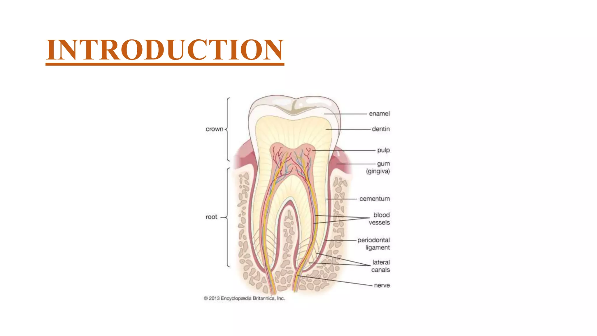
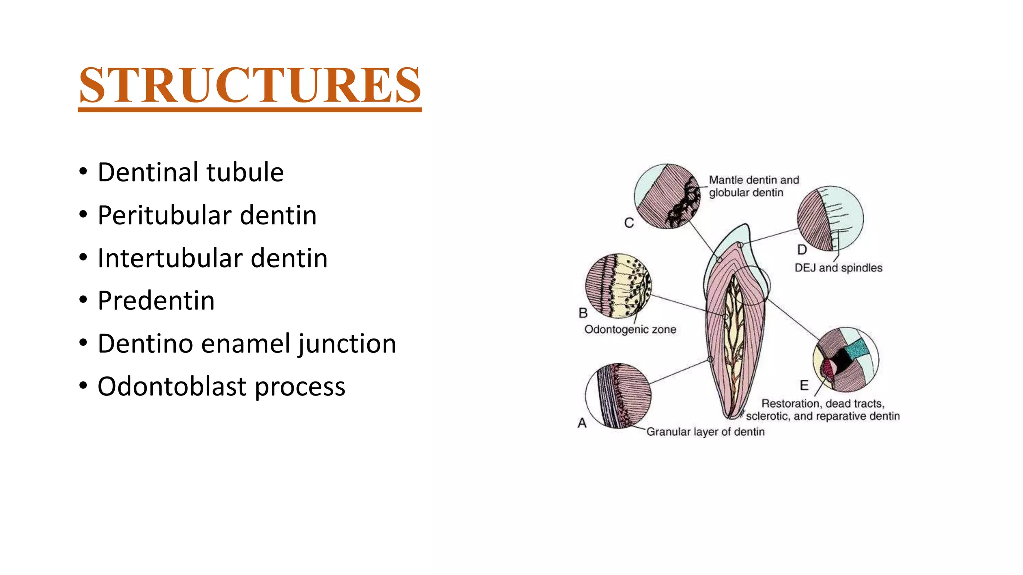
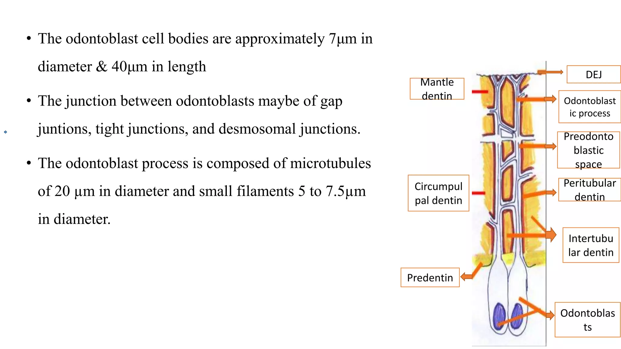
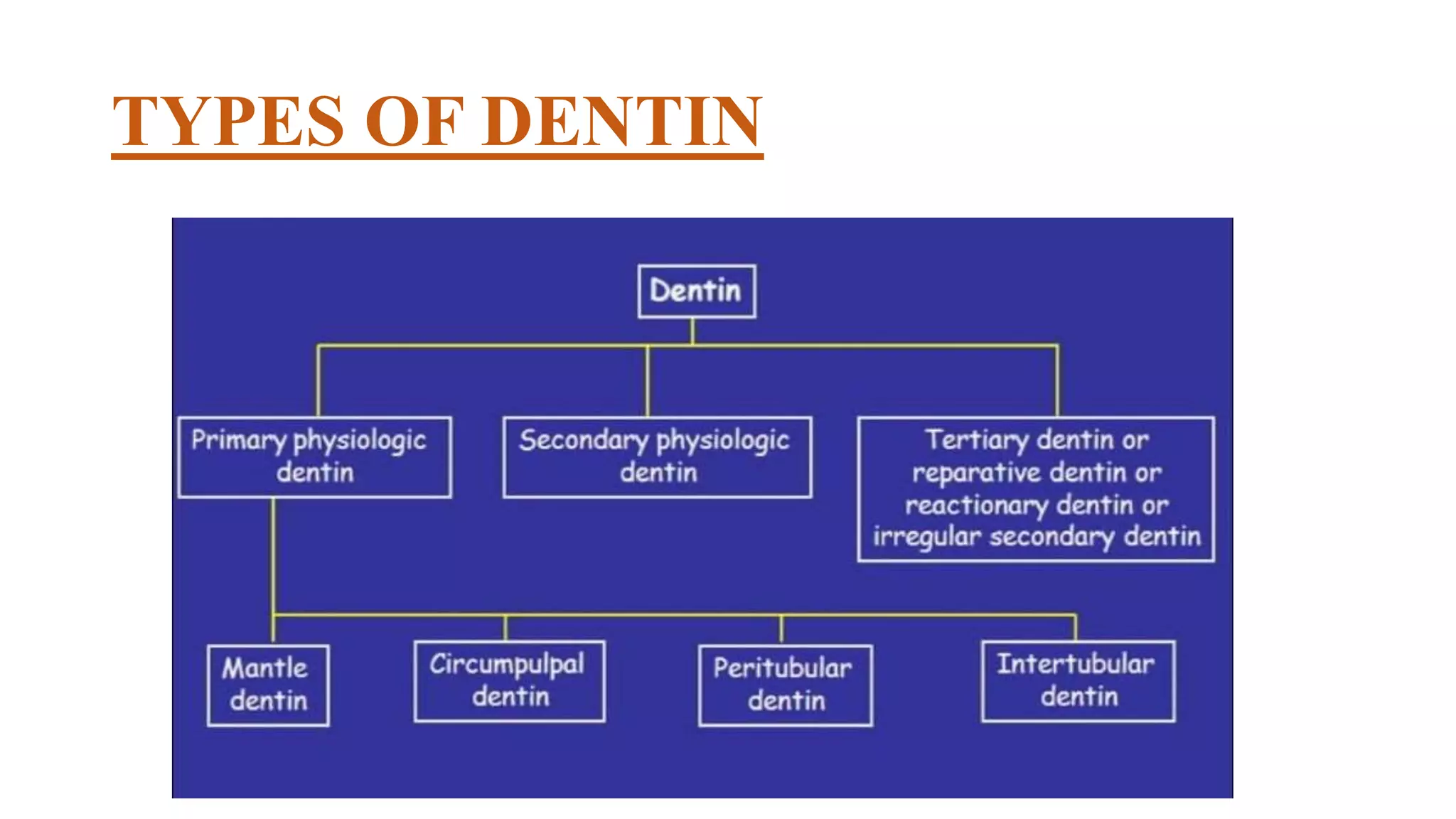
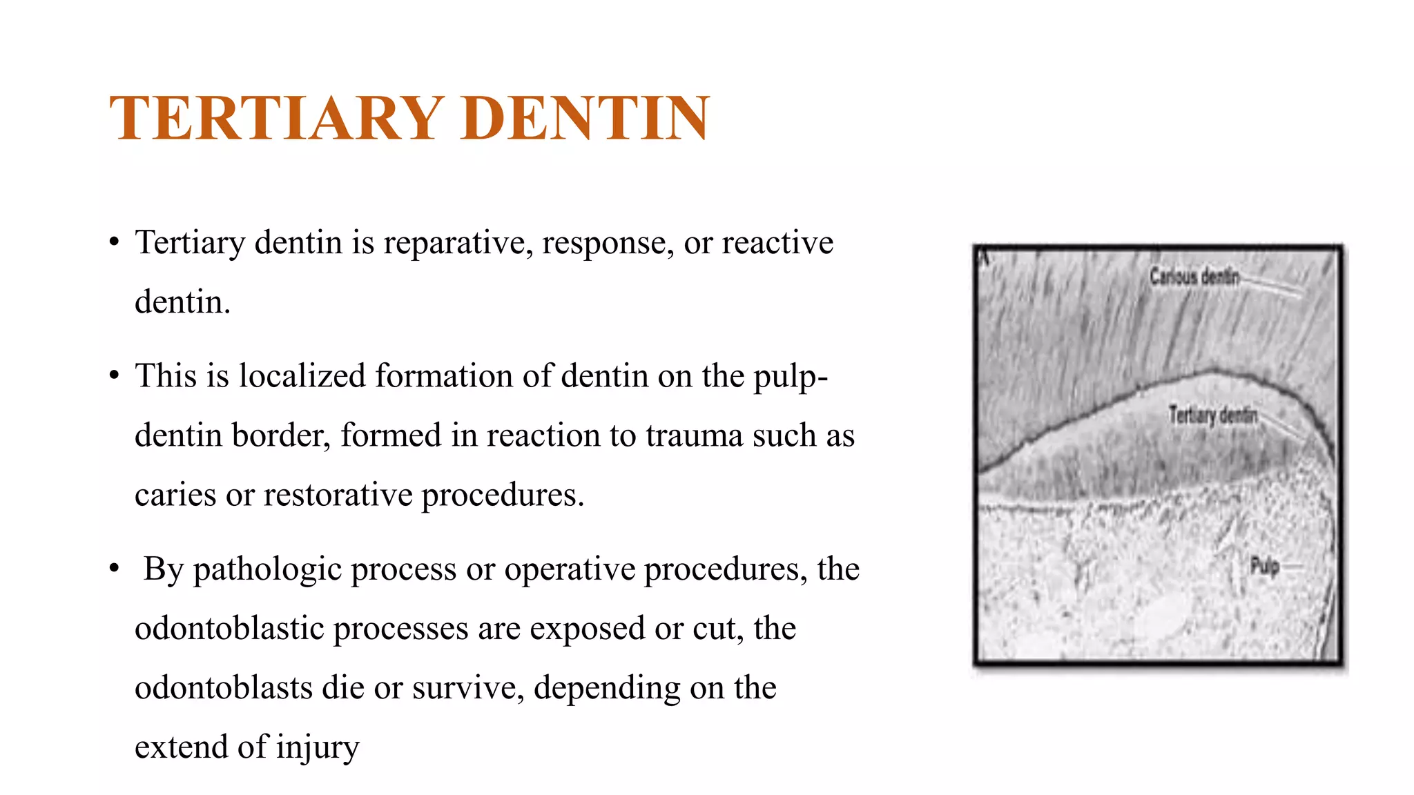
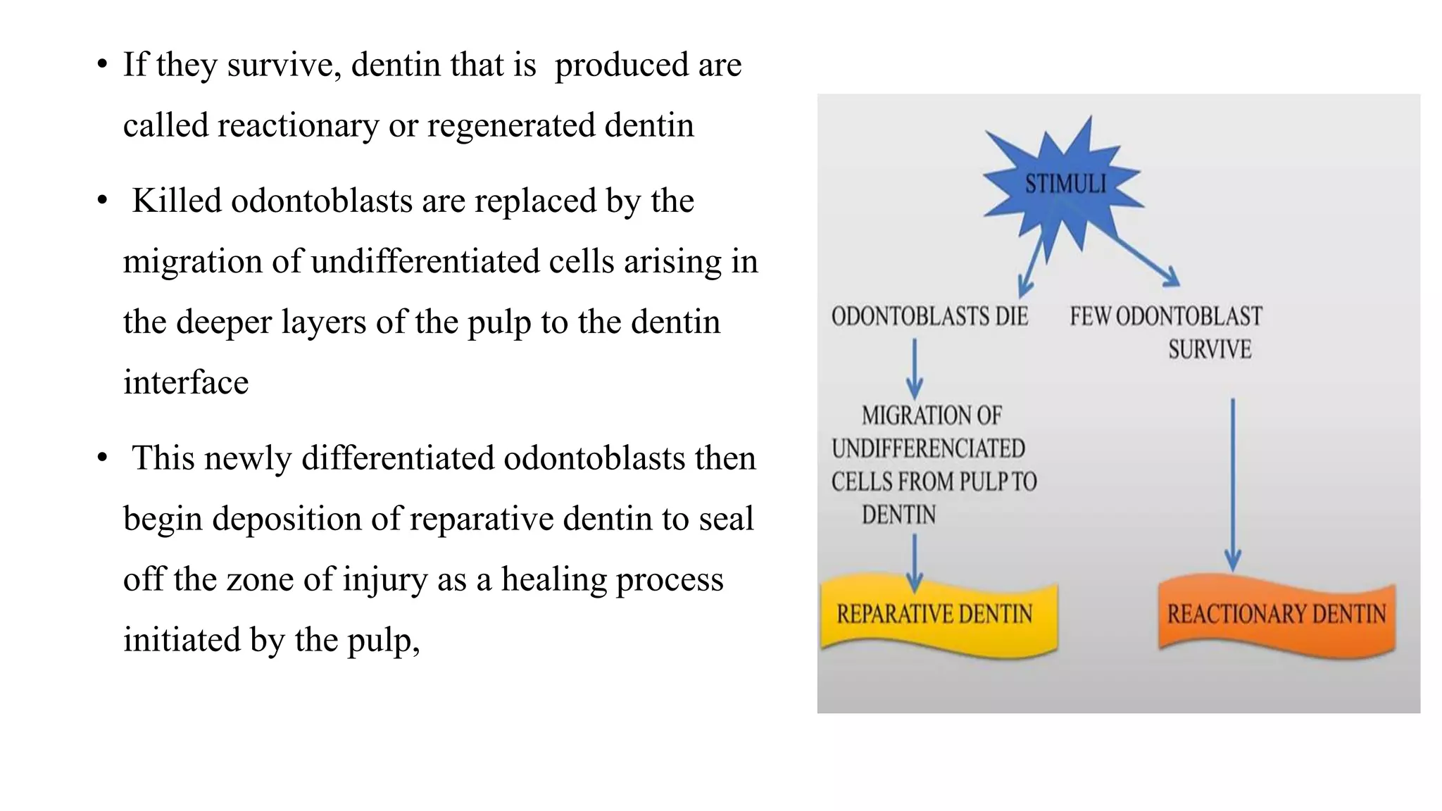
DENTIN
DEPT. OF PEDIATRIC AND PREVENTIVE DENTISTRY
DR. SHEFAAMIR
(JR – 1)
CONTENTS
• INTRODUCTION
• PHYSICAL PROPERTIES
• CHEMICAL PROPERTIES
• STRUCTURES
• TYPES OF DENTIN
• INCREMENTAL LINES OF DENTIN
• INNERVATION OF DENTIN
• THEORIES OF PAIN TRANSMISSION THROUGH DENTIN
• AGE AND FUNCTIONAL CHANGES
• DEVELOPMENT OF DENTIN
• CLINICAL CONSIDERATIONS
• DEVELOPMENTAL DEFFECTS
INTRODUCTION
• Dentin is the hard tissue of the tooth that surrounds the central core
of nerves and blood vessels and provides the bulk and general form
of the tooth
• Since it begins to form slightly before the enamel, it determines the
shape of the crown, including the cusps and ridges and the number
and size of roots
• Said to be a living tissue since the tubules present in it contains
processes of specialised cells, the odontoblast
PHYSICAL PROPERTIES
• In teeth of young individuals, the dentin is usually light yellowish in colour,
becoming darker with age.
• Dentin is viscoelastic and subject to slight deformation
• It is harder than bone but considerably softer than enamel
• The dentin of primary teeth is slightly less hard than that of permanent
teeth.
• The dentin is more radiolucent than enamel
PROPERTY VALUE
Colour Pale yellow
Thickness 3-10 mm
Modulus of Elasticity 15-20 GPa
Hardness 68 KHN
Carious Dentin 25 KHN
Sclerotic Dentin 80 KHN
Compressive strength 266 MPa
Tensile strength 50 MPa
Radiopacity Less than enamel
CHEMICAL PROPERTIES
Dentin
Inorganic
(65%)
Organic
(20%)
Water
(15%)
Organic (20%)
• The organic substance consists of collagenous fibrils embedded in the
ground substance of mucopolysaccharides
• Type I collagen is the principal type of collagen found in the dentin
• Ground substance:-
i. proteoglycans- chondroitin sulphates, decorin & biglycans
ii. Glycoproteins- dentin sialoprotein (DSP), osteonectin, osteopontin
iii. Phosphoproteins- dentin phosphoprotien (DPP), gamma
carboxyglutamate containing proteins and phospholipids.
• In addition, matrix contains growth factors like transforming growth factor
(TGF), fibroblast growth factor (FGF), insulin like growth factor (IGFs),
bone morphogenic proteins (BMPs), vascular endothelial growth factor
(VEGF), and angiogenic growth factor (AGF).
• Inorganic (65%)
• The inorganic component consist of hydroxyapatite.
• Each hydroxyapatite crystal is composed of several thousand unit cells.
• The crystals are plate shaped and much smaller than the hydroxyapatite
crystal in enamel.
• Dentin also contain small amounts of phosphates, carbonates, and sulfates.
• The crystals are poor in calcium but rich in carbon when compared to
enamel
STRUCTURES
• Dentinal tubule
• Peritubular dentin
• Intertubular dentin
• Predentin
• Dentino enamel junction
• Odontoblast process
DENTINAL TUBULE
• The course of the dentinal tubules follow a
gentle curve in the crown where it resembles
an S shape
• Starts at right angles at the pulpal surface,
the first convexity of this doubly curved
course is directed towards the apex of the
tooth
• These tubules end perpendicular to the DEJ
& CDJ
• It is almost straight near the root tip and along the incisal edges and cusps
• Dentin thickness ranges from 3-10mm or more
• No. of tubules per square millimetre varies from 15000
at the DEJ to 65000 at the pulp - density and diameter
increases with depth
• There are more tubules per unit area in the crown than in the root
• These dentinal tubules have lateral branches throughout dentin, which are termed
canaliculi or microtubules
• A few odontoblastic processes extend through the DEJ into the enamel several
millimetres. These are called Enamel Spindles
PERITUBULAR DENTIN
• The dentin that immediately surrounds
the dentinal tubules is termed peritubular dentine
• Highly mineralized than intertubular dentin
(about 9%)
• Twice as thick in outer dentin (approx. 0.75μm)
than inner dentin(approx. 0.4μm)
• Studies with electron microscope show the
increased mineral density in the peritubular dentin
• Between the odontoblastic process and the
peritubular dentin, a space known as
periodontoblastic space is present.
• This space contain the dentinal fluid. The normal
flow of the fluid is outward from the pulp.
INTERTUBULAR DENTIN
• The main body of dentin is composed of intertubular dentin.
• Located between the dentinal tubules specifically between the zones of
peritubular dentin
• One half of its volume is organic matrix, specifically collagen fibers
• The fibrils range from 0.5-0.2μm in diameter and exhibit cross banding at
64μm intervals
• HA crystals are formed along the fibers with their long axis oriented parallel to
the collagen fibers
PREDENTIN
• Located adjacent to the pulp tissues
• 2-6μm, depending on the activity of odontoblast
• First formed dentin and is not mineralised
• The collagen fibres undergo mineralization at the
predentin - dentin JUNCTION, the predentin then
becomes dentin and a new layer of predentin forms
circumpulpally
DENTINO ENAMEL JUNCTION
• The DEJ is a complex and critical structure
uniting two dissimilar calcified tissues and acts
to prevent the propagation of cracks from
enamel into dentin.
• The DEJ has three level structure, 25-100 µm
scallops with their convexities directed
towards the dentin and concavities toward the
enamel; 2-5µm micro scallops and a smaller
scale structure.
• The convexities of the scallops are directed
towards the dentin. The concavities are
directed toward the enamel. The surface of the
dentin at DEJ is pitted.
ODONTOBLAST PROCESS
• Cytoplasmic extensions of the odontoblasts
• The odontoblasts reside in the peripheral pulp at
the pulp-predentin border and their processes
extend into the dentinal tubules
• The processes are largest in diameter near the
pulp and taper further into dentin
• The odontoblast cell bodies are approximately 7μm in
diameter & 40μm in length
• The junction between odontoblasts maybe of gap
juntions, tight junctions, and desmosomal junctions.
• The odontoblast process is composed of microtubules
of 20 µm in diameter and small filaments 5 to 7.5µm
in diameter.
DEJ
Odontoblast
ic process
Preodonto
blastic
space
Peritubular
dentin
Mantle
dentin
Circumpul
pal dentin
Predentin
Intertubu
lar dentin
Odontoblas
ts
TYPES OF DENTIN
PRIMARY DENTIN
• Dentin that is formed prior to eruption of a tooth, before root
completion
• Secreted at a relatively higher rate
• Constitutes major part of the dentin in the tooth
• Consist of mantle dentin and circumpulpal dentin.
1) Mantle Dentine
• First formed dentin in crown underlying the DEJ
• 20 μm thick
• Fibrils found in this zone are perpendicular to DEJ
• Organic matrix - von korffs fibres (large diameter
fibrils- type III collagen fibrils)
• Less mineralized compared to circumpulpal dentin
2) Circumpulpal Dentin
• Forms the remaining primary dentin or
bulk of the tooth.
• The fibrils are much smaller in diameter
(0.05micrometer) & are more closely
packed together.
• Slightly more mineral content than mantle
dentin.
SECONDARY DENTIN
• Formed after root completion
• Narrow band of dentin bordering the pulp
• Contains fewer tubules than primary dentin
• There is usually a bend in the tubules where
primary and secondary dentin interface
• Since it is formed after eruption, the odontoblasts
slightly change direction which contributes to
bending of dentinal tubules
• It is a slow continuous deposition of dentin.
• It is formed more slowly than primary dentin.
• Secondary dentin is not formed uniformly and
appears in great amounts on the roof and floor of
pulp chamber, where it protects pulp from exposure.
TERTIARY DENTIN
• Tertiary dentin is reparative, response, or reactive
dentin.
• This is localized formation of dentin on the pulp-
dentin border, formed in reaction to trauma such as
caries or restorative procedures.
• By pathologic process or operative procedures, the
odontoblastic processes are exposed or cut, the
odontoblasts die or survive, depending on the
extend of injury
• If they survive, dentin that is produced are
called reactionary or regenerated dentin
• Killed odontoblasts are replaced by the
migration of undifferentiated cells arising in
the deeper layers of the pulp to the dentin
interface
• This newly differentiated odontoblasts then
begin deposition of reparative dentin to seal
off the zone of injury as a healing process
initiated by the pulp,
• Resulting in resolution of the inflammatory process and removal of
dead cells
• This type dentin produced by a new generation of odontoblast-like
cells in response to appropriate stimulus after the death of original
odontoblasts is called Reparative dentin
• This reparative dentin has fewer and more twisted tubules than normal
dentin
• Histological difference between reactionary and reparative dentin is
that reactionary dentin is deficient in acid proteins so it doesn't stain.
INCREMENTAL LINES
1) Incremental lines of von Ebner
• Also known as Imbrication line or short period line
• The incremental lines of Von Ebner or imbrications
lines appear as fine lines or striations in dentin
• Run at right angles to the dentinal tubules.
• These lines reflect the daily rhythmic, recurrent
deposition of dentin matrix as well as hesitation in the
daily formative process
• The distance between lines varies from 4-8
µm in the crown to much less in the root. The
daily increment decreases after a tooth
reaches a functional occlusion
• The course of the lines indicates the growth
pattern of the dentin
• Cuspal dentin- 4 µm/day
• Root dentin- 2 µm/day
2) Contour lines of Owens
• Some of the incremental lines are
accentuated because of disturbances in the
matrix and remineralisation process. Such
lines are known as contour lines of Owen
• These lines represent hypocalcified bands
3) Neonatal line
• In the deciduous teeth and in the first
permanent molars, the prenatal and
postnatal dentin is separated by an
accentuated contour line, this is termed
the Neonatal line.
• This line reflects the abrupt change in
environment that occurs at birth .
• The dentin matrix formed prior to birth is
usually of better quality than that formed
after birth
4) Andersons line
• Also known as long period line
• The term long period refers to the intrinsic
temporal repeat interval that is greater than one
day (in contrast to daily short period line). 16-
20um apart
• Between each long period line there are 6 - 10
pairs of short period lines.
• Cause for the 6 to 10 day periodicity is unknown.
GRANULAR LAYER
• There is a zone adjacent to the
cementum that appears granular
known as Tome's granular layer
• It slightly increases in amount from
the CEJ to the root apex
• Caused by coalescing and looping of
the terminal portions of the dentinal
tubules
INNERVATION OF DENTIN
•INTRATUBULAR NERVE
• Nerve fibers were shown to accompany 30 to 70% of
the odontoblastic process and these are referred to as
intratubular nerves
• Dentinal tubules contain numerous nerve endings in
the pre dentin and inner dentin no farther than 100 to
150 µm from the pulp
• Most of these small vesiculated endings are located in
tubules in coronal zone, specifically in the pulp horn.
THEORIES OF PAIN
TRANSMISSION THROUGH
DENTIN
Basic theories of pain conduction through dentin
• Direct neural stimulation
• Transduction theory
• Hydrodynamic theory
Direct Neural Stimulation
• According to which nerves in the dentin get stimulated.
Drawbacks:
• The nerves in dentinal tubules are not commonly seen and even if they are
present, they do not extend beyond the inner dentin
• Topical application of local anaesthetic agents do not abolish sensitivity
• Hence this theory is not accepted
Transduction Theory
• According to which the odontoblasts process is the primary structure
excited by the stimulus and that the impulse is transmitted to the nerve
endings in the inner dentin.
Drawback:
• Since there are no neurotransmitter vesicles in the odontoblast process
to facilitate the synapse or synaptic specialization
Hydrodynamic theory
Most accepted theory
• Various stimuli such as heat, cold, airblast
dessication or mechanical or osmotic
pressure affect fluid movement in the
dentinal tubules.
• This fluid movement either inward or
outward, stimulates the pain mechanism
in the tubules by mechanical disturbance
of the nerves closely associated with the
odontoblast and its process
• Thus these endings may act as
mechanoreceptos as they are affected by
mechanical displacement of tubular fluid
AGE AND FUNCTIONAL CHANGES
Vitality of dentin
• Odontoblasts and its processes are an integral part of dentin
• And so vitality is understood to be the capacity of the tissue to react to
physiologic and pathologic stimuli, dentin must be considered a vital
tissue
• Dentinogenesis is a process that continues through out life
• Although after the teeth have erupted and have been functioning for a short
time, dentinogenesis slows and further dentin formation is at a slower rate.
This is secondary dentin
• Pathologic changes in dentin such as dental caries, abrasion, attrition or the
cutting of dentin in operative procedures cause changes in dentin. They are
the dead tracts, sclerosis and the addition of reparative dentin
1) Dead tracts
• In dried ground section the odontoblastic
processes disintegrate and the empty tubules
are filled with air
• Appear black in transmitted light and white
in reflected light
• Degeneration is often observed in areas of
narrow pulp horns because of crowding of
odontoblasts
• These degenerated empty areas
demonstrate decreased sensitivity
• Seen to a greater extend in older
teeth
• Dead tracts are probably the
initial step in the formation of
sclerotic dentin
2) Reparative dentin
3) Sclerotic dentin
• Caries, attrition, abrasion, erosion or cavity
preparation causes collagen fibres and
apatite crystals to begin appearing in the
dentinal tubules
• This blocking of tubules may be considered
as a defensive reaction of dentin
• These apatite crystals are initially only
sporadic in a dentinal tubule but gradually
fill it with a fine meshwork of crystals
• As this continues, the tubule lumen is
obliterated with mineral which appears
very much like the peritubular dentin
• Dentin in such areas become transparent
• Transparent in transmitted and dark in
reflected light
• There is decreased permeability of
dentin
DEVELOPMENT
1. Dentinogenesis
a) Odontoblast differentiation
b) Matrix formation
2. Mineralization
DENTINOGENESIS
• Formation of dentin is called dentinogenesis, it starts before
amelogenesis
• Dentin is formed by odontoblast cell
• It takes place in two phases, first the formation of organic collagen
matrix and second, the deposition of hydroxyapatite crystal
• Dentin formation begins when the tooth germ
has reached bell-stage of development.
• Under the influence of inner enamel
epithelium, the outermost ectomesenchymal
cells of dental papilla differentiate into
odontoblasts.
• Initially, the cells of the dental papilla are
small, undifferentiated and have few
organelles.
• Inner enamel epithelium releases transforming growth factor, insulin growth
factor and bone morphogenetic protein for the differentiation of odontoblasts.
• Differentiation begins adjacent to the deepest invagination of the enamel
organ, the portion of the future cusp tip.
• The cells of dental papilla immediately adjoining the acellular zone enlarge to
become preodontoblasts, which change their shape from ovoid to columnar
and differentiate into odontoblasts.
• The nucleus is oriented towards the base (away from the inner enamel
epithelium) and increased amounts of protein- synthesizing organelles appear
in cytoplasm.
MATRIX FORMATION
• Matrix formation begins with the appearance of large diameter collagen
fibrils (0.1-0.2 micrometres) known as Vonkroff's fibres.
• These fibres are laid down at right angles to the future DEJ.
• Once the mantle dentin is laid, the remaining collagen fibres are laid
parallel to the DEJ.
• As the odontoblast forms the matrix and move towards the pulp, several
fibres join to form the processes that are embedded in the dentinal tubules
• Dentin is formed at a rate of about 4 micrometres/day till the crown is
formed and the tooth erupts. After this the dentin production slows to
about 1µm/day
• As each increment of the matrix is formed, it remains for a day before
it is calcified and the next increment of the matrix forms.
• The rate of matrix formation is slower in the radicular dentin and it
contains collagen fibres which is parallel to the CEJ.
Formation of pre dentin
• The first indication of pre dentin formation is the development of
bundles of fibrils among the fully differentiated odontoblast
• These bundles are known as Von korff’s fibers, that form the major
component of the of the first formed thickness of dentin
• Are attached to the basement membrane of inner enamel epithelim
MINERALIZATION
• First crystal deposition is in form of very fine plates of hydroxyapatite on the surface of
collagen fibrils and in the ground substance.
• The crystal associated with collagen fibrils are arranged in an orderly fashion, with their
long axis paralleling the fibril long axis and in rows conforming to the 64 nm striation
pattern
• Mineralization usually occurs by matrix vesicle in which odontoblast release membrane
bound vesicles into the organic matrix.
• The general calcification process is gradual, but the peritubular region becomes highly
mineralised at very early stage
• The apatite crystal of dentin resemble those found in bone and
cementum. They are 300 times smaller than those formed in
enamel.
• Dentin sialoprotein present in mineralizing dentin affects the
rate of mineral deposition while other proteoglycans present
more in predentin, inhibits premature calcification of the
predentin
• Many genes are implicated in dentinogenesis, the newer ones
being MAP1B for odontoblast differentiation, and PHEX for
dentin mineralization.
• Several proteins are released to regulate the deposition of minerals.
• OSTEONECTIN - it can inhibit hydroxyapatite crystals growth & promote Calcium
& phosphorous binding to collagen
• OSTEOPONTIN - promotes mineralization
• DPP (Dentin phosphoproteins) concentration elevated - inhibits mineralization
• CHONDROITIN SULPHATE - properties vary depending on whether they are in:
PREDENTIN - prevent transport and diffusion of crystals(inhibitors)
MINERALIZING DENTIN -promote hydroxyapatite crystal formation.
CLINICAL CONSIDERATIONS
Exposure of Dentinal Tubules
• tooth wear, fractures, caries, cavity cutting procedures etc. lead to exposure of
dentinal tubules.
• 1 mm exposed dentin damages 30000 living odontoblasts
• The rapid penetration and spread of caries in the dentin is the result of tubule
system in the dentin
• The dentinal tubules provide a passage for invading bacteria and their
products through either thick or thin dentinal layer.
• Advised to seal dentin surface with nonirritating, insulating substance
such as bonding agents, varnishes or restoration
Dentinal Hypersensitivity
• Short, sharp pain arising from exposed dentin in response to a stimuli
typically thermal, evaporative, tactile, osmotic or chemical and which can’t
be ascribed to any other form of dental defect or pathology.
• The sensitivity of the dentin has been explained by the hydrodynamic
theory, that alteration of the fluid and cellular contents of the dentinal
tubules causes stimulation of the nerve endings in contact with these cells.
• Most pain inducing stimuli increase fluid flow within the dentinal tubule,
giving rise to a pressure change throughout the entire dentin
• This in turn activates the intra dentinal nerves at the pulp-dentinal interface,
or within the dentinal tubules thereby generating pain.
• ETIOLOGY-
a) Exposure of dentinal tubules
b) Loss of enamel
c) Attrition, abrasion and erosion
d) Loss of cementum
e) Gingival recession
• MANAGEMENT-
a) Block the dentinal tubule
b) First line of treatment- use of dentifrices containing potassium nitrate/ stannous
fluoride.
c) Lasers have been used in the treatment of hypersensitivity.
Smear layer and smear plugs
• smear layer- term most often used to describe the
grinding debris left on dentin by cavity preparation
• Cutting debris when forced into dentinal tubules, it
forms plugs known as smear plugs
• Smear layer- 1-3 µm; smear plug- 40µm
• Significance- lowers the permeability of dentin
surface and occludes the tubule
• Disadvantage: prevents the adhesion of restorative
materials in the dentin
• Therefore this layer has to be removed by etching and
a rough porous surface should be created for bonding
agent to penetrate
INFECTED DENTIN AND AFFECTED
DENTIN
OPERATIVE INSTRUMENTATION
• Avoid- excessive cutting, heat generation, continuous drying
• Use- air-water coolant, sharp hand instruments
• Tungsten carbide burs to cut vital dentin, less heat generation.
DEVELOPMENTAL DEFFECTS
1. Dentinogenesis imperfect
• Group of hereditary conditions charecterised by abnormal dentin formation
• It is an autosomal dominant condition.
• This condition causes tooth to be discoloured and translucent
• It can affect both primary teeth and permanent teeth
• Divided into:-
1) Type 1 – occurs with osteogenesis imperfect
2) Type 2 – hereditary opalescent dentin
3) Type 3 – brandywine isolate
CLINICAL FEATURE:-
• Tulip shaped teeth, bluish grey- yellow/brown translucent
• Enamel chips away- exposed dentin, rapid attrition
• Amber appearance, excessive wear, multiple pulp exposures.
• The tooth usually involved and more severely affected are deciduous
teeth in type 1whereas in type 2, both the dentition are equally
affected.
• Radiographically, the teeth appears solid, lacking pulp chambers and
root canal.
TREATMENT
• In patient with DI, one must first be certain which type he/she are dealing
with.
• Severe cases of DI type 1 associated Osteogenesis imperfecta can present
significant medical management problems. Careful review of the patient's
medical history will provide clues as to the severity of bone fragility based
on the number of previous fractures and which bones were involved.
• Patients not exhibiting enamel fracturing , rapid wear crown placement and
routine restorative techniques may be used.
• Bonding of veneers may be used to improve the esthetics
• In more severe cases, where there is significant enamel fracturing and rapid
dental wear, the treatment of choice is full coverage crowns.
• However in case of DI 3 with thin roots are not good cases for full coverage
because of cervical fractures
• Occlusal wear with loss of vertical dimension:-
metal casting
newer composite
2. DENTIN DYSPLASIA
• Rootless teeth
• Rare Dental Anomaly
• Normal Enamel, Atypical Dentin, Abnormal Pulp Morphology
• CLASSIFICATION: (Acc. To WHITKOP) -
a) TYPE I- RADICULAR
b) TYPE II - CORONAL
DENTIN DYSPLASIA TYPE 1
• Radicular dentin dysplasia
• Characterized by :-
• Both dentition are affected
• Normal appearing crown
• No or only rudimentary root
development
• Incomplete or total obliteration of pulp
chamber
• Teeth may exhibit extreme mobility
and exfoliate prematurely
DENTIN DYSPLASIA TYPE 2
• Coronal dentin dysplasia
• Characterized by :-
• Deciduous teeth have yellow, brown or
bluish grey appearance.
• Partial pulp obliteration
• Flame shaped coronal pulp chamber
• thread like root canals
• Usually the absence of periapical
radiolucencies
• Teeth roots are of normal shape and
contour
Management
• Preventive care is of most importance
• Directed towards specific symptoms that are apparent in each individual
• Recommended treatment- regular monitoring by dentist and preventive
dental care.
• Conventional endodontic therapy will require mechanical creations of pulp
path
3. DENS EVAGINATUS
• Cusp like elevation of enamel located in
central groove or lingual ridge of the
buccal cusp of premolar or molar
• Consist of enamel and dentin with pulp
present in half of the cases
• Radiographically the occlusal surface
exhibits a tuberculate appearance and
often pulpal extension is seen in cusp.
• Occlusal problem which leads to pulpal
death
• Removal of cusp is indicated
4. DENS IN DENTE
• Dentin and enamel forming tissue
invaginate the whole length of tooth
• Arise as a result of an invagination in
surface of tooth crown before the
calcification.
• Most frequently involved – maxillary
lateral incisor.
• Radiographically – “tooth within tooth”
• Food lodges in the cavity to cause caries
which rapidly penetrates the distorted
pulp chamber
• To prevent caries, pulp infection and
premature loss of tooth, the condition
must be recognized early and the tooth
should be prophylactically restored.
MANAGEMENT OF CHILDREN WITH
DEVELOPMENTAL DEFECT OF DENTIN
• Early diagnosis and preventive care are essential for successful
management of dentin defects
• Children who have family history of dentine defects such as
dentinogenesis imperfecta or those associated with medical conditions
known to be associated with dentin defect should be screened early
for dental problems
• As DI is associated with rapid tooth wear and crown fracture,
protection from tooth wear is recommended soon after eruption.
• Prophylactic coverage of tooth is necessary to protect the pulp soon
after eruption
Seow WK. developmental defects of enamel and dentine: challenges for basic science research and clinical management. Aust
Dent J. 2014 Jun; 59 Suppl 1:143-54. doi: 10.1111/adj.12104. Epub 2013 Oct 27. PMID: 24164394
REFERENCES
• Orban’s oral histology and embryology – G.S Kumar – Thirteenth edition. P93 -
116
• Ten Cate’s oral histology – development, structure and function – Antonio Nanci –
Sixth edition. P67 - 98
• Pathways of pulp – Cohen. Hargreaves – Ninth edition. P113-117
• Shafer’s Textbook of Oral Pathology – Shafer, Hine, Levy – 8th edition. P38 - 61
• Oral and Maxillofacial pathology – Neville – Third edition. P64-78
• The art and science of Operative dentistry – Theodore Sturdevant – Fourth edition
• Manual of Oral Histology and Oral Pathology – Maji Jose – Second edition. P35 –
43.
• Seow WK. developmental defects of enamel and dentine: challenges for basic
science research and clinical management. Aust Dent J. 2014 Jun; 59 Suppl
1:143-54. doi: 10.1111/adj.12104. Epub 2013 Oct 27. PMID: 24164394
THANK YOU

DENTIN.pptx

  • 1.
    DENTIN DEPT. OF PEDIATRICAND PREVENTIVE DENTISTRY DR. SHEFAAMIR (JR – 1)
  • 2.
    CONTENTS • INTRODUCTION • PHYSICALPROPERTIES • CHEMICAL PROPERTIES • STRUCTURES • TYPES OF DENTIN • INCREMENTAL LINES OF DENTIN • INNERVATION OF DENTIN • THEORIES OF PAIN TRANSMISSION THROUGH DENTIN • AGE AND FUNCTIONAL CHANGES • DEVELOPMENT OF DENTIN • CLINICAL CONSIDERATIONS • DEVELOPMENTAL DEFFECTS
  • 3.
  • 4.
    • Dentin isthe hard tissue of the tooth that surrounds the central core of nerves and blood vessels and provides the bulk and general form of the tooth • Since it begins to form slightly before the enamel, it determines the shape of the crown, including the cusps and ridges and the number and size of roots • Said to be a living tissue since the tubules present in it contains processes of specialised cells, the odontoblast
  • 5.
    PHYSICAL PROPERTIES • Inteeth of young individuals, the dentin is usually light yellowish in colour, becoming darker with age. • Dentin is viscoelastic and subject to slight deformation • It is harder than bone but considerably softer than enamel • The dentin of primary teeth is slightly less hard than that of permanent teeth. • The dentin is more radiolucent than enamel
  • 6.
    PROPERTY VALUE Colour Paleyellow Thickness 3-10 mm Modulus of Elasticity 15-20 GPa Hardness 68 KHN Carious Dentin 25 KHN Sclerotic Dentin 80 KHN Compressive strength 266 MPa Tensile strength 50 MPa Radiopacity Less than enamel
  • 7.
  • 8.
    Organic (20%) • Theorganic substance consists of collagenous fibrils embedded in the ground substance of mucopolysaccharides • Type I collagen is the principal type of collagen found in the dentin • Ground substance:- i. proteoglycans- chondroitin sulphates, decorin & biglycans ii. Glycoproteins- dentin sialoprotein (DSP), osteonectin, osteopontin iii. Phosphoproteins- dentin phosphoprotien (DPP), gamma carboxyglutamate containing proteins and phospholipids. • In addition, matrix contains growth factors like transforming growth factor (TGF), fibroblast growth factor (FGF), insulin like growth factor (IGFs), bone morphogenic proteins (BMPs), vascular endothelial growth factor (VEGF), and angiogenic growth factor (AGF).
  • 9.
    • Inorganic (65%) •The inorganic component consist of hydroxyapatite. • Each hydroxyapatite crystal is composed of several thousand unit cells. • The crystals are plate shaped and much smaller than the hydroxyapatite crystal in enamel. • Dentin also contain small amounts of phosphates, carbonates, and sulfates. • The crystals are poor in calcium but rich in carbon when compared to enamel
  • 10.
    STRUCTURES • Dentinal tubule •Peritubular dentin • Intertubular dentin • Predentin • Dentino enamel junction • Odontoblast process
  • 11.
    DENTINAL TUBULE • Thecourse of the dentinal tubules follow a gentle curve in the crown where it resembles an S shape • Starts at right angles at the pulpal surface, the first convexity of this doubly curved course is directed towards the apex of the tooth • These tubules end perpendicular to the DEJ & CDJ
  • 12.
    • It isalmost straight near the root tip and along the incisal edges and cusps • Dentin thickness ranges from 3-10mm or more • No. of tubules per square millimetre varies from 15000 at the DEJ to 65000 at the pulp - density and diameter increases with depth
  • 13.
    • There aremore tubules per unit area in the crown than in the root • These dentinal tubules have lateral branches throughout dentin, which are termed canaliculi or microtubules • A few odontoblastic processes extend through the DEJ into the enamel several millimetres. These are called Enamel Spindles
  • 15.
    PERITUBULAR DENTIN • Thedentin that immediately surrounds the dentinal tubules is termed peritubular dentine • Highly mineralized than intertubular dentin (about 9%) • Twice as thick in outer dentin (approx. 0.75μm) than inner dentin(approx. 0.4μm)
  • 16.
    • Studies withelectron microscope show the increased mineral density in the peritubular dentin • Between the odontoblastic process and the peritubular dentin, a space known as periodontoblastic space is present. • This space contain the dentinal fluid. The normal flow of the fluid is outward from the pulp.
  • 17.
    INTERTUBULAR DENTIN • Themain body of dentin is composed of intertubular dentin. • Located between the dentinal tubules specifically between the zones of peritubular dentin • One half of its volume is organic matrix, specifically collagen fibers
  • 18.
    • The fibrilsrange from 0.5-0.2μm in diameter and exhibit cross banding at 64μm intervals • HA crystals are formed along the fibers with their long axis oriented parallel to the collagen fibers
  • 19.
    PREDENTIN • Located adjacentto the pulp tissues • 2-6μm, depending on the activity of odontoblast • First formed dentin and is not mineralised • The collagen fibres undergo mineralization at the predentin - dentin JUNCTION, the predentin then becomes dentin and a new layer of predentin forms circumpulpally
  • 20.
    DENTINO ENAMEL JUNCTION •The DEJ is a complex and critical structure uniting two dissimilar calcified tissues and acts to prevent the propagation of cracks from enamel into dentin. • The DEJ has three level structure, 25-100 µm scallops with their convexities directed towards the dentin and concavities toward the enamel; 2-5µm micro scallops and a smaller scale structure. • The convexities of the scallops are directed towards the dentin. The concavities are directed toward the enamel. The surface of the dentin at DEJ is pitted.
  • 21.
    ODONTOBLAST PROCESS • Cytoplasmicextensions of the odontoblasts • The odontoblasts reside in the peripheral pulp at the pulp-predentin border and their processes extend into the dentinal tubules • The processes are largest in diameter near the pulp and taper further into dentin
  • 22.
    • The odontoblastcell bodies are approximately 7μm in diameter & 40μm in length • The junction between odontoblasts maybe of gap juntions, tight junctions, and desmosomal junctions. • The odontoblast process is composed of microtubules of 20 µm in diameter and small filaments 5 to 7.5µm in diameter. DEJ Odontoblast ic process Preodonto blastic space Peritubular dentin Mantle dentin Circumpul pal dentin Predentin Intertubu lar dentin Odontoblas ts
  • 24.
  • 25.
    PRIMARY DENTIN • Dentinthat is formed prior to eruption of a tooth, before root completion • Secreted at a relatively higher rate • Constitutes major part of the dentin in the tooth • Consist of mantle dentin and circumpulpal dentin.
  • 26.
    1) Mantle Dentine •First formed dentin in crown underlying the DEJ • 20 μm thick • Fibrils found in this zone are perpendicular to DEJ • Organic matrix - von korffs fibres (large diameter fibrils- type III collagen fibrils) • Less mineralized compared to circumpulpal dentin
  • 27.
    2) Circumpulpal Dentin •Forms the remaining primary dentin or bulk of the tooth. • The fibrils are much smaller in diameter (0.05micrometer) & are more closely packed together. • Slightly more mineral content than mantle dentin.
  • 28.
    SECONDARY DENTIN • Formedafter root completion • Narrow band of dentin bordering the pulp • Contains fewer tubules than primary dentin • There is usually a bend in the tubules where primary and secondary dentin interface
  • 29.
    • Since itis formed after eruption, the odontoblasts slightly change direction which contributes to bending of dentinal tubules • It is a slow continuous deposition of dentin. • It is formed more slowly than primary dentin. • Secondary dentin is not formed uniformly and appears in great amounts on the roof and floor of pulp chamber, where it protects pulp from exposure.
  • 30.
    TERTIARY DENTIN • Tertiarydentin is reparative, response, or reactive dentin. • This is localized formation of dentin on the pulp- dentin border, formed in reaction to trauma such as caries or restorative procedures. • By pathologic process or operative procedures, the odontoblastic processes are exposed or cut, the odontoblasts die or survive, depending on the extend of injury
  • 31.
    • If theysurvive, dentin that is produced are called reactionary or regenerated dentin • Killed odontoblasts are replaced by the migration of undifferentiated cells arising in the deeper layers of the pulp to the dentin interface • This newly differentiated odontoblasts then begin deposition of reparative dentin to seal off the zone of injury as a healing process initiated by the pulp,
  • 32.
    • Resulting inresolution of the inflammatory process and removal of dead cells • This type dentin produced by a new generation of odontoblast-like cells in response to appropriate stimulus after the death of original odontoblasts is called Reparative dentin • This reparative dentin has fewer and more twisted tubules than normal dentin • Histological difference between reactionary and reparative dentin is that reactionary dentin is deficient in acid proteins so it doesn't stain.
  • 35.
  • 36.
    1) Incremental linesof von Ebner • Also known as Imbrication line or short period line • The incremental lines of Von Ebner or imbrications lines appear as fine lines or striations in dentin • Run at right angles to the dentinal tubules. • These lines reflect the daily rhythmic, recurrent deposition of dentin matrix as well as hesitation in the daily formative process
  • 37.
    • The distancebetween lines varies from 4-8 µm in the crown to much less in the root. The daily increment decreases after a tooth reaches a functional occlusion • The course of the lines indicates the growth pattern of the dentin • Cuspal dentin- 4 µm/day • Root dentin- 2 µm/day
  • 38.
    2) Contour linesof Owens • Some of the incremental lines are accentuated because of disturbances in the matrix and remineralisation process. Such lines are known as contour lines of Owen • These lines represent hypocalcified bands
  • 39.
    3) Neonatal line •In the deciduous teeth and in the first permanent molars, the prenatal and postnatal dentin is separated by an accentuated contour line, this is termed the Neonatal line. • This line reflects the abrupt change in environment that occurs at birth . • The dentin matrix formed prior to birth is usually of better quality than that formed after birth
  • 40.
    4) Andersons line •Also known as long period line • The term long period refers to the intrinsic temporal repeat interval that is greater than one day (in contrast to daily short period line). 16- 20um apart • Between each long period line there are 6 - 10 pairs of short period lines. • Cause for the 6 to 10 day periodicity is unknown.
  • 41.
    GRANULAR LAYER • Thereis a zone adjacent to the cementum that appears granular known as Tome's granular layer • It slightly increases in amount from the CEJ to the root apex • Caused by coalescing and looping of the terminal portions of the dentinal tubules
  • 42.
    INNERVATION OF DENTIN •INTRATUBULARNERVE • Nerve fibers were shown to accompany 30 to 70% of the odontoblastic process and these are referred to as intratubular nerves • Dentinal tubules contain numerous nerve endings in the pre dentin and inner dentin no farther than 100 to 150 µm from the pulp • Most of these small vesiculated endings are located in tubules in coronal zone, specifically in the pulp horn.
  • 43.
    THEORIES OF PAIN TRANSMISSIONTHROUGH DENTIN Basic theories of pain conduction through dentin • Direct neural stimulation • Transduction theory • Hydrodynamic theory
  • 44.
    Direct Neural Stimulation •According to which nerves in the dentin get stimulated. Drawbacks: • The nerves in dentinal tubules are not commonly seen and even if they are present, they do not extend beyond the inner dentin • Topical application of local anaesthetic agents do not abolish sensitivity • Hence this theory is not accepted
  • 45.
    Transduction Theory • Accordingto which the odontoblasts process is the primary structure excited by the stimulus and that the impulse is transmitted to the nerve endings in the inner dentin. Drawback: • Since there are no neurotransmitter vesicles in the odontoblast process to facilitate the synapse or synaptic specialization
  • 46.
    Hydrodynamic theory Most acceptedtheory • Various stimuli such as heat, cold, airblast dessication or mechanical or osmotic pressure affect fluid movement in the dentinal tubules. • This fluid movement either inward or outward, stimulates the pain mechanism in the tubules by mechanical disturbance of the nerves closely associated with the odontoblast and its process • Thus these endings may act as mechanoreceptos as they are affected by mechanical displacement of tubular fluid
  • 48.
    AGE AND FUNCTIONALCHANGES Vitality of dentin • Odontoblasts and its processes are an integral part of dentin • And so vitality is understood to be the capacity of the tissue to react to physiologic and pathologic stimuli, dentin must be considered a vital tissue
  • 49.
    • Dentinogenesis isa process that continues through out life • Although after the teeth have erupted and have been functioning for a short time, dentinogenesis slows and further dentin formation is at a slower rate. This is secondary dentin • Pathologic changes in dentin such as dental caries, abrasion, attrition or the cutting of dentin in operative procedures cause changes in dentin. They are the dead tracts, sclerosis and the addition of reparative dentin
  • 50.
    1) Dead tracts •In dried ground section the odontoblastic processes disintegrate and the empty tubules are filled with air • Appear black in transmitted light and white in reflected light • Degeneration is often observed in areas of narrow pulp horns because of crowding of odontoblasts
  • 51.
    • These degeneratedempty areas demonstrate decreased sensitivity • Seen to a greater extend in older teeth • Dead tracts are probably the initial step in the formation of sclerotic dentin
  • 52.
  • 53.
    3) Sclerotic dentin •Caries, attrition, abrasion, erosion or cavity preparation causes collagen fibres and apatite crystals to begin appearing in the dentinal tubules • This blocking of tubules may be considered as a defensive reaction of dentin • These apatite crystals are initially only sporadic in a dentinal tubule but gradually fill it with a fine meshwork of crystals
  • 54.
    • As thiscontinues, the tubule lumen is obliterated with mineral which appears very much like the peritubular dentin • Dentin in such areas become transparent • Transparent in transmitted and dark in reflected light • There is decreased permeability of dentin
  • 55.
    DEVELOPMENT 1. Dentinogenesis a) Odontoblastdifferentiation b) Matrix formation 2. Mineralization
  • 56.
    DENTINOGENESIS • Formation ofdentin is called dentinogenesis, it starts before amelogenesis • Dentin is formed by odontoblast cell • It takes place in two phases, first the formation of organic collagen matrix and second, the deposition of hydroxyapatite crystal
  • 57.
    • Dentin formationbegins when the tooth germ has reached bell-stage of development. • Under the influence of inner enamel epithelium, the outermost ectomesenchymal cells of dental papilla differentiate into odontoblasts. • Initially, the cells of the dental papilla are small, undifferentiated and have few organelles.
  • 59.
    • Inner enamelepithelium releases transforming growth factor, insulin growth factor and bone morphogenetic protein for the differentiation of odontoblasts. • Differentiation begins adjacent to the deepest invagination of the enamel organ, the portion of the future cusp tip. • The cells of dental papilla immediately adjoining the acellular zone enlarge to become preodontoblasts, which change their shape from ovoid to columnar and differentiate into odontoblasts. • The nucleus is oriented towards the base (away from the inner enamel epithelium) and increased amounts of protein- synthesizing organelles appear in cytoplasm.
  • 61.
    MATRIX FORMATION • Matrixformation begins with the appearance of large diameter collagen fibrils (0.1-0.2 micrometres) known as Vonkroff's fibres. • These fibres are laid down at right angles to the future DEJ. • Once the mantle dentin is laid, the remaining collagen fibres are laid parallel to the DEJ. • As the odontoblast forms the matrix and move towards the pulp, several fibres join to form the processes that are embedded in the dentinal tubules
  • 62.
    • Dentin isformed at a rate of about 4 micrometres/day till the crown is formed and the tooth erupts. After this the dentin production slows to about 1µm/day • As each increment of the matrix is formed, it remains for a day before it is calcified and the next increment of the matrix forms. • The rate of matrix formation is slower in the radicular dentin and it contains collagen fibres which is parallel to the CEJ.
  • 64.
    Formation of predentin • The first indication of pre dentin formation is the development of bundles of fibrils among the fully differentiated odontoblast • These bundles are known as Von korff’s fibers, that form the major component of the of the first formed thickness of dentin • Are attached to the basement membrane of inner enamel epithelim
  • 65.
    MINERALIZATION • First crystaldeposition is in form of very fine plates of hydroxyapatite on the surface of collagen fibrils and in the ground substance. • The crystal associated with collagen fibrils are arranged in an orderly fashion, with their long axis paralleling the fibril long axis and in rows conforming to the 64 nm striation pattern • Mineralization usually occurs by matrix vesicle in which odontoblast release membrane bound vesicles into the organic matrix. • The general calcification process is gradual, but the peritubular region becomes highly mineralised at very early stage
  • 66.
    • The apatitecrystal of dentin resemble those found in bone and cementum. They are 300 times smaller than those formed in enamel. • Dentin sialoprotein present in mineralizing dentin affects the rate of mineral deposition while other proteoglycans present more in predentin, inhibits premature calcification of the predentin • Many genes are implicated in dentinogenesis, the newer ones being MAP1B for odontoblast differentiation, and PHEX for dentin mineralization.
  • 68.
    • Several proteinsare released to regulate the deposition of minerals. • OSTEONECTIN - it can inhibit hydroxyapatite crystals growth & promote Calcium & phosphorous binding to collagen • OSTEOPONTIN - promotes mineralization • DPP (Dentin phosphoproteins) concentration elevated - inhibits mineralization • CHONDROITIN SULPHATE - properties vary depending on whether they are in: PREDENTIN - prevent transport and diffusion of crystals(inhibitors) MINERALIZING DENTIN -promote hydroxyapatite crystal formation.
  • 69.
    CLINICAL CONSIDERATIONS Exposure ofDentinal Tubules • tooth wear, fractures, caries, cavity cutting procedures etc. lead to exposure of dentinal tubules. • 1 mm exposed dentin damages 30000 living odontoblasts • The rapid penetration and spread of caries in the dentin is the result of tubule system in the dentin
  • 70.
    • The dentinaltubules provide a passage for invading bacteria and their products through either thick or thin dentinal layer. • Advised to seal dentin surface with nonirritating, insulating substance such as bonding agents, varnishes or restoration
  • 71.
    Dentinal Hypersensitivity • Short,sharp pain arising from exposed dentin in response to a stimuli typically thermal, evaporative, tactile, osmotic or chemical and which can’t be ascribed to any other form of dental defect or pathology. • The sensitivity of the dentin has been explained by the hydrodynamic theory, that alteration of the fluid and cellular contents of the dentinal tubules causes stimulation of the nerve endings in contact with these cells. • Most pain inducing stimuli increase fluid flow within the dentinal tubule, giving rise to a pressure change throughout the entire dentin • This in turn activates the intra dentinal nerves at the pulp-dentinal interface, or within the dentinal tubules thereby generating pain.
  • 72.
    • ETIOLOGY- a) Exposureof dentinal tubules b) Loss of enamel c) Attrition, abrasion and erosion d) Loss of cementum e) Gingival recession • MANAGEMENT- a) Block the dentinal tubule b) First line of treatment- use of dentifrices containing potassium nitrate/ stannous fluoride. c) Lasers have been used in the treatment of hypersensitivity.
  • 74.
    Smear layer andsmear plugs • smear layer- term most often used to describe the grinding debris left on dentin by cavity preparation • Cutting debris when forced into dentinal tubules, it forms plugs known as smear plugs • Smear layer- 1-3 µm; smear plug- 40µm • Significance- lowers the permeability of dentin surface and occludes the tubule • Disadvantage: prevents the adhesion of restorative materials in the dentin • Therefore this layer has to be removed by etching and a rough porous surface should be created for bonding agent to penetrate
  • 75.
    INFECTED DENTIN ANDAFFECTED DENTIN
  • 76.
    OPERATIVE INSTRUMENTATION • Avoid-excessive cutting, heat generation, continuous drying • Use- air-water coolant, sharp hand instruments • Tungsten carbide burs to cut vital dentin, less heat generation.
  • 77.
    DEVELOPMENTAL DEFFECTS 1. Dentinogenesisimperfect • Group of hereditary conditions charecterised by abnormal dentin formation • It is an autosomal dominant condition. • This condition causes tooth to be discoloured and translucent • It can affect both primary teeth and permanent teeth • Divided into:- 1) Type 1 – occurs with osteogenesis imperfect 2) Type 2 – hereditary opalescent dentin 3) Type 3 – brandywine isolate
  • 79.
    CLINICAL FEATURE:- • Tulipshaped teeth, bluish grey- yellow/brown translucent • Enamel chips away- exposed dentin, rapid attrition • Amber appearance, excessive wear, multiple pulp exposures. • The tooth usually involved and more severely affected are deciduous teeth in type 1whereas in type 2, both the dentition are equally affected. • Radiographically, the teeth appears solid, lacking pulp chambers and root canal.
  • 80.
    TREATMENT • In patientwith DI, one must first be certain which type he/she are dealing with. • Severe cases of DI type 1 associated Osteogenesis imperfecta can present significant medical management problems. Careful review of the patient's medical history will provide clues as to the severity of bone fragility based on the number of previous fractures and which bones were involved. • Patients not exhibiting enamel fracturing , rapid wear crown placement and routine restorative techniques may be used.
  • 81.
    • Bonding ofveneers may be used to improve the esthetics • In more severe cases, where there is significant enamel fracturing and rapid dental wear, the treatment of choice is full coverage crowns. • However in case of DI 3 with thin roots are not good cases for full coverage because of cervical fractures • Occlusal wear with loss of vertical dimension:- metal casting newer composite
  • 82.
    2. DENTIN DYSPLASIA •Rootless teeth • Rare Dental Anomaly • Normal Enamel, Atypical Dentin, Abnormal Pulp Morphology • CLASSIFICATION: (Acc. To WHITKOP) - a) TYPE I- RADICULAR b) TYPE II - CORONAL
  • 83.
    DENTIN DYSPLASIA TYPE1 • Radicular dentin dysplasia • Characterized by :- • Both dentition are affected • Normal appearing crown • No or only rudimentary root development • Incomplete or total obliteration of pulp chamber • Teeth may exhibit extreme mobility and exfoliate prematurely DENTIN DYSPLASIA TYPE 2 • Coronal dentin dysplasia • Characterized by :- • Deciduous teeth have yellow, brown or bluish grey appearance. • Partial pulp obliteration • Flame shaped coronal pulp chamber • thread like root canals • Usually the absence of periapical radiolucencies • Teeth roots are of normal shape and contour
  • 84.
    Management • Preventive careis of most importance • Directed towards specific symptoms that are apparent in each individual • Recommended treatment- regular monitoring by dentist and preventive dental care. • Conventional endodontic therapy will require mechanical creations of pulp path
  • 85.
    3. DENS EVAGINATUS •Cusp like elevation of enamel located in central groove or lingual ridge of the buccal cusp of premolar or molar • Consist of enamel and dentin with pulp present in half of the cases • Radiographically the occlusal surface exhibits a tuberculate appearance and often pulpal extension is seen in cusp. • Occlusal problem which leads to pulpal death • Removal of cusp is indicated
  • 86.
    4. DENS INDENTE • Dentin and enamel forming tissue invaginate the whole length of tooth • Arise as a result of an invagination in surface of tooth crown before the calcification. • Most frequently involved – maxillary lateral incisor. • Radiographically – “tooth within tooth” • Food lodges in the cavity to cause caries which rapidly penetrates the distorted pulp chamber • To prevent caries, pulp infection and premature loss of tooth, the condition must be recognized early and the tooth should be prophylactically restored.
  • 87.
    MANAGEMENT OF CHILDRENWITH DEVELOPMENTAL DEFECT OF DENTIN • Early diagnosis and preventive care are essential for successful management of dentin defects • Children who have family history of dentine defects such as dentinogenesis imperfecta or those associated with medical conditions known to be associated with dentin defect should be screened early for dental problems • As DI is associated with rapid tooth wear and crown fracture, protection from tooth wear is recommended soon after eruption. • Prophylactic coverage of tooth is necessary to protect the pulp soon after eruption Seow WK. developmental defects of enamel and dentine: challenges for basic science research and clinical management. Aust Dent J. 2014 Jun; 59 Suppl 1:143-54. doi: 10.1111/adj.12104. Epub 2013 Oct 27. PMID: 24164394
  • 88.
    REFERENCES • Orban’s oralhistology and embryology – G.S Kumar – Thirteenth edition. P93 - 116 • Ten Cate’s oral histology – development, structure and function – Antonio Nanci – Sixth edition. P67 - 98 • Pathways of pulp – Cohen. Hargreaves – Ninth edition. P113-117 • Shafer’s Textbook of Oral Pathology – Shafer, Hine, Levy – 8th edition. P38 - 61 • Oral and Maxillofacial pathology – Neville – Third edition. P64-78 • The art and science of Operative dentistry – Theodore Sturdevant – Fourth edition • Manual of Oral Histology and Oral Pathology – Maji Jose – Second edition. P35 – 43. • Seow WK. developmental defects of enamel and dentine: challenges for basic science research and clinical management. Aust Dent J. 2014 Jun; 59 Suppl 1:143-54. doi: 10.1111/adj.12104. Epub 2013 Oct 27. PMID: 24164394
  • 89.

Editor's Notes