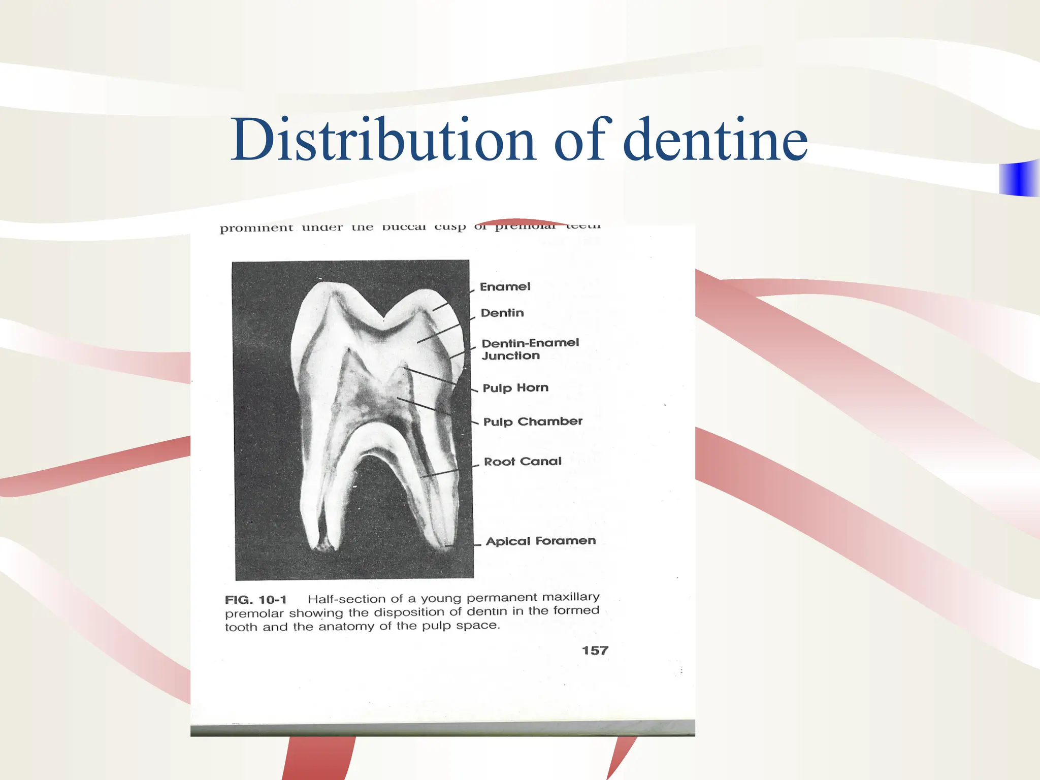
Dr Keya Sircar
Professor
Department of Oral Pathology
Faculty of Dentistry, JMI
DENTIN
Topics to be covered
1. State the composition of mature dentine.
2. Describe the relationship between dentine and
the odontoblast process.
3. Microscopic anatomy of dentine
4. Define: Dentinal tubule
Interglobular dentine/Tomes granular layer
Primary dentine/secondary/tertiary dentine
Mantle dentine/ circumpulpal dentine.
Sclerotic dentine and dead tracts.
5.Age changes and response of dentine to injury.
6.Dentine sensitivity.
7.Dentinogenesis.
What is dentine?
• Dentine forms the bulk of the tooth.
• It is present below enamel in the crown and below
cementum in the root.
• It determines the general form of the tooth.
• It is a hard tissue with tubules present along its
entire thickness.
• It is formed by cells called odontoblasts ,which are
derived from the cells of dental papilla, which is
also the primordium for dental pulp.
Distribution of dentine
Dentine and Bone
• In bone, osteoblasts exist on the surface of
bone, and when these cells get enclosed in
the bone matrix, they are called osteocytes.
• The odontoblast cell remains external to
dentin, but its process remains within
dentine in the dentinal tubules.
• Both are vital tissues as they contain
protoplasm.
Physical Properties
1.Colour:Yellowish.
2.Viscoelastic:It can deform slightly under
occlusal forces. Thus does not fracture
under function.
3.Hardness:More than bone, less than enamel.
Maximum hardness in central part, not near
pulp or at periphery. Deciduous teeth
dentine is less hard than that of permanent
teeth.
Physical Properties
4.Radiodensity:It is more radiolucent as
compared to enamel.
5.Dentine is mechanically bound to enamel at
the scalloped DEJ.
6.The junction of dentine and cementum is
less distinct.
Chemical Composition.
• Dentine contain 65% inorganic salts and
35% organic matter and water.
• Inorganic matter-Crystalline calcium
hydroxyapatite. 3Ca3(PO4)2.Ca(OH)2
• The crystals are plate shaped and smaller
than those of enamel.
• Phosphates, carbonates and sulphates.
Chemical Composition
Organic content
• It is made up of collagen fibrils ( Type I ) and a ground
substance of mucopolysaccharides (proteoglycans and
glycosaminoglycans).
• Proteoglycans: chondroitin sulphates, decorin and
biglycans.
• Glycoproteins: dentine sialoprotein, dentin
phosphoprotein, osteonectin and osteopontin
Chemical Composition
• Growth factors like Transforming growth
factor(TGF),Fibroblast growth factor(FGF), Insulin
like growth factor(IGF) and Bone morphogenic
proteins(BMP),Epidermal growth factor (EGF)etc
• The matrix components play an important role in
mineralization
Chemical Composition
• The organic content (Ground substance) of
bone and dentine are similar but
• Dentine sialoprotein and dentine
phosphoprotein are found only in dentine
and not in bone.
How is dentine studied?
• The structure of the organic part of dentine
is studied in decalcified sections.
• Here the inorganic portion is removed,
while the remaining organic part maintains
the shape of dentine.
How is dentine studied?
• The inorganic structure of dentin is studied
in a ground section.
• The organic matrix (35%) is lost
• The remaining 65%, which is the inorganic
part can be studied
Structure
• The cell bodies of odontoblasts are arranged in a single
layer on the pulpal surface of dentin.
• The cytoplasmic processes of the cells are enclosed in
tubules present in the mineralized dentin.
• Each cell produces one process, which crosses
predentin, calcified dentine and ends in a branching
network at the junction with enamel or cementum.
• Tubules are found throughout dentine and are therefore a
characteristic feature of dentine.
Structure
Dentinal Tubules
• They follow a gentle S-shaped course from the pulpal
surface to the DEJ.
• Starting from the pulpal surface, the first convexity is
directed towards the apex of the root.
• The tubules end at right angles to the DEJ and DCJ.
• At the cusp tips, incisal edges and root apices, the
tubules are almost straight.
S-shaped dentinal tubule
Dentinal Tubules
• The tubules are thus longer than the thickness of dentin.
(3-10mm or more).
• In addition to the S-shaped primary curvature, the
dentinal tubules show regular S-shaped secondary
curvatures along the entire length.
• The tubules show lateral branching throughout dentin,
which start at right angles to the main tubule.
• Lateral branches are present at intervals of 1-2mm.
Dentinal Tubules
• The ratio of the surface area of dentine at the pulpal
surface to that at the DEJ is 1:5.
• Thus the tubules are farther apart at the DEJ and are
closer together at the pulpal surface.
• The diameter of tubules is wider at the pulpal surface
than at the DEJ. It is 3-4 mm at the pulpal surface and
1mm at the DEJ.
Dentinal Tubules
• Ratio of number of tubules per unit area at the pulpal
and outer surface of dentine is 4:1.
• Some tubules extend across the DEJ and into enamel
for a few millimeters. These are enamel spindles.
Types of Dentin
• Primary / Secondary/ Tertiary dentin
• Physiologic / Reactionary dentin
• Peritubular / Intertubular dentin
Types of Dentin
• In a transverse cross-section
of dentine following
structures are seen.
1.Odontoblastic process.
2.Peritubular dentin/
Intratubular dentin.
3.Intertubular dentin.
Peritubular / Intratubular dentin
• It forms the wall of the dentinal tubule.
• It is absent or very thin at the pulpal surface and
twice as thick at the DEJ.
• It is 9% more mineralized than remaining dentine
(intertubular dentine)
Peritubular dentin
• The formation of peritubular dentine is a slow,
continuous process.
• As more and more peritubular or intratubular
dentine is formed, the diameter of the tubule
lumen decreases.
• The lumen of the tubule may also get
obliterated.
Peritubular dentin
• When tubular lumen gets obliterated with the
intratubular dentin, then the dentine has a transparent
or glassy appearance.
• The dentin is called sclerotic or transparent dentine.
• This process by which calcified tissue is laid down
within the dentinal tubule is called sclerosis.
Peritubular dentin
• The amount of sclerotic or peritubular/intratubular
dentin increases with age.
• It is most commonly seen in the apical third of the root,
and in the crown midway between the DEJ and the
pulpal surface.
Peritubular dentin
• Sclerotic dentin acts as a barrier to the
invasion of bacteria into the pulp tissue .
• Thus formation of sclerotic dentin is a
defense mechanism, which helps to prolong
pulp vitality
Intertubular dentin
• This is the main bulk of dentin, which is
present between the zones of peritubular
dentine.
• It is less mineralized than peritubular dentin
Cross-section of dentin
Decalcified section
Peritubular dentine being highly mineralized
is totally lost and seen as empty space
around the odontoblastic process.
Ground section,the empty dentinal tubule is
seen surrounded by well defined collar of
intratubular dentine
Cross section of dentin
Ground section
Ground section with EM
Types of Dentine
• Three Types
• Primary dentine forms the bulk of the tooth.
• It is the dentin formed before root formation is
completed
• It outlines the pulp chamber of the fully formed
tooth.
• Secondary dentine is dentine formed after root
formation is completed.
• Tertiary dentine is produced in reaction to
noxious stimuli such as caries or restorative
dental procedures.
Types of dentin
Primary Dentin
• Mantle dentin: This is the outermost layer
of dentin, which is present just below the
DEJ.
• It is bounded by DEJ and the interglobular
dentin
• Mantle dentin is 4% less mineralized than
rest of primary dentin. (cushioning effect)
• This zone is about 20 micrometers thick
Primary Dentin
• The collagen fibrils in the matrix are
oriented at right angles to the DEJ.
• The diameter of collagen fibres is larger
than that in rest of primary dentin.
• These larger fibres are Type 3 collagen and
are argyrophilic (silver staining positive)
• These fibres are called von Korff’s fibres
Primary Dentin
• Circumpulpal dentine: is the remaining
bulk of primary dentin.
• The collagen fibrils in circumpulpal dentin
are smaller than those in mantle dentin.
• The circumpulpal dentin is more
mineralized as compared to mantle dentin.
Types of dentin
Secondary Dentin
• It is the dentin formed after root formation is
completed.
• It is a narrow band which borders the pulp.
• It forms in erupted/functional as well as unerupted
teeth.
• It is formed at a slower rate.
Secondary Dentin
• It is deposited along the periphery of pulp,
but not evenly on all sides.
• There is greater deposition of the roof and
floor of the pulp chamber.
• This leads to an asymmetric decrease in size
of pulp chamber. This is called pulp
recession.
• Clinical significance?
Secondary dentin
• The tubular structure is less regular and
number of tubules is less than primary
dentin
• Secondary dentin gets sclerosed more easily
than primary dentin.
• This tends to reduce the overall
permeability of dentin, thereby protecting
the pulp from bacterial invasion.
Tertiary Dentin
• It is also called Reactive, reparative or irregular
dentin.
• It is produced in response to noxious stimuli such
as caries, or restorative dental procedures.
• It is produced only by those odontoblasts affected
by the stimulus.
• This is different from primary and secondary
dentine which are formed all along the pulp-
dentine border.
Tertiary dentin
• The quality and structure of this dentin is
related to the intensity and the duration of
the stimulus.
• Ex: An active carious lesion causes
extensive destruction of dentine and
considerable pulp damage.
• In such a case ,the tertiary dentine is
deposited rapidly
• If the stimulus is of low intensity, the
tertiary dentine is formed at a slower rate..
Tertiary dentine
• There is practically no continuity in tubules
of primary or secondary dentine and those
of tertiary dentine.
• Thus dentine permeability is reduced,
which helps to protect the pulp.
Tertiary dentine
Structure differs from primary/secondary
dentine.
1.Number of tubules is reduced.
2.Cells may get included in tertiary
dentine,which forms very rapidly.Such
tertiary dentine is called osteodentine.
3.Tubules are irregular .
Dentin structure
Structures seen in dentin
Predentine
• The layer of predentine or dentine matrix is
located adjacent to pulp.
• It is 2-6 micrometres wide.
• It is secreted by the odontoblasts.
• As mineralization takes place,the predentine
becomes dentine and a new layer of
predentine is secreted by the odontoblasts.
Odontoblastic process
• The odontoblastic process is the
cytoplasmic extension of the odontoblast.
• The process is widest (3-4 microns) at the
pulp border and narrows to 1 microns at the
DEJ.
• The process may extend right upto the DEJ
in some tubules.
• In narrowed tubules or those in which
sclerosis has occurred the process may end
short of the DEJ.
Odontoblastic process
• Some of the odontoblastic processes may
cross the DEJ .
• These terminal ends of the odontoblastic
process may extend into enamel for a short
distance.
• These are called enamel spindles.
Structures seen in dentin
Interglobular dentin
• Mineralization of dentin sometimes begins
as small globules, which then fuse together.
• Some globules may fail to fuse, leaving
hypomineralized or unmineralized areas
within mature dentin.
• These unmineralized areas are called
interglobular dentine.
• In dry ground sections, the hypomineralized
areas are lost, and are seen as black spaces
in transmitted light.
Interglobular dentine
• It forms in circumpulpal dentin just below
the mantle dentin. In this area, the pattern of
mineralization is globular.
• The tubular and incremental patterns
remains unaffected in interglobular dentine.
• This indicates that interglobular dentine is
formed due to defect in mineralization,
while matrix formation is normal.
Interglobular Dentine
The tubular and incremental
pattern remains unaffected.
Interglobular dentine
Tomes Granular Layer
• In ground sections, a granular layer is seen
adjacent to cementum in root dentin.
• The granular area increases from the CEJ
to the apex of the tooth.
• It is produced by looping and joining of the
terminal portions of the dentinal tubules.
• It is similar to Y-shaped branching of
dentinal tubules seen in the crown adjacent
to the DEJ.
Incremental Lines of Dentine
Dentine is formed in an incremental manner.
During formation of dentine,periods of
activity alternate with periods of rest.
These alterations in activity are seen as
incremental lines .
These lines are best seen in longitudinal
ground sections.
Incremental Lines of Dentine
• The incremental lines run at right angles to
the dentinal tubules.
• Their pattern corresponds to the incremental
lines of enamel.
• There are three types of incremental lines in
dentine.
Incremental Lines of Dentine
• Lines marking the daily deposition of
dentine (4 microns) can be demonstrated
with special stains.There is no particular
name for these lines.
• Lines which represent the 5-day
incremental pattern are the lines of Von
Ebner.These are at a distance of 20
micrometres.
Incremental Lines of Dentine
• Some lines are accentuated because of
disturbance in dentine matrix and
mineralization.
• These lines are Contour lines of Owen.
• These have been shown to be hypocalcified
bands.
Incremental Lines of Dentine
• In the deciduous teeth and the first permanent
molars,where the dentine is formed partly before
and partly after birth,an accentuated contour line is
seen.
• It separates the prenatal enamel from the post natal
enamel.
• It is called the neonatal line.
• It represents the sudden change in environment at
birth.
• The dentine matrix formed before birth is of better
quality than that formed after birth.
• The neonatal line may be a hypocalcified band.
Incremental Lines of dentine
Dead Tracts
• Odontoblast processes are lost due to caries,
attrition, cavity preparation etc.
• Degeneration of some odontoblasts occurs
in area of pulp horns due to crowding
• The empty dentinal tubules appear black in
transmitted light and white in reflected light
• These are called dead tracts
• Reparative dentin seals the tubules at the
pulpal end,
• Such areas are less sensitive than normal
dentin
Dentine Sensitivity
• Dentine is very sensitve.
• The sensation is that of pain.
• The sensation is diffuse.
• So clinical localization is difficult.
• Dentine is most sensitive at the the DEJ and
near the pulp.
• Sensitivity is increased when pulp is
inflamed.
Mechanisms of Dentine
Sensitivity
1.Dentine contains nerves which respond
when dentine is stimulated.
2.The odontoblasts act as nerve receptors.
3.Hydrodyanamic theory-Fluid movement
within the dentinal tubule is registered by
free nerve endings in the pulp close to
dentine.MOST ACCEPTED
Intratubular nerves
• Nerve endings extend into dentine upto a distance
of 100-150microns.
• The nerve endings are single terminals,which are
packed with small vesicles that contain
neurotransmitter substance.
• The nerve endings are the terminal processes of
the myelinated nerves of the dental pulp.
• The nerve endings intertwine with the
odontoblastic process.
Intratubular nerves
• Present experimental evidence indicates that
while nerves are present in dentine,its sensitivity
does not depend on stimulation of these nerves.
1. DEJ most sensitive,while nerves don’t extend to
DEJ.
2. Nerves are not established till some time afetr
eruption,but newly erupted teeth are sensitive.
3. Application of LA to exposed dentine does not
eliminate dentine sensitivity.
Odontoblast as a receptor
As the odontoblast has origin fron the neural
crest,so it was thought that it has the ability
of propogating an impulse.
However,no synaptic junction is present
between odontoblasts and pulpal nerves.
So this theory is not acceptable.
Hydrodyanamic theory
• Fluid movement through the tubule distorts the
local pulp invironment.
• This is sensed by the free nerve endings in the
plexus of Raschkow.
• Incresed sensitivity at the DEJ is due to extensive
branching at the DEJ.
• This theory explains why LA does not block
sensitivity of dentine.
• Also explains why pain is produce by
dehydration,hypertonic solution,thermal change
and mechanical probing of exposed dentine.
Dentinogenesis
• Dentine formation begins in late bell stage
of development in the dental papilla.
• Dentine formation begins in the area of the
future cusp tips and progresses cervically.
• In multicusped teeth,dentine formation
begins at the sites of the future cusp tips.
• The thickness of dentine increases until all
the coronal dentine is formed.
Dentinogenesis
• Cells of IEE express TGF, IGF and BMP
• These cytokines are taken up by
preodontoblast cells in dental papilla
• Under the influence of these factors, there is
reorganization of the cytoskeleton of the
preodontoblast, which then differentiates
into the odontoblast.
Dentinogenesis
• As differentiation occurs, the ovoid cell
becomes columnar and nucleus gets basally
oriented.
• The cell assumes a length of 40 microns
and width of 7 microns
• From the apical end of the cell, a
cytoplasmic process develops and grows
towards the basement membrane
Dentinogenesis
• Dentin phosphoprotein (DPP) is an
important protein secreted by odontoblasts
which is concerned with mineralization.
• DPP binds to calcium, carries it to the
mineralizing front and regulates the growth
of the apatite crystal
Dentinogenesis
• Osteonectin, secreted by odontoblasts also
helps to bind calcium.However it inhibits
the growth of the crystal.
• Osteopontin also aids in mineralization.
• GLA protein acts a seeds or nucleators to
attract and concentrate calcium.
Dentinogenesis
• Chondroitin sulfate has different effects.
• In predentin, it prevents transport of apatite
crystals
• In dentin however, it helps to bind calcium
to collagen
Dentinogenesis
• As the odontoblast recedes pulpally, it
leaves behind a single extension.
• The extension gets enclosed within the
dentinal tubule.
• Initially 4 microns of dentin are formed
everyday till crown formation is completed.
• Dentin formation slows down after the
tooth has erupted
Dentinogenesis
• Secondary dentin formation after root
formation is completed is a much slower
process.
• Reparative dentin can form at the rate of 4
microns per day for several months after a
restoration
• Root dentin formation is slower than
coronal dentin formation
• The collagen fibres in root dentin are
parallel to cemento dentinal junction.
Dentinogenesis
• The first calcium crystals are deposited on
the surface of the collagen fibres and later
within the fibrils.
• The apatite crystal in dentin is 300 times
smaller than the crystal in enamel.
• Genes like MAP1B for odontoblast
differentiation and PHEX for dentin
mineralization have been identified
Clinical Consideration
• The rapid spread of caries in dentin is due
to its tubular structure.
• Lateral spread at the DEJ leads to
undermining of enamel
• Concept of backward caries?
Clinical Consideration
• The basic principle of treating
hypersensitivity is to block patent tubules or
to prevent pulpal nerve response
• Dentifrices containing Potassium nitrate,
stannous fluoride etc are used
• Lasers have also been used
Clinical Consideration
• Root dentin is less sensitive than coronal
dentin
• Sensitivity of outer radicular dentin is 2 %
that of coronal dentin
• At the pulpal side, root dentin is 20% as
sensitive as coronal dentin
• Thus outer radicular dentin prevents fluid
movement across dentin
• Root planing is followed by increased
sensitivity?
Clinical Consideration
• What is smear layer?
• The smear layer is a surface accumulation
of debris formed on dentine during
instrumentation.
• It is composed of organic and inorganic
components.
Clinical Consideration
• Should the smear layer should be
removed or not?
• Removal will eliminate the bacteria
contained within it and will expose the
dentinal tubules allowing closer adaptation
of the obturating material.
• Sealer and gutta-percha have been shown
to flow into the tubules and bonding agents
can form tags in the dentine.
Clinical Consideration
• But, opening the tubules may provide a
passage for microorganisms into the body
of the dentin
• Dentin formation is not affected by Vit D
deficiency states
• Fluoride incorporated during dentinigenesis
helps to increase the hardness of dentin

dentin formation , calcification, types , and dentinogenesis explained thoroughly

  • 1.
    Dr Keya Sircar Professor Departmentof Oral Pathology Faculty of Dentistry, JMI DENTIN
  • 2.
    Topics to becovered 1. State the composition of mature dentine. 2. Describe the relationship between dentine and the odontoblast process. 3. Microscopic anatomy of dentine 4. Define: Dentinal tubule Interglobular dentine/Tomes granular layer Primary dentine/secondary/tertiary dentine Mantle dentine/ circumpulpal dentine. Sclerotic dentine and dead tracts. 5.Age changes and response of dentine to injury. 6.Dentine sensitivity. 7.Dentinogenesis.
  • 3.
    What is dentine? •Dentine forms the bulk of the tooth. • It is present below enamel in the crown and below cementum in the root. • It determines the general form of the tooth. • It is a hard tissue with tubules present along its entire thickness. • It is formed by cells called odontoblasts ,which are derived from the cells of dental papilla, which is also the primordium for dental pulp.
  • 4.
  • 5.
    Dentine and Bone •In bone, osteoblasts exist on the surface of bone, and when these cells get enclosed in the bone matrix, they are called osteocytes. • The odontoblast cell remains external to dentin, but its process remains within dentine in the dentinal tubules. • Both are vital tissues as they contain protoplasm.
  • 6.
    Physical Properties 1.Colour:Yellowish. 2.Viscoelastic:It candeform slightly under occlusal forces. Thus does not fracture under function. 3.Hardness:More than bone, less than enamel. Maximum hardness in central part, not near pulp or at periphery. Deciduous teeth dentine is less hard than that of permanent teeth.
  • 7.
    Physical Properties 4.Radiodensity:It ismore radiolucent as compared to enamel. 5.Dentine is mechanically bound to enamel at the scalloped DEJ. 6.The junction of dentine and cementum is less distinct.
  • 8.
    Chemical Composition. • Dentinecontain 65% inorganic salts and 35% organic matter and water. • Inorganic matter-Crystalline calcium hydroxyapatite. 3Ca3(PO4)2.Ca(OH)2 • The crystals are plate shaped and smaller than those of enamel. • Phosphates, carbonates and sulphates.
  • 9.
    Chemical Composition Organic content •It is made up of collagen fibrils ( Type I ) and a ground substance of mucopolysaccharides (proteoglycans and glycosaminoglycans). • Proteoglycans: chondroitin sulphates, decorin and biglycans. • Glycoproteins: dentine sialoprotein, dentin phosphoprotein, osteonectin and osteopontin
  • 10.
    Chemical Composition • Growthfactors like Transforming growth factor(TGF),Fibroblast growth factor(FGF), Insulin like growth factor(IGF) and Bone morphogenic proteins(BMP),Epidermal growth factor (EGF)etc • The matrix components play an important role in mineralization
  • 11.
    Chemical Composition • Theorganic content (Ground substance) of bone and dentine are similar but • Dentine sialoprotein and dentine phosphoprotein are found only in dentine and not in bone.
  • 12.
    How is dentinestudied? • The structure of the organic part of dentine is studied in decalcified sections. • Here the inorganic portion is removed, while the remaining organic part maintains the shape of dentine.
  • 13.
    How is dentinestudied? • The inorganic structure of dentin is studied in a ground section. • The organic matrix (35%) is lost • The remaining 65%, which is the inorganic part can be studied
  • 14.
    Structure • The cellbodies of odontoblasts are arranged in a single layer on the pulpal surface of dentin. • The cytoplasmic processes of the cells are enclosed in tubules present in the mineralized dentin. • Each cell produces one process, which crosses predentin, calcified dentine and ends in a branching network at the junction with enamel or cementum. • Tubules are found throughout dentine and are therefore a characteristic feature of dentine.
  • 15.
  • 16.
    Dentinal Tubules • Theyfollow a gentle S-shaped course from the pulpal surface to the DEJ. • Starting from the pulpal surface, the first convexity is directed towards the apex of the root. • The tubules end at right angles to the DEJ and DCJ. • At the cusp tips, incisal edges and root apices, the tubules are almost straight.
  • 17.
  • 18.
    Dentinal Tubules • Thetubules are thus longer than the thickness of dentin. (3-10mm or more). • In addition to the S-shaped primary curvature, the dentinal tubules show regular S-shaped secondary curvatures along the entire length. • The tubules show lateral branching throughout dentin, which start at right angles to the main tubule. • Lateral branches are present at intervals of 1-2mm.
  • 19.
    Dentinal Tubules • Theratio of the surface area of dentine at the pulpal surface to that at the DEJ is 1:5. • Thus the tubules are farther apart at the DEJ and are closer together at the pulpal surface. • The diameter of tubules is wider at the pulpal surface than at the DEJ. It is 3-4 mm at the pulpal surface and 1mm at the DEJ.
  • 20.
    Dentinal Tubules • Ratioof number of tubules per unit area at the pulpal and outer surface of dentine is 4:1. • Some tubules extend across the DEJ and into enamel for a few millimeters. These are enamel spindles.
  • 21.
    Types of Dentin •Primary / Secondary/ Tertiary dentin • Physiologic / Reactionary dentin • Peritubular / Intertubular dentin
  • 22.
    Types of Dentin •In a transverse cross-section of dentine following structures are seen. 1.Odontoblastic process. 2.Peritubular dentin/ Intratubular dentin. 3.Intertubular dentin.
  • 23.
    Peritubular / Intratubulardentin • It forms the wall of the dentinal tubule. • It is absent or very thin at the pulpal surface and twice as thick at the DEJ. • It is 9% more mineralized than remaining dentine (intertubular dentine)
  • 24.
    Peritubular dentin • Theformation of peritubular dentine is a slow, continuous process. • As more and more peritubular or intratubular dentine is formed, the diameter of the tubule lumen decreases. • The lumen of the tubule may also get obliterated.
  • 25.
    Peritubular dentin • Whentubular lumen gets obliterated with the intratubular dentin, then the dentine has a transparent or glassy appearance. • The dentin is called sclerotic or transparent dentine. • This process by which calcified tissue is laid down within the dentinal tubule is called sclerosis.
  • 26.
    Peritubular dentin • Theamount of sclerotic or peritubular/intratubular dentin increases with age. • It is most commonly seen in the apical third of the root, and in the crown midway between the DEJ and the pulpal surface.
  • 27.
    Peritubular dentin • Scleroticdentin acts as a barrier to the invasion of bacteria into the pulp tissue . • Thus formation of sclerotic dentin is a defense mechanism, which helps to prolong pulp vitality
  • 28.
    Intertubular dentin • Thisis the main bulk of dentin, which is present between the zones of peritubular dentine. • It is less mineralized than peritubular dentin
  • 29.
    Cross-section of dentin Decalcifiedsection Peritubular dentine being highly mineralized is totally lost and seen as empty space around the odontoblastic process. Ground section,the empty dentinal tubule is seen surrounded by well defined collar of intratubular dentine
  • 30.
    Cross section ofdentin Ground section
  • 31.
  • 32.
    Types of Dentine •Three Types • Primary dentine forms the bulk of the tooth. • It is the dentin formed before root formation is completed • It outlines the pulp chamber of the fully formed tooth. • Secondary dentine is dentine formed after root formation is completed. • Tertiary dentine is produced in reaction to noxious stimuli such as caries or restorative dental procedures.
  • 33.
  • 34.
    Primary Dentin • Mantledentin: This is the outermost layer of dentin, which is present just below the DEJ. • It is bounded by DEJ and the interglobular dentin • Mantle dentin is 4% less mineralized than rest of primary dentin. (cushioning effect) • This zone is about 20 micrometers thick
  • 35.
    Primary Dentin • Thecollagen fibrils in the matrix are oriented at right angles to the DEJ. • The diameter of collagen fibres is larger than that in rest of primary dentin. • These larger fibres are Type 3 collagen and are argyrophilic (silver staining positive) • These fibres are called von Korff’s fibres
  • 36.
    Primary Dentin • Circumpulpaldentine: is the remaining bulk of primary dentin. • The collagen fibrils in circumpulpal dentin are smaller than those in mantle dentin. • The circumpulpal dentin is more mineralized as compared to mantle dentin.
  • 37.
  • 38.
    Secondary Dentin • Itis the dentin formed after root formation is completed. • It is a narrow band which borders the pulp. • It forms in erupted/functional as well as unerupted teeth. • It is formed at a slower rate.
  • 39.
    Secondary Dentin • Itis deposited along the periphery of pulp, but not evenly on all sides. • There is greater deposition of the roof and floor of the pulp chamber. • This leads to an asymmetric decrease in size of pulp chamber. This is called pulp recession. • Clinical significance?
  • 40.
    Secondary dentin • Thetubular structure is less regular and number of tubules is less than primary dentin • Secondary dentin gets sclerosed more easily than primary dentin. • This tends to reduce the overall permeability of dentin, thereby protecting the pulp from bacterial invasion.
  • 41.
    Tertiary Dentin • Itis also called Reactive, reparative or irregular dentin. • It is produced in response to noxious stimuli such as caries, or restorative dental procedures. • It is produced only by those odontoblasts affected by the stimulus. • This is different from primary and secondary dentine which are formed all along the pulp- dentine border.
  • 42.
    Tertiary dentin • Thequality and structure of this dentin is related to the intensity and the duration of the stimulus. • Ex: An active carious lesion causes extensive destruction of dentine and considerable pulp damage. • In such a case ,the tertiary dentine is deposited rapidly • If the stimulus is of low intensity, the tertiary dentine is formed at a slower rate..
  • 43.
    Tertiary dentine • Thereis practically no continuity in tubules of primary or secondary dentine and those of tertiary dentine. • Thus dentine permeability is reduced, which helps to protect the pulp.
  • 44.
    Tertiary dentine Structure differsfrom primary/secondary dentine. 1.Number of tubules is reduced. 2.Cells may get included in tertiary dentine,which forms very rapidly.Such tertiary dentine is called osteodentine. 3.Tubules are irregular .
  • 45.
  • 46.
    Structures seen indentin Predentine • The layer of predentine or dentine matrix is located adjacent to pulp. • It is 2-6 micrometres wide. • It is secreted by the odontoblasts. • As mineralization takes place,the predentine becomes dentine and a new layer of predentine is secreted by the odontoblasts.
  • 47.
    Odontoblastic process • Theodontoblastic process is the cytoplasmic extension of the odontoblast. • The process is widest (3-4 microns) at the pulp border and narrows to 1 microns at the DEJ. • The process may extend right upto the DEJ in some tubules. • In narrowed tubules or those in which sclerosis has occurred the process may end short of the DEJ.
  • 48.
    Odontoblastic process • Someof the odontoblastic processes may cross the DEJ . • These terminal ends of the odontoblastic process may extend into enamel for a short distance. • These are called enamel spindles.
  • 49.
    Structures seen indentin Interglobular dentin • Mineralization of dentin sometimes begins as small globules, which then fuse together. • Some globules may fail to fuse, leaving hypomineralized or unmineralized areas within mature dentin. • These unmineralized areas are called interglobular dentine. • In dry ground sections, the hypomineralized areas are lost, and are seen as black spaces in transmitted light.
  • 50.
    Interglobular dentine • Itforms in circumpulpal dentin just below the mantle dentin. In this area, the pattern of mineralization is globular. • The tubular and incremental patterns remains unaffected in interglobular dentine. • This indicates that interglobular dentine is formed due to defect in mineralization, while matrix formation is normal.
  • 51.
  • 52.
    Interglobular Dentine The tubularand incremental pattern remains unaffected.
  • 53.
  • 54.
    Tomes Granular Layer •In ground sections, a granular layer is seen adjacent to cementum in root dentin. • The granular area increases from the CEJ to the apex of the tooth. • It is produced by looping and joining of the terminal portions of the dentinal tubules. • It is similar to Y-shaped branching of dentinal tubules seen in the crown adjacent to the DEJ.
  • 55.
    Incremental Lines ofDentine Dentine is formed in an incremental manner. During formation of dentine,periods of activity alternate with periods of rest. These alterations in activity are seen as incremental lines . These lines are best seen in longitudinal ground sections.
  • 56.
    Incremental Lines ofDentine • The incremental lines run at right angles to the dentinal tubules. • Their pattern corresponds to the incremental lines of enamel. • There are three types of incremental lines in dentine.
  • 57.
    Incremental Lines ofDentine • Lines marking the daily deposition of dentine (4 microns) can be demonstrated with special stains.There is no particular name for these lines. • Lines which represent the 5-day incremental pattern are the lines of Von Ebner.These are at a distance of 20 micrometres.
  • 58.
    Incremental Lines ofDentine • Some lines are accentuated because of disturbance in dentine matrix and mineralization. • These lines are Contour lines of Owen. • These have been shown to be hypocalcified bands.
  • 59.
    Incremental Lines ofDentine • In the deciduous teeth and the first permanent molars,where the dentine is formed partly before and partly after birth,an accentuated contour line is seen. • It separates the prenatal enamel from the post natal enamel. • It is called the neonatal line. • It represents the sudden change in environment at birth. • The dentine matrix formed before birth is of better quality than that formed after birth. • The neonatal line may be a hypocalcified band.
  • 60.
  • 61.
    Dead Tracts • Odontoblastprocesses are lost due to caries, attrition, cavity preparation etc. • Degeneration of some odontoblasts occurs in area of pulp horns due to crowding • The empty dentinal tubules appear black in transmitted light and white in reflected light • These are called dead tracts
  • 62.
    • Reparative dentinseals the tubules at the pulpal end, • Such areas are less sensitive than normal dentin
  • 63.
    Dentine Sensitivity • Dentineis very sensitve. • The sensation is that of pain. • The sensation is diffuse. • So clinical localization is difficult. • Dentine is most sensitive at the the DEJ and near the pulp. • Sensitivity is increased when pulp is inflamed.
  • 64.
    Mechanisms of Dentine Sensitivity 1.Dentinecontains nerves which respond when dentine is stimulated. 2.The odontoblasts act as nerve receptors. 3.Hydrodyanamic theory-Fluid movement within the dentinal tubule is registered by free nerve endings in the pulp close to dentine.MOST ACCEPTED
  • 65.
    Intratubular nerves • Nerveendings extend into dentine upto a distance of 100-150microns. • The nerve endings are single terminals,which are packed with small vesicles that contain neurotransmitter substance. • The nerve endings are the terminal processes of the myelinated nerves of the dental pulp. • The nerve endings intertwine with the odontoblastic process.
  • 66.
    Intratubular nerves • Presentexperimental evidence indicates that while nerves are present in dentine,its sensitivity does not depend on stimulation of these nerves. 1. DEJ most sensitive,while nerves don’t extend to DEJ. 2. Nerves are not established till some time afetr eruption,but newly erupted teeth are sensitive. 3. Application of LA to exposed dentine does not eliminate dentine sensitivity.
  • 67.
    Odontoblast as areceptor As the odontoblast has origin fron the neural crest,so it was thought that it has the ability of propogating an impulse. However,no synaptic junction is present between odontoblasts and pulpal nerves. So this theory is not acceptable.
  • 68.
    Hydrodyanamic theory • Fluidmovement through the tubule distorts the local pulp invironment. • This is sensed by the free nerve endings in the plexus of Raschkow. • Incresed sensitivity at the DEJ is due to extensive branching at the DEJ. • This theory explains why LA does not block sensitivity of dentine. • Also explains why pain is produce by dehydration,hypertonic solution,thermal change and mechanical probing of exposed dentine.
  • 69.
    Dentinogenesis • Dentine formationbegins in late bell stage of development in the dental papilla. • Dentine formation begins in the area of the future cusp tips and progresses cervically. • In multicusped teeth,dentine formation begins at the sites of the future cusp tips. • The thickness of dentine increases until all the coronal dentine is formed.
  • 70.
    Dentinogenesis • Cells ofIEE express TGF, IGF and BMP • These cytokines are taken up by preodontoblast cells in dental papilla • Under the influence of these factors, there is reorganization of the cytoskeleton of the preodontoblast, which then differentiates into the odontoblast.
  • 71.
    Dentinogenesis • As differentiationoccurs, the ovoid cell becomes columnar and nucleus gets basally oriented. • The cell assumes a length of 40 microns and width of 7 microns • From the apical end of the cell, a cytoplasmic process develops and grows towards the basement membrane
  • 72.
    Dentinogenesis • Dentin phosphoprotein(DPP) is an important protein secreted by odontoblasts which is concerned with mineralization. • DPP binds to calcium, carries it to the mineralizing front and regulates the growth of the apatite crystal
  • 73.
    Dentinogenesis • Osteonectin, secretedby odontoblasts also helps to bind calcium.However it inhibits the growth of the crystal. • Osteopontin also aids in mineralization. • GLA protein acts a seeds or nucleators to attract and concentrate calcium.
  • 74.
    Dentinogenesis • Chondroitin sulfatehas different effects. • In predentin, it prevents transport of apatite crystals • In dentin however, it helps to bind calcium to collagen
  • 75.
    Dentinogenesis • As theodontoblast recedes pulpally, it leaves behind a single extension. • The extension gets enclosed within the dentinal tubule. • Initially 4 microns of dentin are formed everyday till crown formation is completed. • Dentin formation slows down after the tooth has erupted
  • 76.
    Dentinogenesis • Secondary dentinformation after root formation is completed is a much slower process. • Reparative dentin can form at the rate of 4 microns per day for several months after a restoration • Root dentin formation is slower than coronal dentin formation • The collagen fibres in root dentin are parallel to cemento dentinal junction.
  • 77.
    Dentinogenesis • The firstcalcium crystals are deposited on the surface of the collagen fibres and later within the fibrils. • The apatite crystal in dentin is 300 times smaller than the crystal in enamel. • Genes like MAP1B for odontoblast differentiation and PHEX for dentin mineralization have been identified
  • 78.
    Clinical Consideration • Therapid spread of caries in dentin is due to its tubular structure. • Lateral spread at the DEJ leads to undermining of enamel • Concept of backward caries?
  • 79.
    Clinical Consideration • Thebasic principle of treating hypersensitivity is to block patent tubules or to prevent pulpal nerve response • Dentifrices containing Potassium nitrate, stannous fluoride etc are used • Lasers have also been used
  • 80.
    Clinical Consideration • Rootdentin is less sensitive than coronal dentin • Sensitivity of outer radicular dentin is 2 % that of coronal dentin • At the pulpal side, root dentin is 20% as sensitive as coronal dentin • Thus outer radicular dentin prevents fluid movement across dentin • Root planing is followed by increased sensitivity?
  • 81.
    Clinical Consideration • Whatis smear layer? • The smear layer is a surface accumulation of debris formed on dentine during instrumentation. • It is composed of organic and inorganic components.
  • 82.
    Clinical Consideration • Shouldthe smear layer should be removed or not? • Removal will eliminate the bacteria contained within it and will expose the dentinal tubules allowing closer adaptation of the obturating material. • Sealer and gutta-percha have been shown to flow into the tubules and bonding agents can form tags in the dentine.
  • 83.
    Clinical Consideration • But,opening the tubules may provide a passage for microorganisms into the body of the dentin
  • 84.
    • Dentin formationis not affected by Vit D deficiency states • Fluoride incorporated during dentinigenesis helps to increase the hardness of dentin