Downloaded 474 times
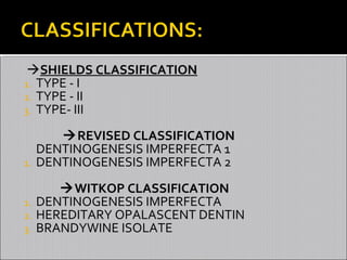


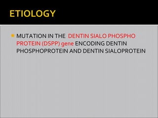


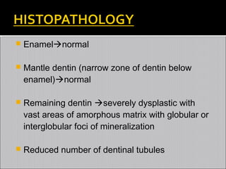
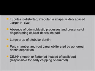


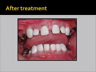


This document provides an overview of dentinogenesis imperfecta. It begins with an introduction to dentin formation and classification systems for dentinogenesis imperfecta. It then describes the etiology as a genetic disorder involving mutations in the DSPP gene. Clinical features include opalescent teeth that become gray/brown with age and obliterated pulp chambers. Radiographic features include bulbous crowns and thin roots. Histopathology shows abnormal dentin formation. Treatment aims to prevent attrition and involves restorations like crowns. The document provides details on different subtypes and clinical management.


