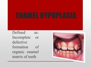
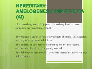
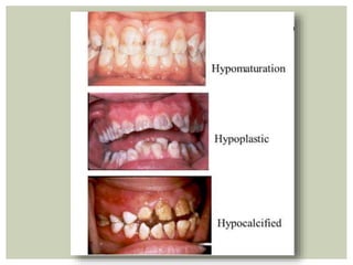
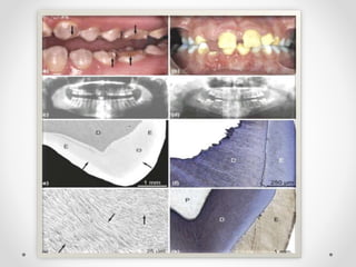
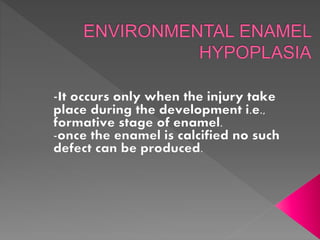
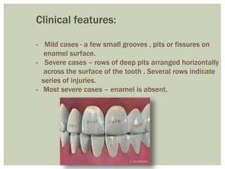
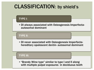
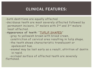
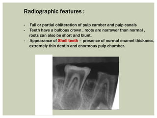
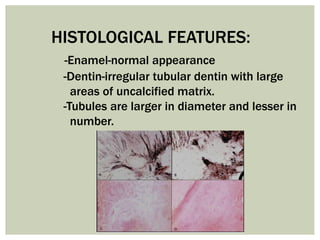
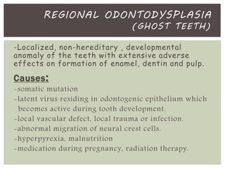
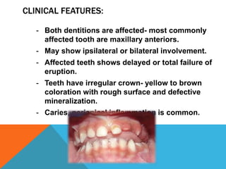
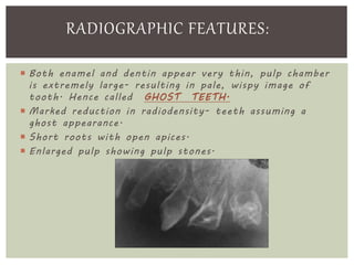
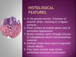
STRUCTURAL
ANOMALIES OF TEETH
B Y : A N K I T A
B A N S A L
Structural
anomalies
Enamel
• Enamel hypoplasia
• Hereditary -Amelogenesis
Imperfecta
• Environmental Enamel
hypoplasia
Dentin
• Dentinogenesis Imperfecta
• Dentin Dysplasia
• Regional Odontodysplasia
ENAMEL HYPOPLASIA
Defined as:
Incomplete or
defective
formation of
organic enamel
matrix of teeth
a.k.a. hereditary enamel dysplasia , hereditary brown enamel ,
hereditary brown opalescent teeth
-It represents a group of hereditary defects of enamel unassociated
with any other generalized defects.
-It is entirely an ectodermal disturbance and the mesodermal
components of teeth are completely normal.
-It is inherited as an autosomal dominant ,autosomal recessive or x-
linked disorder.
 FORMATIVE STAGE: Deposition of organic matrix
Defect- Hypoplastic Amelogenesis Imperfecta
 CALCIFICATION STAGE: Matrix mineralization
Defect- Hypocalcification Amelogenesis Imperfecta
 MATURATION STAGE: Crystallites mature and enlarge
Defect- Hypomaturation Amelogenesis Imperfecta
• IA: HYPOPLASTIC PITTED AUTOSOMAL DOMINANT
• IB: HYPOPLASTIC LOCAL AUTOSOMAL DOMINANT
• IC: HYPOPLASTIC LOCAL AUTOSOMAL RECESSIVE
• ID: HYPOPLATIC SMOOTH AUTOSOMAL DOMINANT
• IE: HYPOPLASTIC SMOOTH X-LINKED DOMINANT
• IF: HYPOPLASTIC ROUGH AUTOSOMAL DOMINANT
• IG: ENAMEL AGENESIS AUTOSOMAL RECESSIVE
TYPE I: HYPOPLASTIC
• IIA: HYPOMATURATION PIGMENTED AUTOSOMAL DOMINANT
• IIB: HYPOMATURATION X-LINKED RECESSIVE
• IIC: SNOW CAPPED TEETH AUTOSOMAL DOMINANT
TYPE II: HYPOMATURATIVE
• IIIA: AUTOSOMAL DOMINANT
• IIIB: AUTOSOMAL RECESSIVE
TYPE III: HYPCALCIFICATION
CLASSIFICATION OF AMELOGENESIS IMPERFECTA
Acc. to Witkop 1989
• IVA: HYPOMATURATION - HYPOPLASTIC WITH
TAURODONTISM , AUTOSOMAL DOMINANT
• IVB: HYPOPALSTIC- HYPOMATURATION WITH
TAURODONTISM , AUTOSOMAL DOMINANT
TYPE IV: HYPOMATURATION-
HYPOPLASTIC WITH TAURODONTISM
 HYPOPLASTIC: Enamel is not formed to the full
normal thickness on the newly erupted
teeth.
 HYPOCALCIFICATION: Enamel is so soft that it
can be removed by prophylaxis treatment.
 HYPOMATURTION: Enamel can be pierced by
an
explorer under firm press and can be
lost by
CLINICAL FEATURES:
-Given by witkop and sauk
CLINICAL FEATURES ACCORDING TO
CLASSIFICATION:
1. HYPOPLASTIC AI:
Occurs due to inadequate deposition of enamel matrix.
i. Generalized pattern
ii. Localized pattern
iii. Autosomal dominant smooth pattern
iv. X-linked smooth pattern
v. Rough pattern
vi. Enamel agenesis
(i) Generalized pattern:
- pin point to head size pits scattered across
the surface of teeth in rows or column.
- buccal surface affected more severely.
- staining of pits
- enamel between the pits have normal thickness,
hardness , coloration.
(ii) Localized pattern:
- affected teeth shows horizontal rows of pits , linear
depressions.
- altered area is located on the middle third o the buccal surfaces,
incisal or occlusal surfaces are not affected.
- both dentitions or primary teeth are affected.
- all teeth affected or only scattered teeth affected
(iii) Autosomal dominant smooth pattern:
- enamel of the teeth exhibit smooth surface-thin, hard and glossy.
- absence of appropriate enamel- cone shaped crown with open
contact points.
- color of the teeth-opaque white to translucent brown
- radiographically -thin peripheral outline of radio opaque enamel.
(iv) x-linked smooth pattern:
- FEMALE- alternating zones of normal and abnormal pattern- because
one x-inherits normal formation of enamel and other x-inherits defective
formation.
- MALE- exhibit diffuse, thin, smooth, shiny enamel in both dentitions.
Teeth are cones shaped, contact points open.
Color-very strong brown to yellow brown.
unerupted tooth shows resorption.
- HETEROZYGOUS FEMALE- shows vertical furrows of thin hypoplastic
enamel, alternating with bands of normal thickness.
(v) Rough pattern:
- enamel is thin, hard, rough surface, teeth taper towards incisal edge,
occlusal surfaces with open contact points.
- color- white to yellow white.
- anterior open bite
- teeth are less vulnerable to attrition.
(vi) Enamel agenesis:
- total lack of enamel formation.
- color-shape end color of the tooth are same as dentin with yellow brown
hue, open contacts.
- surface of dentin- rough , anterior open bite
- x-ray- no peripheral enamel overlying dentin.
2. HYPOMATURATION AI
- Enamel matrix laid down is appropriate, begins to mineralize bu
there is the defect in maturation of enamel’s crystal structure.
- affected teeth- normal in shape but exhibit mottled, opaque whi
brown, yellow discoloration.
- enamel is softer, tends to chip off from the underlying dentin.
- x-ray- the affected enamel exhibit radiolucency similar to dentin
It is shown in 3 patterns:
1. pigmented pattern
2. x-linked pattern
3. snow capped pattern
1.PIGMENTED PATTERN:
-surface- mottled brown.
-enamel fractures from underlying dentin and is soft enough to be
punctured by explorer.
-anterior open bite .
2. X-LINKED PATTERN
-male-deciduous teeth- opaque white with translucent mottling,
permanent teeth-opaque yellow white, darkens with age.
-enamel tends to chip off, can be pierced with explorer.
-degree of enamel loss is more rapid.
-focal area of brown discoloration seen with white opaque enamel
-x-ray shows decreased contrast between enamel and dentin.
-heterozygous female-teeth shows vertical ands of a white opaque
enamel which are random and symmetrical.
3.SNOW CAPPED PATTERN
-zone of white opaque enamel on incisal or occlusal one quarter to
1/3rd of crown.
-affected teeth show an anterior to posterior disturbance.
3. HYPOCALCIFICATION AI:
-Enamel is laid down appropriately with no significant mineralization.
-shape of the tooth is normal but enamel is soft, easily lost.
-color- yellow ,brown or orange but becomes stain-brown to black.
-rapid calculus deposition.
-loss of coronal enamel except for cervical portion-shows calcification.
-before eruption of teeth-normal in shape , later cuspal enamel is lost
with irregular occlusal surface.
HISTOPATHOLOGICAL FEATURES:
-Disturbance in the differentiation and viability o
ameloblast in hypoplastic type seen as defect
matrix formation.
-Hypocalcification type shows defect in matrix
structure and mineral deposition.
-Hypomaturation type shows alteration in enam
rods and rod sheath structure.
Causes:
- Nutritional deficiency-vitamin a, c, d
- Exanthematous diseases- measles, chicken
pox, scarlet fever
- Congenital syphilis – hypocalcaemia
- Birth injury, prematurity, Rh hemolytic
diseases
- Local infection or trauma
- Ingestion of chemicals such as fluorides
- Idiopathic causes
Clinical features:
- Mild cases - a few small grooves , pits or fissures on
enamel surface.
- Severe cases – rows of deep pits arranged horizontally
across the surface of the tooth . Several rows indicate
series of injuries.
- Most severe cases – enamel is absent.
Causes:
1. Hypoplasia due to nutritional factors and
Exanthematous fever
-rickets at the time of tooth formation is the most common
cause.
-exanthematous disease- measels chicken pox, scarlet
fever
can also cause hypoplasia.
-nutritional deficiency or systemic disease- potentially
capable
of producing enamel hypoplasia- as ameloblast are one of
the most sensitive cells in terms of metabolic function.
-hypoplastic teeth- exhibit pitting variety staining of pits.
-teeth- frequently involved central & lateral incisors, 2nd &
3rd
molars are seldom affected.
-hypoplastic teeth appear to decay rapidly once caries is
initiated.
2. Hypoplasia and congenital syphilis
-it present a characteristic and a pathognomic.
-this hypoplasia involves the maxillary and mandibular
permanent
incisors and 1st molars.
-anterior teeth affected are called “Hutchinson's teeth”.
-posterior teeth are affected called mulberry molars, moon’s
molars, Fournier's molars.
-
3. Enamel hypoplasia due to hypocalcaemia
-tetani occurs due to reduction in blood levels of Calcium-
due to vitamin d deficiency and hypoparathyroidism.
-calcium levels falls to 6-8mg/dl and can affect the developing
tooth.
-hypoplasia is of pitting variety.
4. Hypoplasia due to birth injury
-neonatal line or rings are present in the deciduous teeth &
1st permanent molar.
-it is considered as a hypoplastic defect in the enamel due to
trauma or environmental changes at the time of birth.
-in traumatic births- formation of enamel may cease at one
time.
5. Hypoplasia due to local infection and
trauma
(turner’s teeth, turner’s hypoplasia)
-a type of hypoplasia involving single tooth caused due to
infection on periapical inflammatory disease of overlying
deciduous teeth.
-most commonly affected- permanent maxillary and
mandibular premolars, common in maxillary incisors.
single tooth are involved due to infection and is referred to
turner's teeth and the condition is called turners
hypoplasia.
TURNER’S TEETH
6. Enamel hypoplasia due to fluoride
(mottled enamel)
-it is a variety of enamel hypoplasia described by GV
BLACK &
FEDRIC S. MYADY in 1916(fluoride brown stain)
-it exhibit a geographic distribution and due to presence of
some substances in water supply.
-later the causative agent was found to be fluoride.
Etiology:
-ingestion of fluoride containing drinking water during the
time
of tooth formation-mottled enamel.
-severity of hypoplasia increases with concentration of fl
in
water.
PATHOGENESIS:
-due to disturbance of ameloblast during formative stage of tooth
development
-high levels of fluoride interferes with the calcification of the matrix.
-exact nature of cell injury is not known but the enamel matrix is
defective of deficient.
-finding may be related to individual’s fluoride intake.
CLINICAL FEATURES:
-Questionable change: white plaque on spotting of enamel
-Mild change: white plaque areas involving more of teeth surface.
-Moderate and severe change: pitting and brownish changes on
surface, tendency of enamel to wear or fracture, corroded surface of
affected teeth.
TREATMENT:
-For cosmetic reasons-bleaching of affected teeth with agents such
as H2O2.
-it is hereditary disturbance
of dentin in absence of any
other systemic disease.
-only the mesodermal
portion of odontogenic
apparatus is affected.
-also associated with
Osteogenesis Imperfecta.
-associated with maturation
of DSPP(dentin
sialophosphoprotein) gene.
• DI always associated with Osteogenesis Imperfecta-
autosomal dominant
TYPE I:
• DI never associated with Osteogenesis Imperfecta-
hereditary opalescent dentin- autosomal dominant
TYPE II:
• “Brandy Wine type” similar to type I and II along
with multiple pulpal exposures in deciduous teeth
TYPE III:
CLASSIFICATION: by shield’s
-both dentitions are equally affected
-deciduous teeth are most severely affected followed by
permanent incisors, 1st molars with 2nd and 3rd molars
least affected.
-Appearance of teeth: “TUPLiP SHAPED”
-gray to yellowish brown with broad crown,
constriction of cervical area resulting in tulip shape.
-the teeth shows characteristic translucent or
opalescent hue.
-enamel may be lost early as a result, attrition of dentin
is rapid.
-occlusal surface of affected teeth are severely
flattened.
CLINICAL FEATURES:
Radiographic features :
- Full or partial obliteration of pulp camber and pulp canals
- Teeth have a bulbous crown , roots are narrower than normal ,
roots can also be short and blunt.
- Appearance of Shell teeth – presence of normal enamel thickness,
extremely thin dentin and enormous pulp chamber.
HISTOLOGICAL FEATURES:
-Enamel-normal appearance
-Dentin-irregular tubular dentin with large
areas of uncalcified matrix.
-Tubules are larger in diameter and lesser in
number.
-RARE DISTURBANCE IN THE DENTIN
FORMATION CHARACTERIZED BY
NORMAL FORMATION BUT ATYPICAL
DENTIN FORMATION & ABNORMAL PULP
MORPHOLOGY.
ETIOLOGY:
-HEREDITARY: AUTOSOMAL DOMINANT
INHERITANCE.
DENTIN DYSPLASIA /
ROOTLESS DENTIN
By Witkop:
1. Type I: Radicular Dentin Dysplasia
-DDIa
-DDIb
-DDIc
-DDId
2. Type II: coronal dentin dysplasia
CLASSIFICATION:
TYPE: I
-DDIa: no pulp chamber, no root formation,
frequent periapical radiolucency.
-DDIb: single small horizontally oriented and
crescent shaped pulp, roots few m in length,
frequent periapical radiolucency.
-DDIc: two horizontally oriented and crescent
shaped pulpal remnants surrounding a central
island of dentin, significant but shortened root
length, variable periapical radiolucencies.
-DDId: visible pulp chamber and canals, nearly
normal root length , enlarged pulp stone located
in the coronal portion of canal, creating a
localized bulging of the canal and roots,
constriction of pulp canal apical to stones , few
periapical radiolucencies.
Clinical features :
By Witkop
TYPE I: RADICULAR
DENTIN DYSPLASIA
- Both dentitions are affected.
 Enamel and coronal dentin appear
clinically normal in morphology
and color.
 Slight translucency
 Extreme mobility and premature
exfoliation due to due to
abnormally short root.
 Root dentin loses organization
with wide variation in root
formation marked deficient root.
TYPE II – CORONAL DENTIN
DYSPLASISA
-Both dentitions are affected.
-Deciduous teeth are yellowish brown or bluish grey,
opalescent.
-Clinical appearance of permanent dentition is
normal.
-Root length is normal in contrast to RDD.
TYPE I : RADICULAR DENTIN DYSPLASIA
- Roots are short , blunt , conical or malformed.
- Pulp cambers and root canals are completely
obliterated in deciduous dentition.
- Crescent shaped pulpal remnants in the pulp
chamber of permanent dentition.
- Obliteration of permanent teeth occurs pre-
eruptively.
- Periapical radiolucency of the apparently intact
teeth.
RADIOGRAPHIC FEATURES
TYPE II : CORONAL DENTIN DISPLASIA
- Pulp chambers of deciduous teeth are obliterated.
- Permanent teeth shows abnormally large pulp cambers
in coronal portion of tooth – giving rise to Thistle Tube
Appearance – bulbous crown , cervical constriction, thin
root.
- Radio-opaque foci resembling pulp stones may be seen.
- Periapical radiolucencies do not occur.
HISTOLOGICAL FEATURES:
TYPE I: RADICULAR DENTIN DYSPLASIA
- A portion of coronal dentin is usually normal.
- apical to it, areas of tubular dentin- osteodentin is present.
- normal dentinal tubule formation, appears to be block and dentin
forms around the obstacles giving rise to- lava flowing from boulders
appearance.
- electron microscope shows cascades of dentin due to repeated attempt
of root formation.
TYPE II: CORONAL DENTIN DYSPLASIA
-Coronal dentin is relatively normal.
-Deciduous teeth shows amorphous and atubular dentin
in the radicular portion.
-Permanent teeth has multiple pulp stones or denticles.
-Localized, non-hereditary , developmental
anomaly of the teeth with extensive adverse
effects on formation of enamel, dentin and pulp.
Causes:
-somatic mutation
-latent virus residing in odontogenic epithelium which
becomes active during tooth development.
-local vascular defect, local trauma or infection.
-abnormal migration of neural crest cells.
-hyperpyrexia, malnutrition
-medication during pregnancy, radiation therapy.
REGIONAL ODONTODYSPLASIA
(GHOST TEETH)
CLINICAL FEATURES:
- Both dentitions are affected- most commonly
affected tooth are maxillary anteriors.
- May show ipsilateral or bilateral involvement.
- Affected teeth shows delayed or total failure of
eruption.
- Teeth have irregular crown- yellow to brown
coloration with rough surface and defective
mineralization.
- Caries, periapical inflammation is common.
 Both enamel and dentin appear very thin, pulp chamber
is extremely large- resulting in pale, wispy image of
tooth. Hence called GHOST TEETH.
 Marked reduction in radiodensity- teeth assuming a
ghost appearance.
 Short roots with open apices.
 Enlarged pulp showing pulp stones.
RADIOGRAPHIC FEATURES:
1. In the ground section- thickness of
enamel varies- resulting in irregular
surfaces.
2. Prism surface of enamel shows lack of
laminated appearance.
3. Dentin contains clefts through mixture
of interglobular dentin and amorphous
material.
4. Globular areas shows poorly organized
tubular dentin.
5. Pulp tissue contain pulp stones
exhibiting laminated calcification.
TREATMENT:
- Retention of the altered teeth –
for proper development and
preservation of alveolar ridge.
- Endodontic therapy with crown ,
osteointegerated implants, etc.
structural anomalies of teeth

structural anomalies of teeth

  • 1.
    STRUCTURAL ANOMALIES OF TEETH BY : A N K I T A B A N S A L
  • 2.
    Structural anomalies Enamel • Enamel hypoplasia •Hereditary -Amelogenesis Imperfecta • Environmental Enamel hypoplasia Dentin • Dentinogenesis Imperfecta • Dentin Dysplasia • Regional Odontodysplasia
  • 3.
    ENAMEL HYPOPLASIA Defined as: Incompleteor defective formation of organic enamel matrix of teeth
  • 4.
    a.k.a. hereditary enameldysplasia , hereditary brown enamel , hereditary brown opalescent teeth -It represents a group of hereditary defects of enamel unassociated with any other generalized defects. -It is entirely an ectodermal disturbance and the mesodermal components of teeth are completely normal. -It is inherited as an autosomal dominant ,autosomal recessive or x- linked disorder.
  • 5.
     FORMATIVE STAGE:Deposition of organic matrix Defect- Hypoplastic Amelogenesis Imperfecta  CALCIFICATION STAGE: Matrix mineralization Defect- Hypocalcification Amelogenesis Imperfecta  MATURATION STAGE: Crystallites mature and enlarge Defect- Hypomaturation Amelogenesis Imperfecta
  • 6.
    • IA: HYPOPLASTICPITTED AUTOSOMAL DOMINANT • IB: HYPOPLASTIC LOCAL AUTOSOMAL DOMINANT • IC: HYPOPLASTIC LOCAL AUTOSOMAL RECESSIVE • ID: HYPOPLATIC SMOOTH AUTOSOMAL DOMINANT • IE: HYPOPLASTIC SMOOTH X-LINKED DOMINANT • IF: HYPOPLASTIC ROUGH AUTOSOMAL DOMINANT • IG: ENAMEL AGENESIS AUTOSOMAL RECESSIVE TYPE I: HYPOPLASTIC • IIA: HYPOMATURATION PIGMENTED AUTOSOMAL DOMINANT • IIB: HYPOMATURATION X-LINKED RECESSIVE • IIC: SNOW CAPPED TEETH AUTOSOMAL DOMINANT TYPE II: HYPOMATURATIVE • IIIA: AUTOSOMAL DOMINANT • IIIB: AUTOSOMAL RECESSIVE TYPE III: HYPCALCIFICATION CLASSIFICATION OF AMELOGENESIS IMPERFECTA Acc. to Witkop 1989
  • 7.
    • IVA: HYPOMATURATION- HYPOPLASTIC WITH TAURODONTISM , AUTOSOMAL DOMINANT • IVB: HYPOPALSTIC- HYPOMATURATION WITH TAURODONTISM , AUTOSOMAL DOMINANT TYPE IV: HYPOMATURATION- HYPOPLASTIC WITH TAURODONTISM
  • 8.
     HYPOPLASTIC: Enamelis not formed to the full normal thickness on the newly erupted teeth.  HYPOCALCIFICATION: Enamel is so soft that it can be removed by prophylaxis treatment.  HYPOMATURTION: Enamel can be pierced by an explorer under firm press and can be lost by CLINICAL FEATURES: -Given by witkop and sauk
  • 10.
    CLINICAL FEATURES ACCORDINGTO CLASSIFICATION: 1. HYPOPLASTIC AI: Occurs due to inadequate deposition of enamel matrix. i. Generalized pattern ii. Localized pattern iii. Autosomal dominant smooth pattern iv. X-linked smooth pattern v. Rough pattern vi. Enamel agenesis
  • 11.
    (i) Generalized pattern: -pin point to head size pits scattered across the surface of teeth in rows or column. - buccal surface affected more severely. - staining of pits - enamel between the pits have normal thickness, hardness , coloration. (ii) Localized pattern: - affected teeth shows horizontal rows of pits , linear depressions. - altered area is located on the middle third o the buccal surfaces, incisal or occlusal surfaces are not affected. - both dentitions or primary teeth are affected. - all teeth affected or only scattered teeth affected (iii) Autosomal dominant smooth pattern: - enamel of the teeth exhibit smooth surface-thin, hard and glossy. - absence of appropriate enamel- cone shaped crown with open contact points. - color of the teeth-opaque white to translucent brown - radiographically -thin peripheral outline of radio opaque enamel.
  • 12.
    (iv) x-linked smoothpattern: - FEMALE- alternating zones of normal and abnormal pattern- because one x-inherits normal formation of enamel and other x-inherits defective formation. - MALE- exhibit diffuse, thin, smooth, shiny enamel in both dentitions. Teeth are cones shaped, contact points open. Color-very strong brown to yellow brown. unerupted tooth shows resorption. - HETEROZYGOUS FEMALE- shows vertical furrows of thin hypoplastic enamel, alternating with bands of normal thickness. (v) Rough pattern: - enamel is thin, hard, rough surface, teeth taper towards incisal edge, occlusal surfaces with open contact points. - color- white to yellow white. - anterior open bite - teeth are less vulnerable to attrition. (vi) Enamel agenesis: - total lack of enamel formation. - color-shape end color of the tooth are same as dentin with yellow brown hue, open contacts. - surface of dentin- rough , anterior open bite - x-ray- no peripheral enamel overlying dentin.
  • 13.
    2. HYPOMATURATION AI -Enamel matrix laid down is appropriate, begins to mineralize bu there is the defect in maturation of enamel’s crystal structure. - affected teeth- normal in shape but exhibit mottled, opaque whi brown, yellow discoloration. - enamel is softer, tends to chip off from the underlying dentin. - x-ray- the affected enamel exhibit radiolucency similar to dentin It is shown in 3 patterns: 1. pigmented pattern 2. x-linked pattern 3. snow capped pattern
  • 14.
    1.PIGMENTED PATTERN: -surface- mottledbrown. -enamel fractures from underlying dentin and is soft enough to be punctured by explorer. -anterior open bite . 2. X-LINKED PATTERN -male-deciduous teeth- opaque white with translucent mottling, permanent teeth-opaque yellow white, darkens with age. -enamel tends to chip off, can be pierced with explorer. -degree of enamel loss is more rapid. -focal area of brown discoloration seen with white opaque enamel -x-ray shows decreased contrast between enamel and dentin. -heterozygous female-teeth shows vertical ands of a white opaque enamel which are random and symmetrical. 3.SNOW CAPPED PATTERN -zone of white opaque enamel on incisal or occlusal one quarter to 1/3rd of crown. -affected teeth show an anterior to posterior disturbance.
  • 15.
    3. HYPOCALCIFICATION AI: -Enamelis laid down appropriately with no significant mineralization. -shape of the tooth is normal but enamel is soft, easily lost. -color- yellow ,brown or orange but becomes stain-brown to black. -rapid calculus deposition. -loss of coronal enamel except for cervical portion-shows calcification. -before eruption of teeth-normal in shape , later cuspal enamel is lost with irregular occlusal surface.
  • 16.
    HISTOPATHOLOGICAL FEATURES: -Disturbance inthe differentiation and viability o ameloblast in hypoplastic type seen as defect matrix formation. -Hypocalcification type shows defect in matrix structure and mineral deposition. -Hypomaturation type shows alteration in enam rods and rod sheath structure.
  • 19.
    Causes: - Nutritional deficiency-vitamina, c, d - Exanthematous diseases- measles, chicken pox, scarlet fever - Congenital syphilis – hypocalcaemia - Birth injury, prematurity, Rh hemolytic diseases - Local infection or trauma - Ingestion of chemicals such as fluorides - Idiopathic causes
  • 20.
    Clinical features: - Mildcases - a few small grooves , pits or fissures on enamel surface. - Severe cases – rows of deep pits arranged horizontally across the surface of the tooth . Several rows indicate series of injuries. - Most severe cases – enamel is absent.
  • 21.
    Causes: 1. Hypoplasia dueto nutritional factors and Exanthematous fever -rickets at the time of tooth formation is the most common cause. -exanthematous disease- measels chicken pox, scarlet fever can also cause hypoplasia. -nutritional deficiency or systemic disease- potentially capable of producing enamel hypoplasia- as ameloblast are one of the most sensitive cells in terms of metabolic function. -hypoplastic teeth- exhibit pitting variety staining of pits. -teeth- frequently involved central & lateral incisors, 2nd & 3rd molars are seldom affected. -hypoplastic teeth appear to decay rapidly once caries is initiated.
  • 22.
    2. Hypoplasia andcongenital syphilis -it present a characteristic and a pathognomic. -this hypoplasia involves the maxillary and mandibular permanent incisors and 1st molars. -anterior teeth affected are called “Hutchinson's teeth”. -posterior teeth are affected called mulberry molars, moon’s molars, Fournier's molars. -
  • 23.
    3. Enamel hypoplasiadue to hypocalcaemia -tetani occurs due to reduction in blood levels of Calcium- due to vitamin d deficiency and hypoparathyroidism. -calcium levels falls to 6-8mg/dl and can affect the developing tooth. -hypoplasia is of pitting variety. 4. Hypoplasia due to birth injury -neonatal line or rings are present in the deciduous teeth & 1st permanent molar. -it is considered as a hypoplastic defect in the enamel due to trauma or environmental changes at the time of birth. -in traumatic births- formation of enamel may cease at one time.
  • 24.
    5. Hypoplasia dueto local infection and trauma (turner’s teeth, turner’s hypoplasia) -a type of hypoplasia involving single tooth caused due to infection on periapical inflammatory disease of overlying deciduous teeth. -most commonly affected- permanent maxillary and mandibular premolars, common in maxillary incisors. single tooth are involved due to infection and is referred to turner's teeth and the condition is called turners hypoplasia. TURNER’S TEETH
  • 25.
    6. Enamel hypoplasiadue to fluoride (mottled enamel) -it is a variety of enamel hypoplasia described by GV BLACK & FEDRIC S. MYADY in 1916(fluoride brown stain) -it exhibit a geographic distribution and due to presence of some substances in water supply. -later the causative agent was found to be fluoride. Etiology: -ingestion of fluoride containing drinking water during the time of tooth formation-mottled enamel. -severity of hypoplasia increases with concentration of fl in water.
  • 26.
    PATHOGENESIS: -due to disturbanceof ameloblast during formative stage of tooth development -high levels of fluoride interferes with the calcification of the matrix. -exact nature of cell injury is not known but the enamel matrix is defective of deficient. -finding may be related to individual’s fluoride intake. CLINICAL FEATURES: -Questionable change: white plaque on spotting of enamel -Mild change: white plaque areas involving more of teeth surface. -Moderate and severe change: pitting and brownish changes on surface, tendency of enamel to wear or fracture, corroded surface of affected teeth. TREATMENT: -For cosmetic reasons-bleaching of affected teeth with agents such as H2O2.
  • 27.
    -it is hereditarydisturbance of dentin in absence of any other systemic disease. -only the mesodermal portion of odontogenic apparatus is affected. -also associated with Osteogenesis Imperfecta. -associated with maturation of DSPP(dentin sialophosphoprotein) gene.
  • 29.
    • DI alwaysassociated with Osteogenesis Imperfecta- autosomal dominant TYPE I: • DI never associated with Osteogenesis Imperfecta- hereditary opalescent dentin- autosomal dominant TYPE II: • “Brandy Wine type” similar to type I and II along with multiple pulpal exposures in deciduous teeth TYPE III: CLASSIFICATION: by shield’s
  • 30.
    -both dentitions areequally affected -deciduous teeth are most severely affected followed by permanent incisors, 1st molars with 2nd and 3rd molars least affected. -Appearance of teeth: “TUPLiP SHAPED” -gray to yellowish brown with broad crown, constriction of cervical area resulting in tulip shape. -the teeth shows characteristic translucent or opalescent hue. -enamel may be lost early as a result, attrition of dentin is rapid. -occlusal surface of affected teeth are severely flattened. CLINICAL FEATURES:
  • 31.
    Radiographic features : -Full or partial obliteration of pulp camber and pulp canals - Teeth have a bulbous crown , roots are narrower than normal , roots can also be short and blunt. - Appearance of Shell teeth – presence of normal enamel thickness, extremely thin dentin and enormous pulp chamber.
  • 32.
    HISTOLOGICAL FEATURES: -Enamel-normal appearance -Dentin-irregulartubular dentin with large areas of uncalcified matrix. -Tubules are larger in diameter and lesser in number.
  • 33.
    -RARE DISTURBANCE INTHE DENTIN FORMATION CHARACTERIZED BY NORMAL FORMATION BUT ATYPICAL DENTIN FORMATION & ABNORMAL PULP MORPHOLOGY. ETIOLOGY: -HEREDITARY: AUTOSOMAL DOMINANT INHERITANCE. DENTIN DYSPLASIA / ROOTLESS DENTIN
  • 34.
    By Witkop: 1. TypeI: Radicular Dentin Dysplasia -DDIa -DDIb -DDIc -DDId 2. Type II: coronal dentin dysplasia CLASSIFICATION:
  • 35.
    TYPE: I -DDIa: nopulp chamber, no root formation, frequent periapical radiolucency. -DDIb: single small horizontally oriented and crescent shaped pulp, roots few m in length, frequent periapical radiolucency. -DDIc: two horizontally oriented and crescent shaped pulpal remnants surrounding a central island of dentin, significant but shortened root length, variable periapical radiolucencies. -DDId: visible pulp chamber and canals, nearly normal root length , enlarged pulp stone located in the coronal portion of canal, creating a localized bulging of the canal and roots, constriction of pulp canal apical to stones , few periapical radiolucencies.
  • 36.
    Clinical features : ByWitkop TYPE I: RADICULAR DENTIN DYSPLASIA - Both dentitions are affected.  Enamel and coronal dentin appear clinically normal in morphology and color.  Slight translucency  Extreme mobility and premature exfoliation due to due to abnormally short root.  Root dentin loses organization with wide variation in root formation marked deficient root.
  • 37.
    TYPE II –CORONAL DENTIN DYSPLASISA -Both dentitions are affected. -Deciduous teeth are yellowish brown or bluish grey, opalescent. -Clinical appearance of permanent dentition is normal. -Root length is normal in contrast to RDD.
  • 38.
    TYPE I :RADICULAR DENTIN DYSPLASIA - Roots are short , blunt , conical or malformed. - Pulp cambers and root canals are completely obliterated in deciduous dentition. - Crescent shaped pulpal remnants in the pulp chamber of permanent dentition. - Obliteration of permanent teeth occurs pre- eruptively. - Periapical radiolucency of the apparently intact teeth. RADIOGRAPHIC FEATURES
  • 39.
    TYPE II :CORONAL DENTIN DISPLASIA - Pulp chambers of deciduous teeth are obliterated. - Permanent teeth shows abnormally large pulp cambers in coronal portion of tooth – giving rise to Thistle Tube Appearance – bulbous crown , cervical constriction, thin root. - Radio-opaque foci resembling pulp stones may be seen. - Periapical radiolucencies do not occur.
  • 40.
    HISTOLOGICAL FEATURES: TYPE I:RADICULAR DENTIN DYSPLASIA - A portion of coronal dentin is usually normal. - apical to it, areas of tubular dentin- osteodentin is present. - normal dentinal tubule formation, appears to be block and dentin forms around the obstacles giving rise to- lava flowing from boulders appearance. - electron microscope shows cascades of dentin due to repeated attempt of root formation.
  • 41.
    TYPE II: CORONALDENTIN DYSPLASIA -Coronal dentin is relatively normal. -Deciduous teeth shows amorphous and atubular dentin in the radicular portion. -Permanent teeth has multiple pulp stones or denticles.
  • 42.
    -Localized, non-hereditary ,developmental anomaly of the teeth with extensive adverse effects on formation of enamel, dentin and pulp. Causes: -somatic mutation -latent virus residing in odontogenic epithelium which becomes active during tooth development. -local vascular defect, local trauma or infection. -abnormal migration of neural crest cells. -hyperpyrexia, malnutrition -medication during pregnancy, radiation therapy. REGIONAL ODONTODYSPLASIA (GHOST TEETH)
  • 43.
    CLINICAL FEATURES: - Bothdentitions are affected- most commonly affected tooth are maxillary anteriors. - May show ipsilateral or bilateral involvement. - Affected teeth shows delayed or total failure of eruption. - Teeth have irregular crown- yellow to brown coloration with rough surface and defective mineralization. - Caries, periapical inflammation is common.
  • 44.
     Both enameland dentin appear very thin, pulp chamber is extremely large- resulting in pale, wispy image of tooth. Hence called GHOST TEETH.  Marked reduction in radiodensity- teeth assuming a ghost appearance.  Short roots with open apices.  Enlarged pulp showing pulp stones. RADIOGRAPHIC FEATURES:
  • 45.
    1. In theground section- thickness of enamel varies- resulting in irregular surfaces. 2. Prism surface of enamel shows lack of laminated appearance. 3. Dentin contains clefts through mixture of interglobular dentin and amorphous material. 4. Globular areas shows poorly organized tubular dentin. 5. Pulp tissue contain pulp stones exhibiting laminated calcification.
  • 46.
    TREATMENT: - Retention ofthe altered teeth – for proper development and preservation of alveolar ridge. - Endodontic therapy with crown , osteointegerated implants, etc.