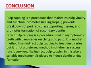
This document summarizes different techniques for pulp capping including direct and indirect pulp capping. It discusses the history of various materials used for pulp capping such as calcium hydroxide and mineral trioxide aggregate. Key points covered include the indications and contraindications for pulp capping, the ideal properties of capping materials, and the histological response after pulp capping including zone formation. The document provides an overview of pulp capping procedures and factors influencing their success.
a detailed account of the principles of tooth preparation with main reference from Shillingburg
The presentation is available on request. Mail me at apurvathampi@gmail.com
a detailed account of the principles of tooth preparation with main reference from Shillingburg
The presentation is available on request. Mail me at apurvathampi@gmail.com
A concise and brief presentation on cleaning and shaping of root canals. Colorful and well pictured. Ideal for UG students and PG students to get a good understanding of BMP techniques.
An inlay may cap none, or may cap all but one cusp.
Sturdevant’s 4th ed. page579
Inlays may be used as single-tooth restorations for proximo-occlusal or gingival lesions with minimal to moderate extensions
Shillingburg page 1
An inlay may be defined as a restoration which has been constructed out of mouth from gold, porcelain, or other material & then cemented into the prepared cavity of a tooth.
William McGehee pg410
Splinting is one of the oldest forms of aids to periodontal therapy. By redistribution of forces on the affected teeth the splint minimizes the effects caused by loss of support. Splinting teeth to each other allows weakened teeth to be supported by neighbouring teeth. This presentation reviews the rationale, techniques, advantages and ill effects of stabilization of teeth by splinting as an aid to periodontal therapy. With the acceptance and clinical predictability of adhesive procedures, the use of conservative bonding techniques to splint teeth offers a useful alternative to more invasive restorative procedures. Loss of tooth-supporting structures results in tooth mobility. Increased tooth mobility adversely affects function, aesthetics, and the patient’s comfort. Splints are used to overcome all these problems. When faced with the dilemma of how to manage periodontally compromised teeth, splinting of mobile teeth to stronger adjacent teeth is a viable option. This prolongs the life expectancy of loose teeth, gives stability for the periodontium to reattach, and improves comfort, function and aesthetics.
This lecture, which oriented to the level of mind of undergraduate students, discuss the topic of pulpectomy, its indications, contraindications, and procedural steps.
Visit us on Facebook:
https://www.facebook.com/iraqi.Dental.Academy
Deep caries management /certified fixed orthodontic courses by Indian dental ...Indian dental academy
Welcome to Indian Dental Academy
The Indian Dental Academy is the Leader in continuing dental education , training dentists in all aspects of dentistry and offering a wide range of dental certified courses in different formats.
Indian dental academy has a unique training program & curriculum that provides students with exceptional clinical skills and enabling them to return to their office with high level confidence and start treating patients
State of the art comprehensive training-Faculty of world wide repute &Very affordable.
How to Make a Field invisible in Odoo 17Celine George
It is possible to hide or invisible some fields in odoo. Commonly using “invisible” attribute in the field definition to invisible the fields. This slide will show how to make a field invisible in odoo 17.
Welcome to TechSoup New Member Orientation and Q&A (May 2024).pdfTechSoup
In this webinar you will learn how your organization can access TechSoup's wide variety of product discount and donation programs. From hardware to software, we'll give you a tour of the tools available to help your nonprofit with productivity, collaboration, financial management, donor tracking, security, and more.
The French Revolution, which began in 1789, was a period of radical social and political upheaval in France. It marked the decline of absolute monarchies, the rise of secular and democratic republics, and the eventual rise of Napoleon Bonaparte. This revolutionary period is crucial in understanding the transition from feudalism to modernity in Europe.
For more information, visit-www.vavaclasses.com
Synthetic Fiber Construction in lab .pptxPavel ( NSTU)
Synthetic fiber production is a fascinating and complex field that blends chemistry, engineering, and environmental science. By understanding these aspects, students can gain a comprehensive view of synthetic fiber production, its impact on society and the environment, and the potential for future innovations. Synthetic fibers play a crucial role in modern society, impacting various aspects of daily life, industry, and the environment. ynthetic fibers are integral to modern life, offering a range of benefits from cost-effectiveness and versatility to innovative applications and performance characteristics. While they pose environmental challenges, ongoing research and development aim to create more sustainable and eco-friendly alternatives. Understanding the importance of synthetic fibers helps in appreciating their role in the economy, industry, and daily life, while also emphasizing the need for sustainable practices and innovation.
Model Attribute Check Company Auto PropertyCeline George
In Odoo, the multi-company feature allows you to manage multiple companies within a single Odoo database instance. Each company can have its own configurations while still sharing common resources such as products, customers, and suppliers.
Instructions for Submissions thorugh G- Classroom.pptxJheel Barad
This presentation provides a briefing on how to upload submissions and documents in Google Classroom. It was prepared as part of an orientation for new Sainik School in-service teacher trainees. As a training officer, my goal is to ensure that you are comfortable and proficient with this essential tool for managing assignments and fostering student engagement.
The Roman Empire A Historical Colossus.pdfkaushalkr1407
The Roman Empire, a vast and enduring power, stands as one of history's most remarkable civilizations, leaving an indelible imprint on the world. It emerged from the Roman Republic, transitioning into an imperial powerhouse under the leadership of Augustus Caesar in 27 BCE. This transformation marked the beginning of an era defined by unprecedented territorial expansion, architectural marvels, and profound cultural influence.
The empire's roots lie in the city of Rome, founded, according to legend, by Romulus in 753 BCE. Over centuries, Rome evolved from a small settlement to a formidable republic, characterized by a complex political system with elected officials and checks on power. However, internal strife, class conflicts, and military ambitions paved the way for the end of the Republic. Julius Caesar’s dictatorship and subsequent assassination in 44 BCE created a power vacuum, leading to a civil war. Octavian, later Augustus, emerged victorious, heralding the Roman Empire’s birth.
Under Augustus, the empire experienced the Pax Romana, a 200-year period of relative peace and stability. Augustus reformed the military, established efficient administrative systems, and initiated grand construction projects. The empire's borders expanded, encompassing territories from Britain to Egypt and from Spain to the Euphrates. Roman legions, renowned for their discipline and engineering prowess, secured and maintained these vast territories, building roads, fortifications, and cities that facilitated control and integration.
The Roman Empire’s society was hierarchical, with a rigid class system. At the top were the patricians, wealthy elites who held significant political power. Below them were the plebeians, free citizens with limited political influence, and the vast numbers of slaves who formed the backbone of the economy. The family unit was central, governed by the paterfamilias, the male head who held absolute authority.
Culturally, the Romans were eclectic, absorbing and adapting elements from the civilizations they encountered, particularly the Greeks. Roman art, literature, and philosophy reflected this synthesis, creating a rich cultural tapestry. Latin, the Roman language, became the lingua franca of the Western world, influencing numerous modern languages.
Roman architecture and engineering achievements were monumental. They perfected the arch, vault, and dome, constructing enduring structures like the Colosseum, Pantheon, and aqueducts. These engineering marvels not only showcased Roman ingenuity but also served practical purposes, from public entertainment to water supply.
TESDA TM1 REVIEWER FOR NATIONAL ASSESSMENT WRITTEN AND ORAL QUESTIONS WITH A...
Pulp capping
1. Submitted by:
Shekhar kumar mandal
BDS IV
Roll no:26
SUBMITTED TO:
Dr. Deepika Kapoor
Department of Pedodontics and
Preventive Dentistry
College of Medical Sciences,
Bharatpur, Nepal
2. 1. INTRODUCTION
2. TREATMENT MODALITIES
3. TYPES OF PULP CAPPING
4. INDIRECT PULP CAPPING
5. PATENT DENTIN MEASURING DEVICE
6. DIRECT PULP CAPPING
7. FEATURES OF SUCCESSFUL PULP CAPPING
8. PULP CAPPING AGENTS
9. CONCLUSION
10.REFERENCES
3. PULP is defined as soft
tissue forming inner
structure of tooth and
containing nerve and blood
vessel , also called as tooth
pulp.
The dental pulp occupies the
center of each tooth and
shapes itself to a maturation
of the tooth .
12. Large carious lesion
Absence of lymphadenopathy
Normal appearance of adjacent
gingiva
Normal colour of tooth
Clinical examination
13. Large carious lesion in
close proximity to the
pulp
Normal lamina dura
Normal periodontal
ligament space
No interradicular or
periapical radiolucency
17. Large carious lesion with apparent
pulp exposure
Interrupted or broken lamina dura
Widened periodontal ligament space
Radiolucency at the root apices or furcation areas
21. First appointment
• Tooth is isolated with a rubber dam
• The gross caries is removed with a large round bur (6 or
8) or sharp spoon exclavator .Care must be taken
removing the caries to prevent exposure of pulp .Care
must be taken to eliminate all the caries at the DEJ .
Because of its closeness to the surface caries left in the
areas will likely cause failure .
• The remaining thin layer of caries is covered
with a radiopaque biocompatible base
material and sealed with a durable interim
restoration
• Wait for 6-8 weeks.
• During this time the caries process
in the deeper layer is arrested.
22. .
• If the tooth is asymptomatic, the surrounding
soft tissues are free from swelling and the
temporary filling is intact , the bitewing
radiograph at the treated tooth should be taken
for the presence of reparative dentin .
• Carefully remove all the temporary filling material ,
especially base over the deep portion of the cavity
floor.The remaining affected carious dentin should
appear dehydrated and flaky and should be easily
removed
•The cavity preparation should be irrigated and gently dried.
•Cover entire floor with Hard setting Ca(OH)2 dressing
• Base should be placed with reinforced GIC or ZOE
• Final restoration with composite or Amalgam
23.
24. Avoid unintentional pulp exposure
Dentist get chance to assess reaction of tooth and
caries activity
Helps to remove slowly progressing lesion
Final excavation of caries is safer in second sitting.
26. If the tooth remains asymptomatic there is no need for reentry.
The removal of bacteria and substrate together with an effective seal of restoration
provides the means whereby the pulp can recover the laying down secondary dentin .
Assure retention and sealing of temporary restoration
A protective radiopaque biocompatible base material must be placed prior to
temporary restoration
Remove all the soft moist and leathery texture of dentin
The DEJ must be free from all softened material and stain even if the stain is firm .
All margins are left adequately supported and peripheral caries is removed with a
large round bur .
The tooth is isolated with rubber dam ,the cavity outline form is made .
28. Electronically measures the
thickness of dentin layer above
the pulp chamber during crown
preparation with a simple touch
of probe
GREEN LIGHT:safe zone
ORANGE LIGHT:limit of safe
zone
RED LIGHT:danger of
penetration through dentin.
30. First 30 days
•1/5th Tubular dentin is formed
First 2
months
•Cellular fibrillar dentin is formed
Third
month
•Globular dentin is formed
More than
3 months
•0.1mm Tubular dentin is formed
31. Infected Dentin
▪Highly demineralized
▪Unremineralizable
▪Superficial layer
▪Lacking sensation
▪Stained by 0.5% fuschin or i.e. 1.0% acid
red solution
▪Ultrastructure- intertubular dentin
greately demineralized, with irregular
scattered crystals.
Presence of deteriorated collagen fibers
that have only distinct cross bands and
no interbands.
▪Should be excavated
Affected Dentin
Intermediately demineralized
Remineralizable
Deeper layer
Sensitive
Does not stain
Ultrasyructure: intertubular dentin
partially demineralized, but
apatitie crystals bound like fringes
Sound collagen fibers with distinct
cross bands and interbands.
Should be left remineralized.
Difference between affected and infected dentin
32.
33.
34. One year later, Rebel performed the first animal experiments with disastrous results, so he regarded the
exposed pulp as a doomed organ.
The first scientific clinical study to compare different capping materials was made by Dätwyler in 1921,
whereupon zinc oxide-eugenol showed the best results.
Hunter(1883) suggested 1st pulp capping materials. He recommended covering an exposure with a mixture
of Sorghum molasses and the droppings of the English sparrow and claimed 98% success rate
Until the end of the 19th century, most materials were used empirically with the idea that the pulp tissue
must be irritated by etching or cauterization to heal.
The first method of capping exposed pulps, using gold foils, was described by Pfaff in 1756. Thereafter,
numerous agents for direct pulp capping have been recommended.
35. Placement of a medicated or a nonmedicated material on a pulp that
has been exposed in the course of preparing a cavity in a carious
tooth or as the result of trauma.[Kopel, 1997]
Since then, calcium hydroxide has been recommended by several
authors for direct pulp capping, but it took until the middle of 20th
century until it was regarded as the standard of care.
In 1920 Hermann, introduced calcium hydroxide for root canal fillings.
Between 1928 and 1930 he studied the reaction of vital pulp tissue to
calcium hydroxide to prove that it was a biocompatible material.
36. Encouragement of young , healthy pulps to initiate a
dentin bridge, thus walling of the exposure site
OBJECTIVES
a) preservation of vitality of the radicular pulp.
b) relief of pain in patients with acute pulpagia.
c) ensuring the continuity of normal apexogenesis in immature
permanent teeth
37. Accidental pin point exposure of pulp when
excavating deep caries, less than 1 sq. mm.
surrounded by clean dentin for (<24hours)
Traumatic fracture of tooth(<24 hours) with
pin point exposure
Iatrogenic exposure during cavity
preparation & crown preparation
38. Bleed if touched but not excessively and
controlled easily with cotton pellet
Normal vitality tests without tender to
percussion
No radiographic evidence of periradicular
pathology
Young patient
39. History of severe spontaneous tooth aches at night
Excessive tooth mobility
Periodontal ligament thickening
Intraradicular radiolucency
Excessive bleeding at exposure site
Purulent , serous exudate from exposure
External or internal resorption
Swelling and fistula with associated tooth
40. Localization of infection & inflammation in primary teeth is poorer than
in permanent teeth. [Mc Donalds,1956]
Incidence of reparative dentin formation in primary teeth is more
extensive than permanent Teeth. [Sayegh , 1968]
Primary pulp contain high cellular content which might be responsible
for failures. Primary pulp responds more rapidly to the effects of
dentinal caries then the perm. Teeth. [Rayner & Southam, 1979]
Undifferentiated mesenchymal cells may differentiate into osteoclasts
in response to caries or pulp capping material which could lead to
internal resorption. [Kennedy,1985]
41. Primary Pulp are more closer to outer enamel surface & are rapidly
infected by the carious lesion. Once exposed pulpal inflammation is so
involved that the DPC proves unfavorable. [Kennedy & Kopel,1985]
Increased resorption in primary teeth is because already root resorption
is in progress. [Stanley, 1985]
Wide apical foramina in pri. teeth leads to abundant blood supply which
results in more typical and faster inflammation response to irritation than
in permanent teeth. [Kopel,1992]
Acc. To finn, in primary teeth pulp capping is best carried out in teeth
where dental pulp has been mechanically exposed.
Pulp capping not recommended. Internal resorption or acute
dentoalveolar abscess may result . [Pinkham]
42. Rubber Dam placement
Deep carious dentin
excavation:inhibit infected matter
from entering pulp. Necrotic &
infected dentin chips will invariably be
pushed into the exposed pulp during
last stages of caries removal.
Bleeding controlled with sterile cotton
wool (blast of air not used).No
instruments should be inserted into
exposure site.
Layer of hard setting calcium flowed IRM
and a permanent restoration. In small teeth,
Ca(OH)2 can act as base. Blood clot-not
allowed to be formed after cessation of
bleeding from exposure as it impedes pulpal
healing. Kopel,1992 a more adequate seal of
pulp capping is needed (Stainless steel crown
-best).
43. Dentin bridging
Maintenance of pulp vitality
Lack of undue sensitivity or pain
Minimum pulpal inflammation response
Ability of pulp to maintain itself without progressive degeneration
Lack of internal resorption and/ or interradicular pathosis
In Accordance with Kennedy & Kopel (1985):
44.
45.
46. IDEAL REQUIREMENTS
Ideal dressing material for pulp therapy in primary teeth does not
exist, but the material should be:
1. Bactericidal
2. Biocompatible
3. Harmless to the pulp, surrounding structures and the
permanent tooth germ.
4. Promote healing
5. Not interfere with physiologic process of resorption.
47. Calcium hydroxide
Zinc oxide eugenol cement
Corticosteroids and antibiotics
Polycarboxylate cement
Isobutyl cyanoacrylate and tri
calcium phosphate ceramic
Collagen
Bonding Agents
Calcium phosphate
Hydroxyapatite
Mineral trioxide aggregate
GIC/RMGIC
Lasers
MTA 1-Calcium
Growth factors
Odontogenic ameloblast
associated protein
Endo sequence root repair material
Castor oil bean cement
Thera Cal
48. It is a colorless crystal or white powder
prepared by reacting calcium oxide with water.
The use of calcium hydroxide in endodontics was introduced by
Hermann in between 1920-1930.
Calcium hydroxide was most favored as a pulpotomy agent in the
1940s and mid- 1950s.
“Calcium hydroxide has the ability to form reparative dentin form
ation”,this rationale was introduced by Teuscher and Zander
in 1938.
Lim and Kirk, in an extensive review of direct pulp capping
literature, found little support for pulp obliteration and
internal resorption being a major complication of pulp
capping
Estrela et al. summarized the antibacterial properties of
calcium hydroxide.
49. ADVANTAGES
Reparative dentin formation
Antibacterial action
Pulp protection
The tissue-dissolving property
Newer preparation shows Improved
strength, essentially no solubility in acid, and
minimal solubility in water and control the
over working time
DISADVANTAGES
Pulp obliteration
Internal resorption
Lack of adhesion to hard tissues
Microleakage
Short working time of self cured
preparation
Base paste –
Glycol salicylate-40%-reacts with
calcium hydroxide and ZnO
Titanium dioxide-Inert fillers
Calcium tungstate - Fillers
Barium sulphate-provide radioopacity
Catalyst paste
Calcium hydroxide-50%-principal
reactive ingredient
Zinc oxide-10%
Zinc stearate-0.55%-accelerator
Sulphonamide-39.5%-oily
compound acts as carrier.
COMPOSITION
•AVAILABLE AS
a)Pulpdent b)Hydrex : two paste system c)Dycal.
50. D: After 8 weeks
A: After 24 hours
B: After 2-3 weeks
C: After 4-5 weeks
51. Three distinct zone can be visualised:
I. Zone of obliteration (early changes: area of superficial debris)
II. Zone of coagulation necrosis (Schroeder’s layer of “firm necrosis”,
Stanley’s “mummified zone”)
III. Line of demarcation
Zone of obliteration
Early changes: area of
superficial debris
Drug’s caustic effect
Tissue in immediate contact
becomes deranged and
distorted.
This Zone consists of-
i. Debris
ii. Dentinal fragments
iii. Blood clot
iv. Blood pigment
v. Calcium hydroxide
particles
Zone of coagulation necrosis
Weaker chemical reaction from 1st
zone reaches the subjacent , more
apical tissues & results in this zone of
coagulation & necrosis.
Thickness- 0.3-0.7 mm (Acc. to
Craig: 1mm thick)
Represents devitalised tissue
without complete obliteration of its
structural architecture
Cellular details-greatly diminished
Capillaries outlines, nerve bundles
& pyknotic nuclei can be recognized.
Stimulates subjacent vital pulp
Vascular changes occur
Line of demarcation
Develops between Zone
of coagulation necrosis
and vital tissues
This zone results from
reaction of Calcium
hydroxide with the
tissue protein to form
proteinate globules.
52. It is a new remarkable biocompatible material with exciting clinical
applications pioneered by Dr. Mahmoud Torabinejad, Loma
Linda University, in 1993
COMPOSITION
MTA is a mechanical mixture of 3 powder ingredients:
• Portland cement (75%)
• Bismuth oxide (20%)
• Gypsum (5%)
Composition includes :
• Tricalcium silicate
• Dicalcium silicate
• Tricalcium aluminate
• Tetracalcium aluminoferrite
• Calcium sulfate
• Bismuth oxide (provides radio-opacity)
53. CONTRAINDIACTION
Irreversible pulpitis
INDICATION
•To preserve pulp vitality
• Prevent pathological changes in the
periradicular tissues
• Mechanical pulp exposures
• Carious pulp exposures with
immature apices.
PROPERTIES OF MTA
•Mixing MTA:
Powder: Water = 3: 1
Glass SLABor paper slab used
•SETTING TIME:
Hydration of MTA powder results in a
colloidal gel that solidifies to a hard
structure in 3~ 4 hrs which has a long
setting time with less shrinkage.
•pH=12.5
ADVANTAGES
• Antimicrobial Activity
• Prevents MicroLeakage over vital pulp
• Cementoconductive
• Non toxic and Non-mutagenic
• Cell adherence & growth
• Alkaline phosphotase/ osteocalcin
• Interleukin production
• Periodontal ligament attachment to
cementum growth
• Dentinal bridge formation
DISADVANTAGES
• More difficult to manipulate
• Longer setting time
55. Ca(OH)2 MTA
Hard tissue formation Not much Root end induction
Calcific bridge Not continuous Continuous with dentin
Biocompatibility Low High
Degree of Inflammation Low High
Sets Not Hard Hard
pH High High
Solubility Partially disolve Less soluble
Permeable to fluids Non permeable
Viscosity Poor Good
Application Not easy to apply in RC Easy
Resorption Rate vary with density Non-resorbable
Appical barrier formation Change rate/ initial narrow
appical width
Less/wide
Patient follow up More Less
Treatment Delay shortens
56. Germicidal agent
Used in indirect pulp capping due to its
This gives the pulp the chance for
healing & regeneration
Direct contact →chronic inflammatiom ,abscess formation and
liquefaction necrosis.
After 24Hr of capping →a mass of red blood cells &PNLs. Demarcated
from the underlying tissue by zone of fibrin and inflammatory cells.
After 2W of capping → pulp degeneration &chronic inflammation
extends deep to the apex.
Palliative affect
Excellent initial seal
Kills bacteria present in
carious lesions
So arrests the caries process
57. Different studies were led on laser energy
to overcome the histological deficits of electrosurgery.
Used in Direct pulp capping & pulpotomy.
Co2 Laser , Argon Laser, Diode Laser, Erbium:Yttrium-Aluminum Garnet (Er.YAG).
Laser radiation has been proposed for pulp treatment based on its haemostatic,
coagulative and sterilizing effects.
Laser irradiation creates a superficial zone of coagulation necrosis that remains
compatible with the underlying tissue and isolate pulp from effects of the subbase.
Mortiz et al., reported that the thermal effects of laser radiation caused
sterilization and scar formation in the irradiated area, which in turn preserves the
pulp from bacterial invasion.
58. Alpha-tricalcium phosphate & Tetracalcium phosphate (4CP)
set & convert to hydroxyapatite.
Stimulate the pulp to form hard tissue.
No finding of necrotic pulp tissue in direct contact with 4CP
cement compared to calcium hydroxide slight acidity after
mixing
4CP cement has mechanical strengths so it could be used as
so called “dentin substitute”. Pulp capping agent
lining material
59. There were suggested as direct pulp capping and pulpotomy
agents with the introduction of adhesive dentistry in both
primary and permanent dentition.
Adhesive material forms:
- A complete marginal seal
- Prevents bacterial intrusion
- Allowed pulp repair, characterized by a new odontoblast cell
layer underlying the dentin bridge formation.
Many studies have indicated that composite & resin-modified
glass-ionomer are compatible with pulp tissue.
60. Propolis, a resinous material collected by
honey bees, has been used as a traditional anti-infalmmatory
and anti-bacterial medicine for many centuries.
Used as indirect pulp capping paste when mixed with ZnO
powder and this showed similar effect of ZnO and Eugenol as
secondary dentin formation.
In direct capping with this paste showed no pulp
. degeneration and formation of protective layer.
61.
62. PULP CAPPING AGENT ADVANTAGES DISADVANTAGES
Zinc oxide eugenol cement. 1)Reduces inflammation. 1) Lack of calcific bridge
formation.
2) Releases eugenol in high
concentration which is
cytotoxic.
3) Demonstrates interfacial
leakage.
Corticosteroids and
antibiotics.
1) Reduces pulp inflammation.
2) Vanomycin and calcium hydroxide
stimulated a more regular reparative
dentin.
1) Should not be used in patients
with risk from bacteremia.
Polycarboxylate cement. 1)Chemically bond to tooth structure. 1) Lack of antibacterial effect.
2) Fail to stimulate calcific bridge
formation.
Inert materials( Isobutyl
cyanoacrylate and tri
calcium phosphate ceramic)
1) Reduces pulp inflammation.
2) Stimulate dentin bridge formation.
1) NONE of these materials have
been promoted in dentist
profession as a viable technique
Collagen 1) Less irritating than calcium hydroxide
and promotes mineralization.
1) Does not help in thick dentin
bridge formation.
Bonding Agents 1) Superior adhesion to hard tissues.
2) Effective seal against micro leakage.
1) Has cytotoxic effect.
2) Absence of calcific bridge
formation.
63. PULP CAPPING
AGENT
ADVANTAGES DISADVANTAGES.
Calcium phosphate. 1) Helps in bridge formation with no
superficial tissue necrosis.
2) Significant absence of pulp
inflammation.
3) Good physical properties.
1) Clinical trials are necessary to
evaluate this material.
Hydroxyapatite. 1) Biocompatible.
2) Act as a scaffold for the newly formed
mineralized tissue.
1) Mild inflammation with superficial
necrosis of pulp.
Carbon dioxide lasers 1) Formation of secondary dentin.
2) Bactericidal effects.
1) Technique sensitive.
2) Causes thermal damage to pulp at
high doses.
Glass ionomer/ Resin
modified glass
ionomer.
1) Excellent bacterial seal.
2) Fluoride release, coeffient of thermal
expansion and modulus of elasticity
similar to dentin.
3) Good biocompatibility.
1) Cause chronic inflammation.
2) Lack of dentin bridge formation.
3) Cytotoxic when in direct cell
contact.
4) High solubility and slow setting
rate.
MTA 1-Calcium 1) Helps in dentin bridge formation
without formation of necrotic layer.
2) Shear bond strength is higher than
conventional GIC and similar to RMGIC.
1) Presence of 10% calcium hydroxide
interferes with complete curing of the
material, residual monomers causes
cytotoxicity.
Growth factors. 1) Formation of osteodentin and tubular
dentin.
2) Formation of more homogenous
reparative dentin
3) Superior to calcium hydroxide in the
mineralization inducing properties.
1) High concentration is required.
2) Half life is less.
3) Appropriate dose response is
required to avoid uncontrolled
obliteration of pulp chamber.
64. PULP CAPPING
AGENT
ADVANTAGES. DISADVANTAGES.
Odontogenic
ameloblast
associated
protein.
1) Biocompatible.
2) Accelerates reactionary dentin
formation.
3) Normal pulp tissue appearance
without excessive tertiary dentin
formation and obliteration of the
pulp cavity compared to MTA
1) Till now only invitro studies
were conducted.
2) Further studies regarding
this material is required.
Endo sequence
root repair
material
1) Antibacterial property.
2) Less cytotoxic than MTA, Dycal and
light cure calcium hydroxide.
1) Bioactivity of the cells were
decreased gradually when
exposed to this material.
Castor oil bean
cement.
1) Good antibacterial property.
2) Less cytotoxic.
3) Good mechanical properties.
4) Facilitates tissue healing.
5) Better sealing ability than MTA and
GIC.
6) Less cost.
1) Bio inert rather than
bioactive.
2) More clinical trials are
required.
Thera Cal. 1) Act as protectant of the dental pulp
complex.
2) Has strong physical properties, no
solubility, high radiopacity.
3) TheraCal exhibited higher calcium
1) It is opaque and whitish in
color and it should be kept thin
so as not to show through
composite material that are
very translucent affecting final
65. Pulp capping is a procedure that maintains pulp vitality
and function, promotes healing/repair, prevents
breakdown of peri radicular supporting tissues, and
promotes formation of secondary dentin
Direct pulp capping is a procedure used in asymptomatic
teeth with deep caries reaching upto pulp. It is another
method than Indirect pulp capping to treat deep caries
but it is not a preferred method in children as success
rate is very low, like indirect pulp capping in this also a
suitable medicament is placed to induce dentin bridge
formation
66. Teacher’s Note
Textbook of Pedodontics 2e, - Shobha Tandon
Principle and practice of pedodontics, 3e, Aarti Rao
Textbook of Pedodontics, 3e, Nikhil Marwah
Dentisry on Child and Adloscence, Mc Donald
Text book of Endodontics, Grossman
Pathway of Pulp 9e,Cohen
Stewart DJ and Kramer IRH. Effects of calcium hydroxide on the
unexposed pulp, J. Dent
Suneda YT et al . A histopathological study of direct pulp capping with
adhesive resins
Teethanime.com/pulp
Peter. Murray et al. Analysis of pulpal reactions to restorative
procedures, materials, pulp capping, and future therapies crit rev oral
biol med 509-13:5202002