Downloaded 262 times
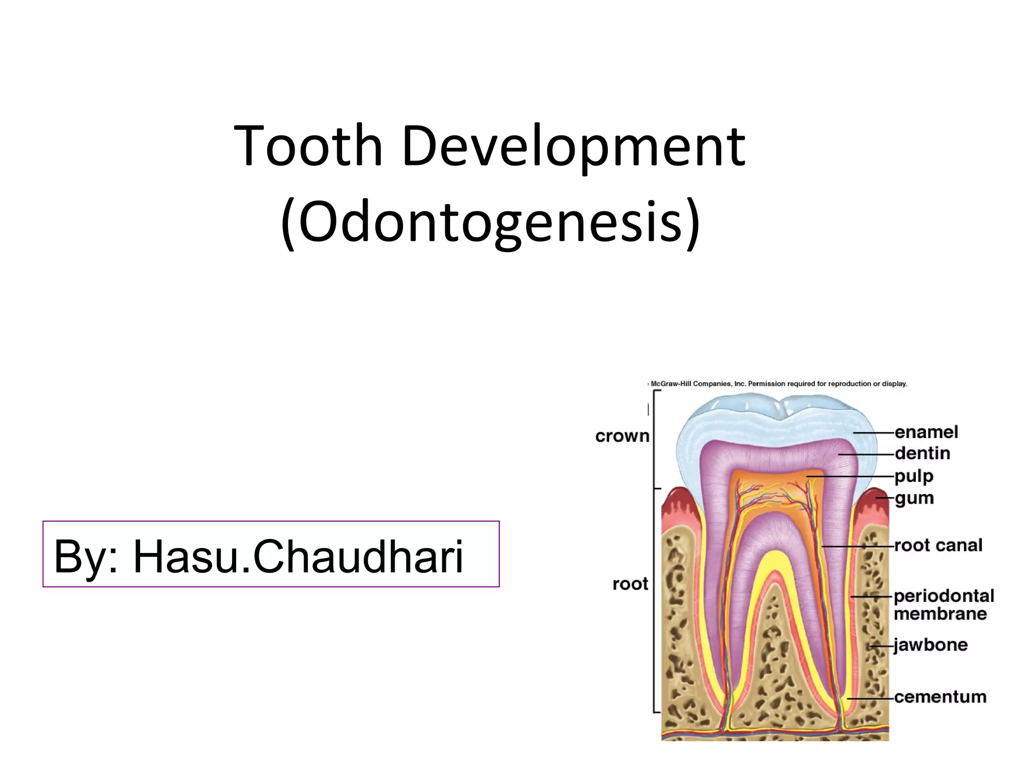


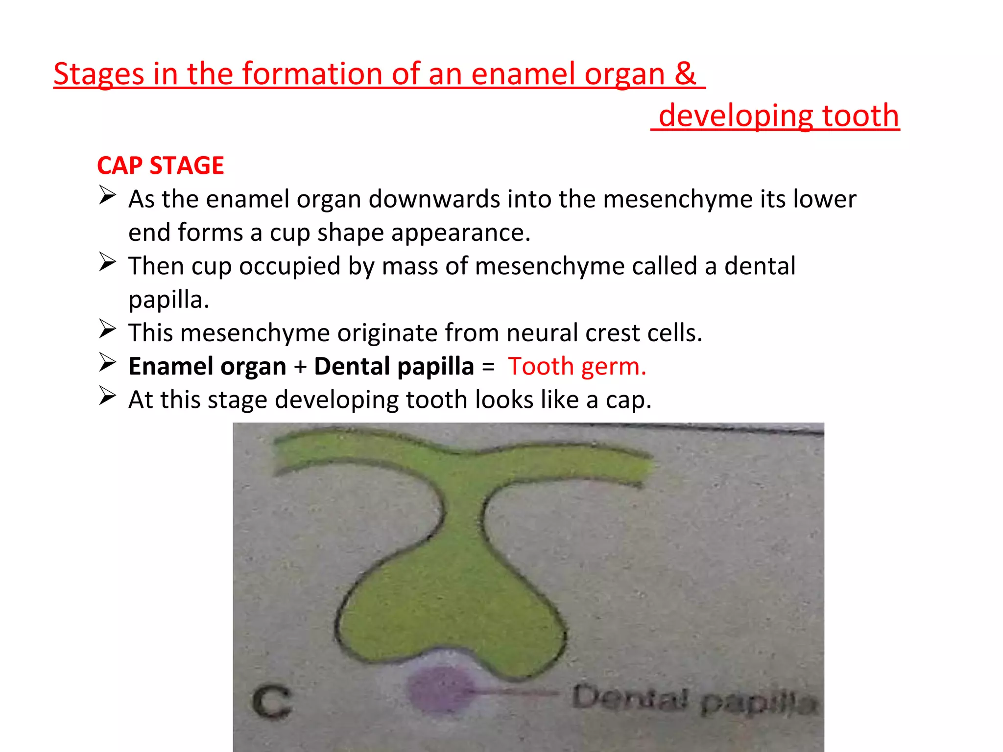


This document summarizes the stages of tooth development from initiation to maturation. It describes the development of both the primary and permanent dentition. Tooth development occurs through multiple stages: initiation, bud, cap, bell, appositional, and maturation. The bud stage involves thickening of the dental lamina. In the cap stage, the enamel organ invaginates into the dental papilla. During the bell stage, ameloblasts and odontoblasts form and begin to secrete enamel and dentine. Permanent teeth develop from dental lamina buds on either side of the primary teeth. Anomalies such as missing teeth, extra teeth, fused teeth, and malocclusion can also occur.


