1. The document describes how to test various superficial and deep reflexes through elicitation techniques and provides clinical significance of reflex assessment.
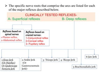
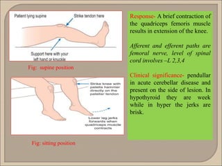
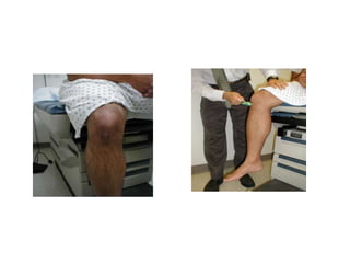
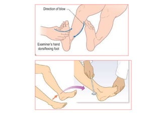
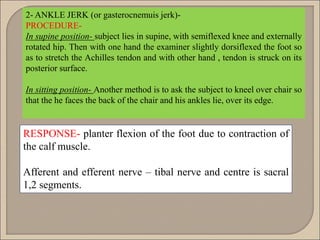
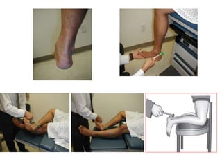
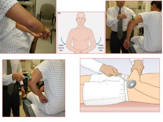
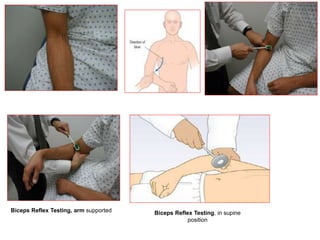
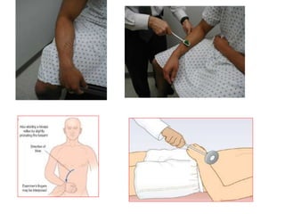
2. Superficial reflexes include plantar, abdominal, and cranial nerve reflexes while deep reflexes tested are knee jerk, ankle jerk, biceps, triceps, and brachioradialis jerks.
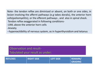
3. Abnormal reflexes can indicate lesions in the sensory, motor or central pathways and provide clues to neurological conditions like diabetes, thyroid disorders, or spinal cord injuries. Precise diagnosis requires correlating reflex findings with the clinical picture.
A neurological examination is the assessment of sensory neuron and motor responses, especially reflexes, to determine whether the nervous system is impaired.
ocular anatomy fluid system glaucoma lens cataract phototransduction field visual acuity ocular movement errors of refraction light reflex accommodation corneal reflex visual pathway and its lesions
Pupillary light reflex (PLR) : that controls the diameter of the pupil, in
response to the intensity of light that falls on the retinal ganglion cells of
the retina in the back of the eye.
- Light reflex
- Corneal reflex.
-Accommodation reflex:
Argyll Robertson pupils
Horner's syndrome:
Holmes–Adie syndrome
BRAINSTEM
The Brainstem lies at the base of the brain and the top of the spinal cord.
The brainstem is located in the posterior cranial fossa.
The brainstem is the structure that connects the cerebrum of the brain to the spinal cord and cerebellum.
Provides a pathway for tracts running between higher and lower neural centers.
Divided into 3 major divisions:
midbrain,
pons, and
medulla oblongata.
It is responsible for many vital functions of life, such as breathing, consciousness, blood pressure, heart rate, and sleep.
It contains many critical collections of white and grey matter.
The grey matter within the brainstem consists of nerve cell bodies and form many important brainstem nuclei. Ten of the twelve cranial nerves arise from their cranial nerve nuclei in the brainstem.
The white matter tracts of the brainstem include axons of nerves traversing their course to different structures. These tracts travel both to the brain (afferent) and from the brain (efferent) such as the somatosensory pathways and the corticospinal tracts, respectively.
Mid Brain
The midbrain is continuous with the cerebral hemisphere.
The upper posterior (i.e. rear) portion of the midbrain is called the tectum, which means "roof."
The surface of the tectum is covered with four bumps representing two paired structures: the superior and inferior colliculi.
The superior colliculi are involved in eye movements and visual processing, while the inferior colliculi are involved in auditory processing.
Another important nucleus, the substantia nigra, is located here.
The substantia nigra is rich in dopamine neurons and is considered part of the basal ganglia.
Pons
An important pathway for tracts that run from the cerebrum down to the medulla and spinal cord, as well as for tracts that travel up into the brain.
It also forms important connections with the cerebellum via fibre bundles known as the cerebellar peduncles.
Posteriorly, the pons and medulla are separated from the cerebellum by the fourth ventricle.
Home to several nuclei for cranial nerves.
Medulla
The point where the brainstem connects to the spinal cord.
Contains a nucleus called the nucleus of the solitary tract that is crucial for our survival (receives information about blood flow, along with information about levels of oxygen and carbon dioxide in the blood, from the heart and major blood vessels).
When this information suggests a discordance with bodily needs (e.g. blood pressure is too low), there are reflexive actions initiated in the nucleus of the solitary tract to bring things back to within the desired range.
Blood Supply
The brain stem receives its blood supply exclusively from the posterior circulation, including the vertebrae and basilar artery.
The medulla receives its blood supply from the vertebral via medial and lateral perforating arteries.
The pons and midbrain receive their blood from the basilar via the medial and lateral perforating arteries.
Human reflexes
Definition: It is involuntary response of an organ to a stimulus.
- It is the arrangement of neurons through which the reflex is carried out.
- It is usually formed of:
Afferent (sensory) neuron.
2) An interneuron (may be absent).
3) nerve center (cell body of the efferent neuron).
4) Efferent (motor) neuron.
Reflexes are important to understand for all medical professional it is an assessment tool for patients with neurological conditions.
a god knowledge of primitive reflexes can be effective for pediatric health care as well. it helps us in identifying any developmental delay in children.
Dear all,
This ppt contains the cause, types, clinical and radiological features, treatment and complication of supra Condylar fracture of Humerus. I hope this is useful to you.
Thank you
Lower limb neurological examination frequently appears in OSCEs. You’ll be expected to pick up the relevant clinical signs using your examination skills. This lower limb neurological examination OSCE guide provides a clear, concise, step-by-step approach to performing a neurological examination of the lower limb
Reflex activity is the response to a peripheral stimulation that occurs without our consciousness.
Is an involuntary response to a stimulus.
It is a type of protective mechanism.
micro teaching on communication m.sc nursing.pdfAnurag Sharma
Microteaching is a unique model of practice teaching. It is a viable instrument for the. desired change in the teaching behavior or the behavior potential which, in specified types of real. classroom situations, tends to facilitate the achievement of specified types of objectives.
These lecture slides, by Dr Sidra Arshad, offer a quick overview of physiological basis of a normal electrocardiogram.
Learning objectives:
1. Define an electrocardiogram (ECG) and electrocardiography
2. Describe how dipoles generated by the heart produce the waveforms of the ECG
3. Describe the components of a normal electrocardiogram of a typical bipolar leads (limb II)
4. Differentiate between intervals and segments
5. Enlist some common indications for obtaining an ECG
Study Resources:
1. Chapter 11, Guyton and Hall Textbook of Medical Physiology, 14th edition
2. Chapter 9, Human Physiology - From Cells to Systems, Lauralee Sherwood, 9th edition
3. Chapter 29, Ganong’s Review of Medical Physiology, 26th edition
4. Electrocardiogram, StatPearls - https://www.ncbi.nlm.nih.gov/books/NBK549803/
5. ECG in Medical Practice by ABM Abdullah, 4th edition
6. ECG Basics, http://www.nataliescasebook.com/tag/e-c-g-basics
The prostate is an exocrine gland of the male mammalian reproductive system
It is a walnut-sized gland that forms part of the male reproductive system and is located in front of the rectum and just below the urinary bladder
Function is to store and secrete a clear, slightly alkaline fluid that constitutes 10-30% of the volume of the seminal fluid that along with the spermatozoa, constitutes semen
A healthy human prostate measures (4cm-vertical, by 3cm-horizontal, 2cm ant-post ).
It surrounds the urethra just below the urinary bladder. It has anterior, median, posterior and two lateral lobes
It’s work is regulated by androgens which are responsible for male sex characteristics
Generalised disease of the prostate due to hormonal derangement which leads to non malignant enlargement of the gland (increase in the number of epithelial cells and stromal tissue)to cause compression of the urethra leading to symptoms (LUTS
HOT NEW PRODUCT! BIG SALES FAST SHIPPING NOW FROM CHINA!! EU KU DB BK substit...GL Anaacs
Contact us if you are interested:
Email / Skype : kefaya1771@gmail.com
Threema: PXHY5PDH
New BATCH Ku !!! MUCH IN DEMAND FAST SALE EVERY BATCH HAPPY GOOD EFFECT BIG BATCH !
Contact me on Threema or skype to start big business!!
Hot-sale products:
NEW HOT EUTYLONE WHITE CRYSTAL!!
5cl-adba precursor (semi finished )
5cl-adba raw materials
ADBB precursor (semi finished )
ADBB raw materials
APVP powder
5fadb/4f-adb
Jwh018 / Jwh210
Eutylone crystal
Protonitazene (hydrochloride) CAS: 119276-01-6
Flubrotizolam CAS: 57801-95-3
Metonitazene CAS: 14680-51-4
Payment terms: Western Union,MoneyGram,Bitcoin or USDT.
Deliver Time: Usually 7-15days
Shipping method: FedEx, TNT, DHL,UPS etc.Our deliveries are 100% safe, fast, reliable and discreet.
Samples will be sent for your evaluation!If you are interested in, please contact me, let's talk details.
We specializes in exporting high quality Research chemical, medical intermediate, Pharmaceutical chemicals and so on. Products are exported to USA, Canada, France, Korea, Japan,Russia, Southeast Asia and other countries.
Explore natural remedies for syphilis treatment in Singapore. Discover alternative therapies, herbal remedies, and lifestyle changes that may complement conventional treatments. Learn about holistic approaches to managing syphilis symptoms and supporting overall health.
MANAGEMENT OF ATRIOVENTRICULAR CONDUCTION BLOCK.pdfJim Jacob Roy
Cardiac conduction defects can occur due to various causes.
Atrioventricular conduction blocks ( AV blocks ) are classified into 3 types.
This document describes the acute management of AV block.
Title: Sense of Taste
Presenter: Dr. Faiza, Assistant Professor of Physiology
Qualifications:
MBBS (Best Graduate, AIMC Lahore)
FCPS Physiology
ICMT, CHPE, DHPE (STMU)
MPH (GC University, Faisalabad)
MBA (Virtual University of Pakistan)
Learning Objectives:
Describe the structure and function of taste buds.
Describe the relationship between the taste threshold and taste index of common substances.
Explain the chemical basis and signal transduction of taste perception for each type of primary taste sensation.
Recognize different abnormalities of taste perception and their causes.
Key Topics:
Significance of Taste Sensation:
Differentiation between pleasant and harmful food
Influence on behavior
Selection of food based on metabolic needs
Receptors of Taste:
Taste buds on the tongue
Influence of sense of smell, texture of food, and pain stimulation (e.g., by pepper)
Primary and Secondary Taste Sensations:
Primary taste sensations: Sweet, Sour, Salty, Bitter, Umami
Chemical basis and signal transduction mechanisms for each taste
Taste Threshold and Index:
Taste threshold values for Sweet (sucrose), Salty (NaCl), Sour (HCl), and Bitter (Quinine)
Taste index relationship: Inversely proportional to taste threshold
Taste Blindness:
Inability to taste certain substances, particularly thiourea compounds
Example: Phenylthiocarbamide
Structure and Function of Taste Buds:
Composition: Epithelial cells, Sustentacular/Supporting cells, Taste cells, Basal cells
Features: Taste pores, Taste hairs/microvilli, and Taste nerve fibers
Location of Taste Buds:
Found in papillae of the tongue (Fungiform, Circumvallate, Foliate)
Also present on the palate, tonsillar pillars, epiglottis, and proximal esophagus
Mechanism of Taste Stimulation:
Interaction of taste substances with receptors on microvilli
Signal transduction pathways for Umami, Sweet, Bitter, Sour, and Salty tastes
Taste Sensitivity and Adaptation:
Decrease in sensitivity with age
Rapid adaptation of taste sensation
Role of Saliva in Taste:
Dissolution of tastants to reach receptors
Washing away the stimulus
Taste Preferences and Aversions:
Mechanisms behind taste preference and aversion
Influence of receptors and neural pathways
Impact of Sensory Nerve Damage:
Degeneration of taste buds if the sensory nerve fiber is cut
Abnormalities of Taste Detection:
Conditions: Ageusia, Hypogeusia, Dysgeusia (parageusia)
Causes: Nerve damage, neurological disorders, infections, poor oral hygiene, adverse drug effects, deficiencies, aging, tobacco use, altered neurotransmitter levels
Neurotransmitters and Taste Threshold:
Effects of serotonin (5-HT) and norepinephrine (NE) on taste sensitivity
Supertasters:
25% of the population with heightened sensitivity to taste, especially bitterness
Increased number of fungiform papillae
Flu Vaccine Alert in Bangalore Karnatakaaddon Scans
As flu season approaches, health officials in Bangalore, Karnataka, are urging residents to get their flu vaccinations. The seasonal flu, while common, can lead to severe health complications, particularly for vulnerable populations such as young children, the elderly, and those with underlying health conditions.
Dr. Vidisha Kumari, a leading epidemiologist in Bangalore, emphasizes the importance of getting vaccinated. "The flu vaccine is our best defense against the influenza virus. It not only protects individuals but also helps prevent the spread of the virus in our communities," he says.
This year, the flu season is expected to coincide with a potential increase in other respiratory illnesses. The Karnataka Health Department has launched an awareness campaign highlighting the significance of flu vaccinations. They have set up multiple vaccination centers across Bangalore, making it convenient for residents to receive their shots.
To encourage widespread vaccination, the government is also collaborating with local schools, workplaces, and community centers to facilitate vaccination drives. Special attention is being given to ensuring that the vaccine is accessible to all, including marginalized communities who may have limited access to healthcare.
Residents are reminded that the flu vaccine is safe and effective. Common side effects are mild and may include soreness at the injection site, mild fever, or muscle aches. These side effects are generally short-lived and far less severe than the flu itself.
Healthcare providers are also stressing the importance of continuing COVID-19 precautions. Wearing masks, practicing good hand hygiene, and maintaining social distancing are still crucial, especially in crowded places.
Protect yourself and your loved ones by getting vaccinated. Together, we can help keep Bangalore healthy and safe this flu season. For more information on vaccination centers and schedules, residents can visit the Karnataka Health Department’s official website or follow their social media pages.
Stay informed, stay safe, and get your flu shot today!
Title: Sense of Smell
Presenter: Dr. Faiza, Assistant Professor of Physiology
Qualifications:
MBBS (Best Graduate, AIMC Lahore)
FCPS Physiology
ICMT, CHPE, DHPE (STMU)
MPH (GC University, Faisalabad)
MBA (Virtual University of Pakistan)
Learning Objectives:
Describe the primary categories of smells and the concept of odor blindness.
Explain the structure and location of the olfactory membrane and mucosa, including the types and roles of cells involved in olfaction.
Describe the pathway and mechanisms of olfactory signal transmission from the olfactory receptors to the brain.
Illustrate the biochemical cascade triggered by odorant binding to olfactory receptors, including the role of G-proteins and second messengers in generating an action potential.
Identify different types of olfactory disorders such as anosmia, hyposmia, hyperosmia, and dysosmia, including their potential causes.
Key Topics:
Olfactory Genes:
3% of the human genome accounts for olfactory genes.
400 genes for odorant receptors.
Olfactory Membrane:
Located in the superior part of the nasal cavity.
Medially: Folds downward along the superior septum.
Laterally: Folds over the superior turbinate and upper surface of the middle turbinate.
Total surface area: 5-10 square centimeters.
Olfactory Mucosa:
Olfactory Cells: Bipolar nerve cells derived from the CNS (100 million), with 4-25 olfactory cilia per cell.
Sustentacular Cells: Produce mucus and maintain ionic and molecular environment.
Basal Cells: Replace worn-out olfactory cells with an average lifespan of 1-2 months.
Bowman’s Gland: Secretes mucus.
Stimulation of Olfactory Cells:
Odorant dissolves in mucus and attaches to receptors on olfactory cilia.
Involves a cascade effect through G-proteins and second messengers, leading to depolarization and action potential generation in the olfactory nerve.
Quality of a Good Odorant:
Small (3-20 Carbon atoms), volatile, water-soluble, and lipid-soluble.
Facilitated by odorant-binding proteins in mucus.
Membrane Potential and Action Potential:
Resting membrane potential: -55mV.
Action potential frequency in the olfactory nerve increases with odorant strength.
Adaptation Towards the Sense of Smell:
Rapid adaptation within the first second, with further slow adaptation.
Psychological adaptation greater than receptor adaptation, involving feedback inhibition from the central nervous system.
Primary Sensations of Smell:
Camphoraceous, Musky, Floral, Pepperminty, Ethereal, Pungent, Putrid.
Odor Detection Threshold:
Examples: Hydrogen sulfide (0.0005 ppm), Methyl-mercaptan (0.002 ppm).
Some toxic substances are odorless at lethal concentrations.
Characteristics of Smell:
Odor blindness for single substances due to lack of appropriate receptor protein.
Behavioral and emotional influences of smell.
Transmission of Olfactory Signals:
From olfactory cells to glomeruli in the olfactory bulb, involving lateral inhibition.
Primitive, less old, and new olfactory systems with different path
Ozempic: Preoperative Management of Patients on GLP-1 Receptor Agonists Saeid Safari
Preoperative Management of Patients on GLP-1 Receptor Agonists like Ozempic and Semiglutide
ASA GUIDELINE
NYSORA Guideline
2 Case Reports of Gastric Ultrasound
Anti ulcer drugs and their Advance pharmacology ||
Anti-ulcer drugs are medications used to prevent and treat ulcers in the stomach and upper part of the small intestine (duodenal ulcers). These ulcers are often caused by an imbalance between stomach acid and the mucosal lining, which protects the stomach lining.
||Scope: Overview of various classes of anti-ulcer drugs, their mechanisms of action, indications, side effects, and clinical considerations.
These simplified slides by Dr. Sidra Arshad present an overview of the non-respiratory functions of the respiratory tract.
Learning objectives:
1. Enlist the non-respiratory functions of the respiratory tract
2. Briefly explain how these functions are carried out
3. Discuss the significance of dead space
4. Differentiate between minute ventilation and alveolar ventilation
5. Describe the cough and sneeze reflexes
Study Resources:
1. Chapter 39, Guyton and Hall Textbook of Medical Physiology, 14th edition
2. Chapter 34, Ganong’s Review of Medical Physiology, 26th edition
3. Chapter 17, Human Physiology by Lauralee Sherwood, 9th edition
4. Non-respiratory functions of the lungs https://academic.oup.com/bjaed/article/13/3/98/278874
- Video recording of this lecture in English language: https://youtu.be/lK81BzxMqdo
- Video recording of this lecture in Arabic language: https://youtu.be/Ve4P0COk9OI
- Link to download the book free: https://nephrotube.blogspot.com/p/nephrotube-nephrology-books.html
- Link to NephroTube website: www.NephroTube.com
- Link to NephroTube social media accounts: https://nephrotube.blogspot.com/p/join-nephrotube-on-social-media.html
2. Reflex –
It is an rapid, predictable, unconscious and
involuntary response (i.e. contraction of a
muscle or secretion of glands) to a specific
stimulus.
3. Diagram showing the pathway of reflex arc with its different components
Stimulus
1-Receptor
In skin
stimulated
2- sensory
neuron
3- integration
center
interneuron
4 –Motor
neuron
5–Effector
5. Parts of knee hammer
1- It consist of a long metallic handle
At one end on which a triangular shaped soft rubber
piece is attached.
This rubber piece is used to give a sharp blow to muscle
tendon, in order to get sudden stretching of its muscle.
On other end of handle is also provided with brush of
nylon hairs and a pointer for testing sensory components
of neurological examination.
6. 2- The rubber piece has two end –
-The broader end
-Pointed or narrow end.
The broader end is used when the muscle belly is
broader and thin (like tendon of triceps muscle),
while the pointed end is used when the tendon is
stout and narrow (as of biceps muscle tendon)
7. The specific nerve roots that comprise the arcs are listed for each
of the major reflexes described below.
CLINICALLY TESTED REFLEXES-
A- Superficial reflexes B- Deep reflexes
1-Planter reflex,
2-Abdominal reflex
1-Conjunctival reflex
2-Corneal reflex
3- Pupillary reflex
1-Knee Jerk
(Or Patellar)
Or Qudericeps
Femoris Jerk
2-Ankle Jerk
(Or
Gasterocnemuis
Jerk)-
3- Triceps Jerk 4- Biceps Jerk
5-Brachioradialis jerk
6-Jaw Jerk
8. Pre- requisites-
1- make sure that the subject is warm and comfortable
2- explain the procedure to the subject before eliciting
the reflex in order to make him relax, reassure and
decrease anxiety and apprehension associated with the
test.
3- subject attention should be diverted from the test to
obtain better response to the stimulus applied.
9. A- Superficial reflexes-
If a stimulus is applied on superficial structures of the body
like skin or mucous membrane, it results in contraction of
underlying muscles. This group of reflexes is polysynaptic.
Following are superficial reflexes -
Reflexes based on cranial nerves-
1-Conjunctival reflex
2-Corneal reflex
3-Ciliospinal reflex
4-Pupillary reflex
Reflexes based on spinal nerves
1-Planter reflex, 2-Abdominal reflex,
Cremestric, Bulbocavernosus, Anal, Scapular reflex
10. CORNEAL REFLEX
• Take a wisp of cotton
and ask the subject to
look to one side and
bring the wisp just to
touch the cornea.
• Obsevation- Closure of
the lids.
• Afferent nerve- Vth
• Efferent nerve- VIIth
11. CONJUCTIVAL REFLEX
• Touch the conjuctiva
with a cotton wool
swab.
• Observation- Closure of
the lids.
• Afferent nerve- Vth
• Efferent nerve- VIIth
13. 1- PLANTER REFLEX- to elicit it the sole should be warm and clean, with leg
completely relaxed.
Procedure –gently scratch the outer edge of the foot with a blunt object (say
key or blunt end of knee hammer handle ) from heel towards the little toe
and then medially along the base of the other toes up to 2nd toe.
Babinski Response Present
14.
15. Interpretation:
In the normal patient, the first movement of the great toe
should be downwards (i.e. plantar flexion). If there is an upper
motor neuron injury (e.g. spinal cord injury, stroke), then the
great toe will dorsiflex and the remainder of the other toes will
fan out.
Normal Response:
Plantar Flexion Of The Foot And Toes.
Babinski sign negative
Abnormal Response:
Babinski sign positive-
Dorsiflexion Of Great Toe and
Fanning Of Other Toes and
Dorsiflexion Of Ankle
16. Level of Spinal cord invovles:
L5, S1,S2 afferent and efferent nerve is tibial nerve
Importance-
The plantar reflex (the Babinski response first described by
Babinski in1896) is important in identifying a UMN lesion;
Examples –
normally in infants below 1-2 years and deep sleep
Pathologically in Upper motor neuron lesion, spinal cord
tumour, coma due to any cause etc.
17. 2- ABDOMINAL REFLEX-
Segmental Innervation : T7-12
PROCEDURE- To elicit the abdominal reflex, patient lies relaxed and
supine, with the abdomen uncovered. A key or the reverse end of the
tendon hammer is dragged quickly and lightly across the abdominal skin
lightly in a medial direction across the upper and lower quadrants of the
abdomen parallel to coastal margin and inguinal ligament.
Fig:- abdominal reflex
18.
19. NORMAL RESPONSE - A ripple of contraction of the
underlying abdominal musculature follows the stimulus.
Abdominal reflexes are difficult to elicit in obese or multiparous
women and in anxious patients.
Abnormal Response: are absent in UMN lesions above their
spinal level, as well as in lesions of the local segmental thoracic
root or the spinal cord.
21. DEEP (OR TENDON) REFLEXES-
A tendon reflex is the involuntary contraction of a muscle in
response to stretch.
It is mediated by a reflex arc consisting of an afferent
(sensory) and an efferent (motor) neuron with one synapse
between: that is, a monosynaptic reflex.
Muscle stretch activates the muscle spindles, which send a
burst of afferent signals that in turn lead to direct efferent
impulses, causing muscle contraction.
These stretch reflex arcs are served by a particular spinal cord
segment which is modified by the influence of descending
upper motor neurons.
22. Following deep reflexes will be examined in the clinical lab-
1- KNEE JERK (OR PATELLAR) OR QUDERICEPS FEMORIS JERK
Procedure-
In supine position – the examiner's hand is passed under
the knee to be tested and placed on the opposite knee or
the test knee rests on the dorsum of the examiners wrist.
Strike the patellar tendon midway between its origin and
insertion.
In sitting position- Alternately, it can be also more easily
elicited with the subjects sitting up , the leg hanging freely
or crossed legs on the edge of bed.
23. Fig: sitting position
Fig: supine position
Response- A brief contraction of
the quadriceps femoris muscle
results in extension of the knee.
Afferent and efferent paths are
femoral nerve, level of spinal
cord involves –L 2,3,4
Clinical significance- pendullar
in acute cerebellar disease and
present on the side of lesion. In
hypothyroid they are week
while in hyper the jerks are
brisk.
24.
25.
26. 2- ANKLE JERK (or gasterocnemuis jerk)-
PROCEDURE-
In supine position- subject lies in supine, with semiflexed knee and externally
rotated hip. Then with one hand the examiner slightly dorsiflexed the foot so
as to stretch the Achilles tendon and with other hand , tendon is struck on its
posterior surface.
In sitting position- Another method is to ask the subject to kneel over chair so
that the he faces the back of the chair and his ankles lie, over its edge.
RESPONSE- planter flexion of the foot due to contraction of
the calf muscle.
Afferent and efferent nerve – tibal nerve and centre is sacral
1,2 segments.
27.
28. 3- TRICEPS JERK-
PROCEDURE-
In standing/sitting-the arm is flexed to right angle and is
supported on the examiner’s arm. the triceps tendon is then
struck just proximal to the point of the elbow with broader end of
patellar hammer.
In supine position-flex the elbow and allow it to rest across the
subject’ chest. Tap the tendon with hammer.
RESPONSE-
Contraction of triceps muscle with extension at elbow.
Afferent and effernt path- radial nerve; centre is C-6,7
29.
30. 4- BICEPS JERK –
PROCEDURE-
In standing position- The subject’s arm is flexed to a right angle and
the fore arm semipronated and supported on the examiner’s hand.
-The examiner then places hi thumb on the biceps tendon and
strikes it with the narrow end of hammer.
In supine position- The subject’s arm is flexed to a right angle and
the fore arm semipronated, rest over the chest of the subject.
-The examiner then places hi thumb on the biceps tendon and
strikes it with the narrow end of hammer.
32. RESPONSE-
Contraction of biceps muscle with flexion at elbow and slightly
pronation of the fore arm in standing position .
If the patient is in supine position then biceps muscle
contraction is observed.
Afferent and efferent path- musculocutaneous nerve; centre is
C-5,6.
33. 5-SUPINATOR JERK OR BRANCIORADIALIS JERK
Procedure – same as the biceps procedure for supine
position.tap the brachioradialis tendon upon styloid process
of radius.
The tendon of the Brachioradialis muscle cannot be seen or
well palpated, which makes this reflex a bit tricky to elicit. The
tendon crosses the radius (thumb side of the lower arm)
approximately 10 cm proximal to the wrist.
Response - Contraction of brachioradialis muscle with
supination of elbow.
Afferent and efferent path- radial nerve; centre is C-5,6.
34.
35. 6-JAW JERK –
Procedure – Ask the subject to open mouth but not too widely.
Place one finger firmly on chin and tap suddenly with the other
hand ( like percussion).
Response-contraction of massater muscle resulting in closing of
jaw. Sometime this jerk cannot elicit in the healthy subjects.
36. Note-
if unable to elicit any type of reflex
then apply reinforcement; Jenderassik’s
manoeuvre.
In this subject is asked to perform
some muscular effort, such as clenching
the teeth or locking the fingers of the
hands as hard as possible and then trying
to pull them apart while the examiner
strikes the tendon foe example patellar
tendon.
It results in increasing excitability of the
anterior horn cells for impulses and also
increases of gamma motor neuron
activity which increase the sensitivity of
the spindle cells to strech.
37. Clinical Sense of Reflexes:
Normal reflexes require that every aspect of the system
function normally. Breakdowns cause specific patterns of
dysfunction. These are interpreted as follows:
•Disorders in the sensory limb will prevent or delay the
transmission of the impulse to the spinal cord. This causes the
resulting reflex to be diminished or completely absent. Diabetes
induced peripheral neuropathy (the most common sensory
neuropathy seen in developed countries), for example, is a
relatively common reason for loss of reflexes.
Abnormal lower motor neuron (LMN) function will result in
decreased or absent reflexes. If, for example, a peripheral motor
neuron is transected as a result of trauma, the reflex dependent
on this nerve will be absent
38. If the upper motor neuron (UMN)is completely transected, as
might occur in traumatic spinal cord injury, the arc receiving
input from this nerve becomes disinhibited, resulting in
hyperactive reflexes.
Note, immediately following such an injury, the reflexes are
actually diminished, with hyper-reflexia developing several
weeks later.
A similar pattern is seen with the death of the cell body of the
UMN (located in the brain), as occurs with a stroke affecting the
motor cortex of the brain.
Primary disease of the neuro-muscular junction or the muscle
itself will result in a loss of reflexes, as disease at the target organ
(i.e. the muscle) precludes movement.
39. •A number of systemic disease states can affect reflexes.
•Some have their impact through direct toxicity to a specific limb of the system.
•Poorly controlled diabetes, as described above, can result in a peripheral sensory
neuropathy.
•Extremes of thyroid disorder can also affect reflexes, though the precise
mechanisms through which this occurs are not clear. Hyperthyroidisim is
associated with hyperreflexia, and hypothyroidism with hyporeflexia.
•Detection of abnormal reflexes (either increased or decreased) does not
necessarily tell you which limb of the system is broken, nor what might be causing
the dysfunction.
•Decreased reflexes could be due to impaired sensory input or abnormal motor
nerve function.
•Only by considering all of the findings, together with their rate of progression,
pattern of distribution (bilateral v unilateral, etc.) and other medical conditions can
the clinician make educated diagnostic inferences about the results generated
during reflex testing.
40. Note- the tendon reflex are diminised or absent, on both or one sides, in
lesion involving the affernt pathways (e.g tabes doralis), the anterior horn
cells(poliomyelitis), or the efferent pathways , and also in spinal shock.
Tendon reflex exaggerated in following conditions-
-UML above the anterior horn cells
-Anxiety
--hyperexcitibility of nervous system, as in hyperthyroidism and tetanus
Observation and result-
Tabulated your result as under:
REFLEXES RIGHT SIDE LEFT SIDE REMARK/
GRADING