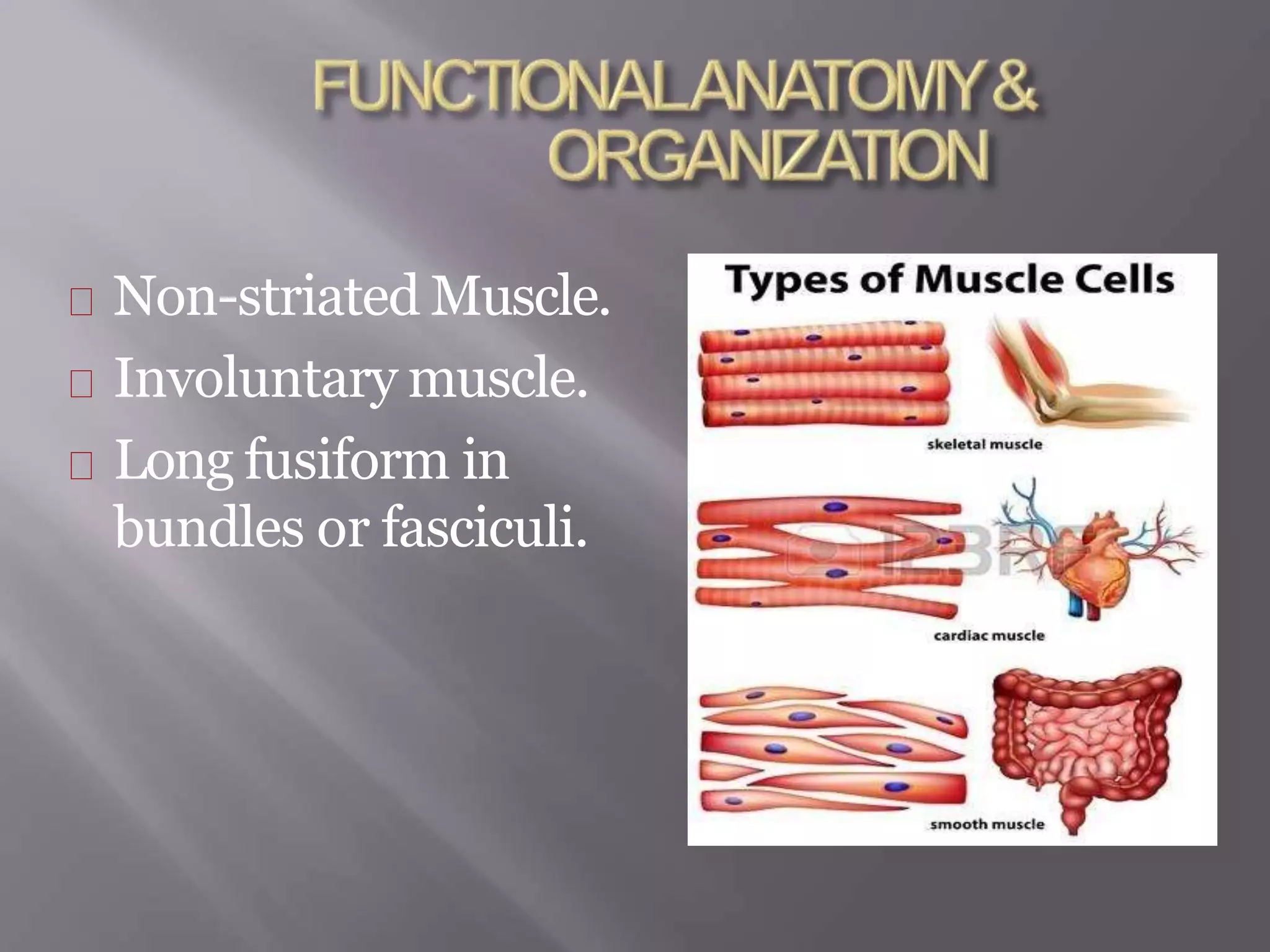
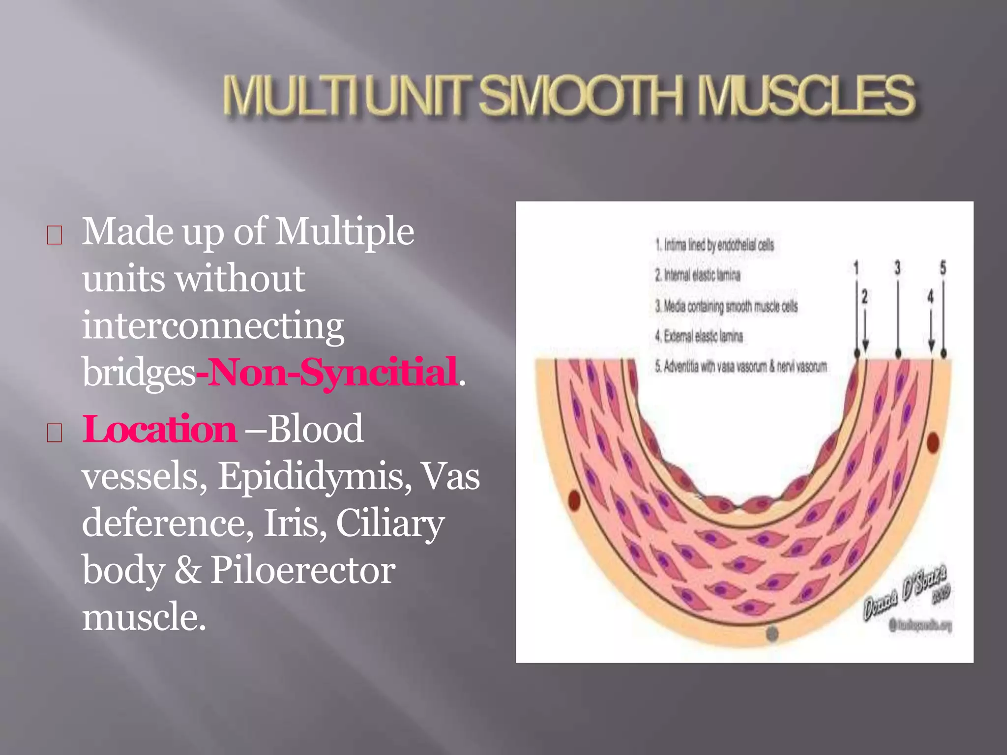
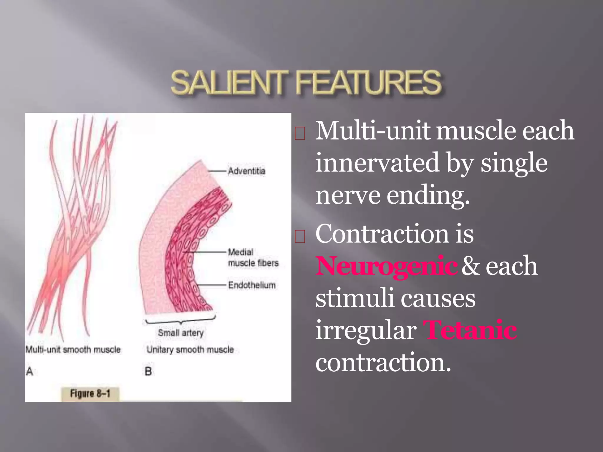
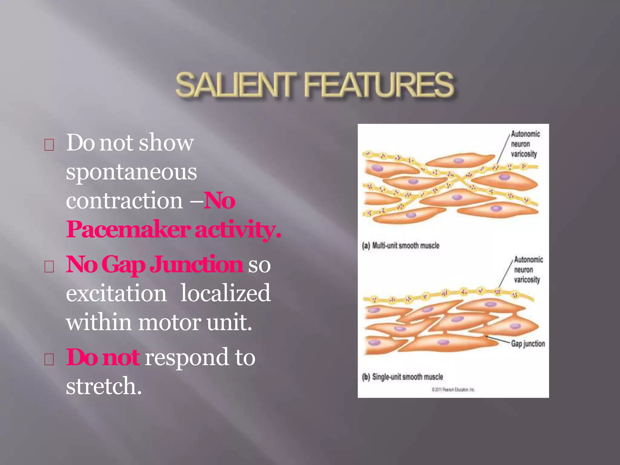
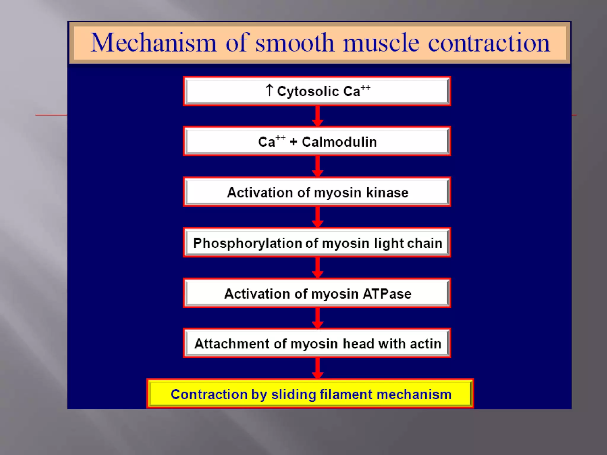
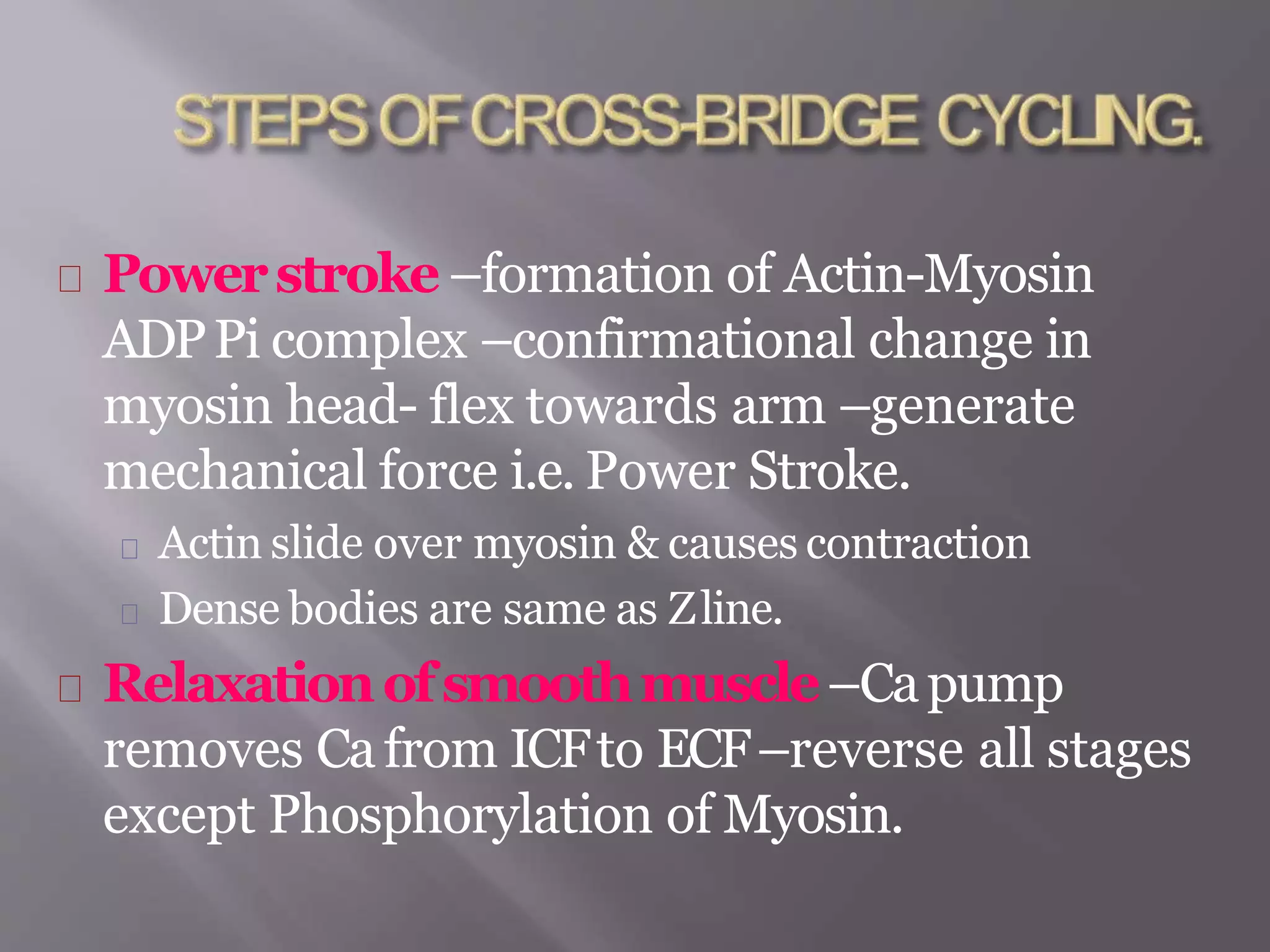
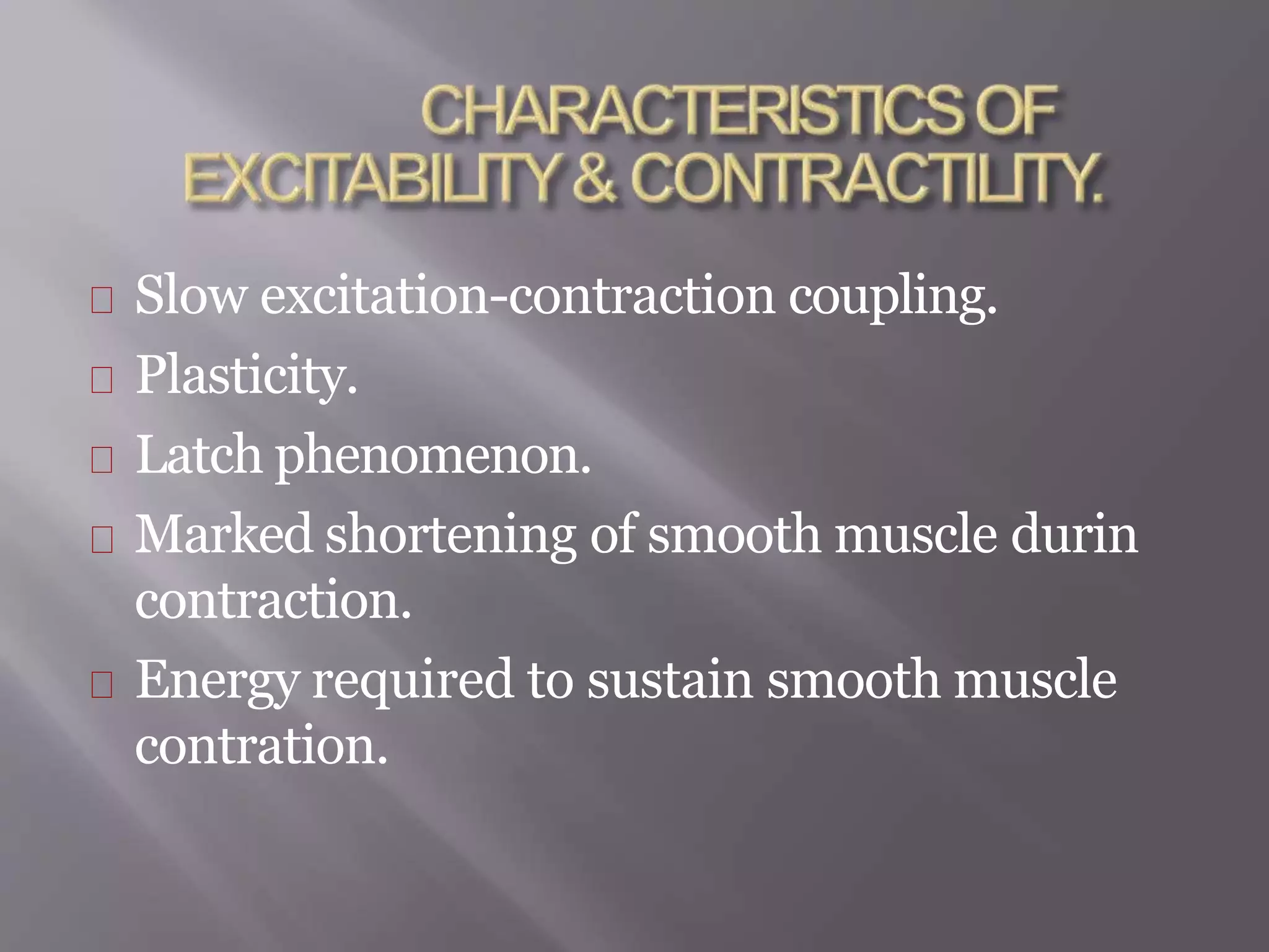
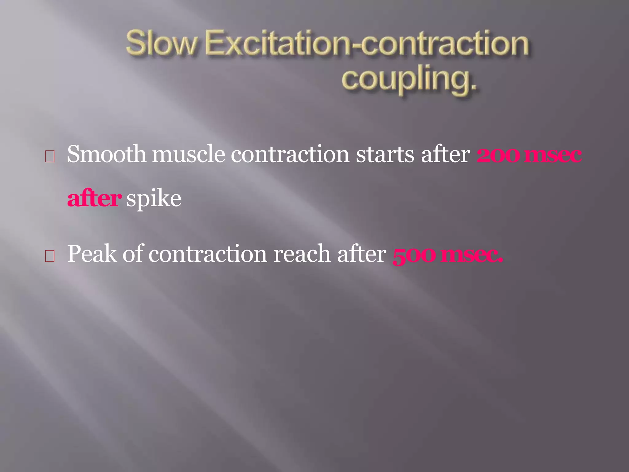
SMOOTH MUSCLE
PHYSIOLOGY
Non-striated Muscle.
Involuntary muscle.
Long fusiform in
bundles or fasciculi.
Single unit smooth
muscle
Muliti unit smooth
muscle.
Visceralsmooth
muscles
Present in hollow
viscera like GIT,
Uterus, Ureter,
Urinary Bladder &
Respiratory tract.
Arranged in large sheet.
Haslow resistance bridges
(GapJunctions)between
individual muscles.
Function in a Syncitial
fashion & contract as a
single unit.
Has their own Rhythmic
contractility ( Myogenic
Tone)
Rate of contraction
determined by
Pacemakerregion &
Nervesupply only
Modulatesits activity.
Contraction is
stimulated by
Stretchinginvolved in
Autoregulationof blood
flow.
Made up of Multiple
units without
interconnecting
bridges-Non-Syncitial.
Location–Blood
vessels, Epididymis, Vas
deference, Iris, Ciliary
body & Piloerector
muscle.
Multi-unit muscle each
innervated by single
nerve ending.
Contraction is
Neurogenic& each
stimuli causes
irregular Tetanic
contraction.
Donot show
spontaneous
contraction –No
Pacemakeractivity.
NoGapJunctionso
excitation localized
within motor unit.
Donot respond to
stretch.
Nervesupply–
Autonomic both
Sympathetic &
Parasympathetic.
Symp–Contraction &
Parasymp–Relaxation.
Preganglionic fibres –
ganglion –postganglionic
fibres –muscle.
Post ganglionic fibres
branch extensively.
Neuronal network has
Beadedappearance
due to varicosities.
Varicosities contains
chemical
neurotransmitter
(Ach/NE)
Nerve fibres donotendin
MotorEndPlatebut releases
its neuro-transmitter in
interstitialfluid near muscle
fibre.
It then diffusesin muscle fibre
& causes activation.
So single stimuli will not cause
activation of entire muscle
MultipleStimuliare required.
Either depolarizing or
repolarizing responses
are recorded in
response to stimuli.
These potentials
Summatewith
repeated stimuli
EJP&IJPare local
responses.
Spindle shaped cells with
broad central part &
tapering ends.
Length & diameter varies
with organ
GIT–30-40 mm/5 mm
diameter.
Bloodvessels–15-20
mm/2-3 mm.
Uterus–300 mm/10 mm
Plasmamembrane–surrounded by Externa
Lamina.
Cell communicate through Gap Junction.
Sarcoplasm
Nucleus–Central/Oval
Contains Mitochondria, Golgi apparatus, Endoplasmic
Reticulum, Ribosomes.
Sarcoplasmicreticulum–similar to skeletal
muscle but not as developed.
t
Myofibrils-Sarcotubular
system & triad not well
developed.
Lessthick filaments &
Morethin filaments.
Zline not well developed.
Actin–Troponin is absen
Myosin–bind only if
phosphorylated.
Densebodies –
attached to thecell
membrane
Actin filaments are
attached to dense
bodies.
When muscle contracts
dense bodies are drawn
close to each other.
PROCESSOFMUSCLEEXCITATION
PROCESSOFEXCITATIONCONTRACTION
COUPLING
PROCESSOFMUSCLECONTRACTION.
Electricalactivity insingleunit(Visceral)
smooth muscles.
Resting membrane potential
Action potential
Spike potential
Spike potential superimposed over slow wave
potentials.
Action potential with plateau.
Rangebetween- -50mv to
-75mv
Peculiarity–Unstability –
keeps on oscillating between -55
to -35mv.
Oscillationsdueto–
Superimposition by pacemaker
potential due to rhythmicchange
in Cachannelpermeability&
activity of Na-KPump.
When depolarization
reaches threshold action
potential begins &
transmitted to other muscle
cells through Gapjunctions.
3 types action potential
Spike potential
Spike potential over
pacemaker potential
Action potential with plateau.
Similartoskeletal
muscleexcept
Duration–10 to 50 msec
Amplitude–very low
Donot reach Iso-electric
base.
Occur in response to
electrical stimulation,
hormone, neurotransmitter
& stretch of smooth muscle.
Slow wave rhythm called
Pacemakerwaves seen in
GIT
These cannot cause muscle
contraction but when
potential rises above -35mv
action potential develop.
Appear rhythmically &
causes contraction of muscle
fibre.
Seen in ureter, uterus &
vascular smooth muscle.
Like in skeletal muscle there
is rapid depolarization but
Repolarization is delayed
by 100-150msec.
This prolonged
depolarization is
responsible for sustained
contraction of certain
smooth muscle.
Depolarization–due to entry
of Carather thanNa.
This muscle has more voltage
gated Cachannels than Na
channels.
These open & close slowly &
responsible for prolonged
action.
Caalong with depolarization
alsocausescontraction of
smooth muscle.
Multi-unitsmooth(Iris&Piloerector)muscle
respond to nerve stimulation which releases Ach&
NE.
These do not generate APbut causes excitatory
Junctional Potential (EJP)& spread to entire fibre.
Molecular mechanism is similarto skeletal muscle
but..
Smooth muscle don’t contain Tropomyosin &
Troponin.
Light chain of Myosin acts as Tropomyosin & called
 Regulatorychain of Myosin.
Ca binding protein Calmodulinacts as Troponin.
Cacombinewith Calmodulin–forms
complex –activates enzymeMyosinLight
ChainKinase (MLCK)
Phosphorylation of the Myosin Regulatory
chain.
Myosin head acquires capability to bind with
actin & forms Cross-bridging.
Powerstroke –formation of Actin-Myosin
ADPPi complex –confirmational change in
myosin head- flex towards arm –generate
mechanical force i.e. Power Stroke.
Actin slide over myosin & causes contraction
Dense bodies are same as Zline.
Relaxationofsmoothmuscle–Capump
removes Cafrom ICFto ECF–reverse all stages
except Phosphorylation of Myosin.
Slow excitation-contraction coupling.
Plasticity.
Latch phenomenon.
Marked shortening of smooth muscle durin
contraction.
Energy required to sustain smooth muscle
contration.
Smooth muscle contraction starts after 200msec
after spike
Peak of contraction reach after 500msec.
CanReadjustits
resting length.
Thus it not follows
length tension
relationship that is valid
for striated muscle.
So length tension
relationship curve is
Jaggedline.
Mechanism by which
smooth muscle maintain
high tension without
actively contracting.
Allows long term
maintenance of Tone.
So muscle can not generate
active tension but Resists
passive stretching.
Since smooth muscle found
mainly in hollow viscera
that should resists
stretching.
CAUSE–Both myosin
kinase & myosin
phosphatase enzyme
strongly activated, cycling
frequency of myosin head &
velocity of contraction
increases.
As this activation &
cycling frequency
decreases –lower
activation of enzymes
causes myosin head to
remain attached to actin
for longer time with less
energy expenditure as
ATP is required for
detachment.
Asmooth muscle can
contract more than 2/3rd
its stretched length
while skeletal muscle
contract up to 1/3rd.
This allows viscera to
change diameter from
largetoalmost zero.
Energyis muchless than skeletal
muscle as attachment cycling of cross
Excitation –
MultiUnitSmoothMuscle–stimulated
through nerves
SingleUnitSmoothMuscle–through
Nerves, Hormones, Pacemakers,
Stretching, Cold Temeprature.
Control movement of material through most
of hollow organs
Propel material in GIT
Control blood flow in arterioles.
Expell material from bladder.
Control Piloerection
Iris control.
Throughnervesby Sympathetic
Stimulation.e.g. –Intestinal smooth
muscles.
ThroughHormones–Progesterone
decrease activity of uterus.
Smooth Muscle.pptx

Smooth Muscle.pptx