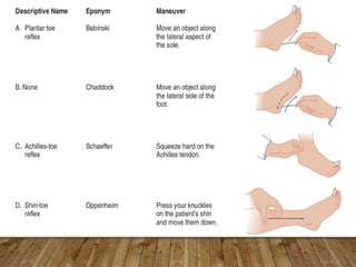
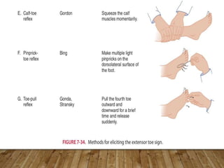
BABINSKI’S SIGN
CHAIRPERSON: DR PRAVEEN KUSUBI
STUDENT: DR VIGNESHWAR M
INTRODUCTION
• Joseph Jules François Félix Babinski(1857-1932)
• Made Hôpital de Salpêtrière in Paris a world famous medical centre
in the late nineteenth and early twentieth century.
• The others being, among others, Jean-Martin Charcot, Claude
Bernard, and Joseph Jules Dejerine
• Babinski took an interest in the pathogenesis of hysteria and was the first to
present acceptable differential diagnostic criteria for separating hysteria
from organic diseases.
METHOD
• Stroking the lateral part of the sole of the foot with a fairly sharp
object produces plantar flexion of the big toe;
• often there is also flexion and adduction of the other toes.
• This normal response is termed the flexor plantar reflex.
TECHNIQUE
• In some patients, stroking the sole produces extension
(dorsiflexion) of the big toe, often with extension and
abduction ("fanning") of the other toes.
• This abnormal response is termed the extensor plantar
reflex, or Babinski reflex.
ANATOMICAL
CONSIDERATIONS
• The reflex arc for the PR comprises of the afferent and
efferent fibres in the tibial nerve and the L4-5 to S1-2
cord segments.
• The reflexogenic area is the first sacral dermatome,
with the receptor nerve endings being located in the
skin on the sole of the foot.
• The afferent fibres travel in the tibial nerve which is a
branch of the sciatic nerve, to relay in the L4-5 to S1-2
cord segments.
• The efferent fibres from the spinal cord travel back in
the sciatic nerve which divides into two large branches
just proximal to the knee.
• Fibres supplying the toe flexors travel in the tibial
nerve while those supplying the toe extensors travel in
the peroneal nerve to reach the foot.
• Injury or transection of the tibial nerve therefore, would
interrupt the afferent and efferent arcs of the normal
plantar response, leaving the toe extensor muscles
innervated.
• Interruption of the reflex arc can cause a diminution or
absence of the reflex.
• Supraspinal influences from the cortex also influence
and modify the spinal reflex or response.
• A lesion anywhere along the corticospinal pathway can
modify the response on the contralateral side of the
body if the lesion is above the pyramidal decussation,
and on the same or homolateral side of the body if the
lesion is below the medullary decussation in the cord
itself.
• The PR being a polysynaptic reflex, the interneurons
in the reflex arc connect with motor neurons at several
segmental levels, leading to a co-ordinated motor
response or movement of the foot and lower limb
following cutaneous stimulation of the sole of the foot.
• Complex reflexes like plantar flexion use more of
inhibitory interneurons, sometimes referred to as the
internuncial pool.
• The axons of the Renshaw cells contact the alpha
motor neuron.
• An action potential down the axon of the alpha motor
neuron also excites the Renshaw cell through the
recurrent collateral.
• The Renshaw cell in turn inhibits the same alpha
motor neuron and other alpha motor neurons that
innervate agonists.
• The Renshaw cell also inhibits the inhibitory
interneuron mediating the reciprocal inhibition.
• In this way, the Renshaw cell shortens the reflex
contraction of the agonist and, at the same time,
shortens the reciprocal inhibition of the antagonist.
• Through this mechanism, the motor neurons can
inhibit their own activity.
• This seems to be important in preventing alpha motor
neurons from sending long trains of action
potentials in response to a brief stimulus.
• The Renshaw cell and other internuncial neurons
receive input from the higher motor centers, which can
modulate the activity of these neurons and fine-tune
the reflex movements.
IN CASE OF PLANTAR REFLEX
• The sensory limb of this reflex arc is mediated by
cutaneous receptors of fast-conducting 1a afferents
that converge on the internuncial pool of inhibitory
interneurons.
• While the motor neurons to the flexor muscles are
excited, the extensor muscles are inhibited through
reciprocal inhibition.
• At the same time, motor neurons to the extensors of
the contralateral leg are activated and the flexors are
relaxed to compensate for the shift of weight to the
contralateral leg while the ipsilateral leg is withdrawn
from the painful stimulus.
• This crossed extensor reflex maintains postural
support during withdrawal from a painful stimulus.
• As with other reflexes, the strength of the response corresponds
to the strength of the stimulus.
• In a normal individual only a painful stimulus elicits the reflex.
• When descending motor pathways that suppress and modulate
the reflex are damaged, a lighter, non-painful stimulus may elicit
the reflex. This was discovered by Babinski when he scratched
the sole of the foot of a patient with central nervous system
lesions.
• With the light non-painful stimulus, the strength of the response
parallels the extent to which the upper motor neuron lesion has
allowed up regulation of the reflex.
THERE ARE THREE RESPONSES POSSIBLE:
• Flexor: the toes curve inward and the foot everts; this is the
response seen in healthy adults (aka a "negative" Babinski).
• Indifferent: there is no response.
• Extensor: the hallux dorsiflexes and the other toes fan out –
the "positive Babinski's sign" indicating damage to the central
nervous system.
TYPES OF BABINSKI SIGN
• True Babinski sign – includes all the components of the
fully developed extensor plantar response.
• Minimal Babinski sign – is characterised by contraction of
the hamstring muscles and the tensor fasciae latae which
can be detected by palpation of the thigh.
• Spontaneous Babinski sign – is encountered in patients with
extensive pyramidal tract lesions. Passive flexion of the hip and
knee or passive extension of the knee may produce a positive
Babinski sign in adults, as may foot manipulation in infants and
children.
• Crossed extensor response/bilateral Babinski sign – may be
encountered in cases with bilateral cerebral or spinal cord
disease. Unilateral foot stimulation elicits a bilateral response in
such cases.
• Tonic Babinski reflex – is characterised by a slow
prolonged contraction of the toe extensors. It is encountered
in patients with combined frontal lobe lesions and
extrapyramidal involvement.
• Exaggerated Babinski sign – may take the form of a flexor
or extensor spasm.
CAUSES
• Pyramidal tract lesions
• Normal children upto one year of age
• Deep sleep
• Coma
• General Anaesthesia
• Post-ictal stage of epilepsy
CAUSES
• Electroconvulsive therapy (ECT)
• Hypoglycaemia
• Alcohol intoxication
• Narcosis
• Hypnosis
• Following severe physical exhaustion
• Head trauma with concussion.
ADDITIONAL MANEUVERS FOR ELICITING
SUPERFICIAL TOE REFLEXES
• These maneuvers, in particular those outside the S1
dermatome, generally are less effective than stimuli within
the S1 dermatome.
• In normal persons, these stimuli usually fail to elicit toe
flexion, but after UMN lesions, they may elicit toe
extension, just as with Babinski’s maneuver.
REFERENCES
• Bickerstaff ER, Spillane A. Neurological Examination in Clinical
Practice. 7th edition.
• Mayo Clinic. Clinical Examination in Neurology. Asian edition.
Fourth edition. W.B. Saunders company, 1976; Reflexes. Chapter
8, pg 169-80.
• DeJong's The Neurologic Examination, 7th edition.
• DeMyer's the Neurologic Examination,6th edition.

Babinski Sign

  • 1.
    BABINSKI’S SIGN CHAIRPERSON: DRPRAVEEN KUSUBI STUDENT: DR VIGNESHWAR M
  • 2.
    INTRODUCTION • Joseph JulesFrançois Félix Babinski(1857-1932) • Made Hôpital de Salpêtrière in Paris a world famous medical centre in the late nineteenth and early twentieth century. • The others being, among others, Jean-Martin Charcot, Claude Bernard, and Joseph Jules Dejerine
  • 3.
    • Babinski tookan interest in the pathogenesis of hysteria and was the first to present acceptable differential diagnostic criteria for separating hysteria from organic diseases.
  • 4.
    METHOD • Stroking thelateral part of the sole of the foot with a fairly sharp object produces plantar flexion of the big toe; • often there is also flexion and adduction of the other toes. • This normal response is termed the flexor plantar reflex.
  • 6.
  • 7.
    • In somepatients, stroking the sole produces extension (dorsiflexion) of the big toe, often with extension and abduction ("fanning") of the other toes. • This abnormal response is termed the extensor plantar reflex, or Babinski reflex.
  • 8.
    ANATOMICAL CONSIDERATIONS • The reflexarc for the PR comprises of the afferent and efferent fibres in the tibial nerve and the L4-5 to S1-2 cord segments. • The reflexogenic area is the first sacral dermatome, with the receptor nerve endings being located in the skin on the sole of the foot.
  • 10.
    • The afferentfibres travel in the tibial nerve which is a branch of the sciatic nerve, to relay in the L4-5 to S1-2 cord segments. • The efferent fibres from the spinal cord travel back in the sciatic nerve which divides into two large branches just proximal to the knee.
  • 12.
    • Fibres supplyingthe toe flexors travel in the tibial nerve while those supplying the toe extensors travel in the peroneal nerve to reach the foot. • Injury or transection of the tibial nerve therefore, would interrupt the afferent and efferent arcs of the normal plantar response, leaving the toe extensor muscles innervated. • Interruption of the reflex arc can cause a diminution or absence of the reflex.
  • 13.
    • Supraspinal influencesfrom the cortex also influence and modify the spinal reflex or response. • A lesion anywhere along the corticospinal pathway can modify the response on the contralateral side of the body if the lesion is above the pyramidal decussation, and on the same or homolateral side of the body if the lesion is below the medullary decussation in the cord itself.
  • 14.
    • The PRbeing a polysynaptic reflex, the interneurons in the reflex arc connect with motor neurons at several segmental levels, leading to a co-ordinated motor response or movement of the foot and lower limb following cutaneous stimulation of the sole of the foot.
  • 15.
    • Complex reflexeslike plantar flexion use more of inhibitory interneurons, sometimes referred to as the internuncial pool. • The axons of the Renshaw cells contact the alpha motor neuron. • An action potential down the axon of the alpha motor neuron also excites the Renshaw cell through the recurrent collateral.
  • 16.
    • The Renshawcell in turn inhibits the same alpha motor neuron and other alpha motor neurons that innervate agonists. • The Renshaw cell also inhibits the inhibitory interneuron mediating the reciprocal inhibition.
  • 18.
    • In thisway, the Renshaw cell shortens the reflex contraction of the agonist and, at the same time, shortens the reciprocal inhibition of the antagonist. • Through this mechanism, the motor neurons can inhibit their own activity.
  • 19.
    • This seemsto be important in preventing alpha motor neurons from sending long trains of action potentials in response to a brief stimulus. • The Renshaw cell and other internuncial neurons receive input from the higher motor centers, which can modulate the activity of these neurons and fine-tune the reflex movements.
  • 20.
    IN CASE OFPLANTAR REFLEX • The sensory limb of this reflex arc is mediated by cutaneous receptors of fast-conducting 1a afferents that converge on the internuncial pool of inhibitory interneurons. • While the motor neurons to the flexor muscles are excited, the extensor muscles are inhibited through reciprocal inhibition.
  • 21.
    • At thesame time, motor neurons to the extensors of the contralateral leg are activated and the flexors are relaxed to compensate for the shift of weight to the contralateral leg while the ipsilateral leg is withdrawn from the painful stimulus. • This crossed extensor reflex maintains postural support during withdrawal from a painful stimulus.
  • 22.
    • As withother reflexes, the strength of the response corresponds to the strength of the stimulus. • In a normal individual only a painful stimulus elicits the reflex. • When descending motor pathways that suppress and modulate the reflex are damaged, a lighter, non-painful stimulus may elicit the reflex. This was discovered by Babinski when he scratched the sole of the foot of a patient with central nervous system lesions. • With the light non-painful stimulus, the strength of the response parallels the extent to which the upper motor neuron lesion has allowed up regulation of the reflex.
  • 23.
    THERE ARE THREERESPONSES POSSIBLE: • Flexor: the toes curve inward and the foot everts; this is the response seen in healthy adults (aka a "negative" Babinski). • Indifferent: there is no response. • Extensor: the hallux dorsiflexes and the other toes fan out – the "positive Babinski's sign" indicating damage to the central nervous system.
  • 24.
    TYPES OF BABINSKISIGN • True Babinski sign – includes all the components of the fully developed extensor plantar response. • Minimal Babinski sign – is characterised by contraction of the hamstring muscles and the tensor fasciae latae which can be detected by palpation of the thigh.
  • 25.
    • Spontaneous Babinskisign – is encountered in patients with extensive pyramidal tract lesions. Passive flexion of the hip and knee or passive extension of the knee may produce a positive Babinski sign in adults, as may foot manipulation in infants and children. • Crossed extensor response/bilateral Babinski sign – may be encountered in cases with bilateral cerebral or spinal cord disease. Unilateral foot stimulation elicits a bilateral response in such cases.
  • 26.
    • Tonic Babinskireflex – is characterised by a slow prolonged contraction of the toe extensors. It is encountered in patients with combined frontal lobe lesions and extrapyramidal involvement. • Exaggerated Babinski sign – may take the form of a flexor or extensor spasm.
  • 27.
    CAUSES • Pyramidal tractlesions • Normal children upto one year of age • Deep sleep • Coma • General Anaesthesia • Post-ictal stage of epilepsy
  • 28.
    CAUSES • Electroconvulsive therapy(ECT) • Hypoglycaemia • Alcohol intoxication • Narcosis • Hypnosis • Following severe physical exhaustion • Head trauma with concussion.
  • 29.
    ADDITIONAL MANEUVERS FORELICITING SUPERFICIAL TOE REFLEXES • These maneuvers, in particular those outside the S1 dermatome, generally are less effective than stimuli within the S1 dermatome. • In normal persons, these stimuli usually fail to elicit toe flexion, but after UMN lesions, they may elicit toe extension, just as with Babinski’s maneuver.
  • 32.
    REFERENCES • Bickerstaff ER,Spillane A. Neurological Examination in Clinical Practice. 7th edition. • Mayo Clinic. Clinical Examination in Neurology. Asian edition. Fourth edition. W.B. Saunders company, 1976; Reflexes. Chapter 8, pg 169-80. • DeJong's The Neurologic Examination, 7th edition. • DeMyer's the Neurologic Examination,6th edition.