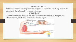
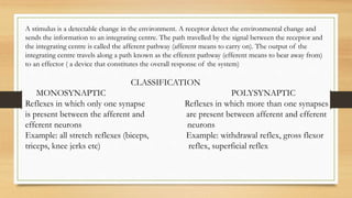
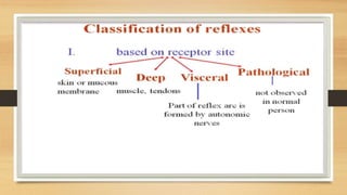
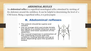
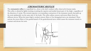
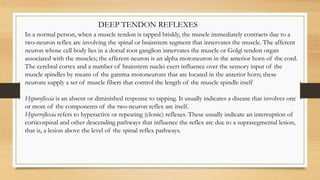
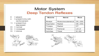
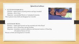
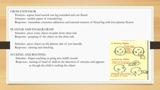
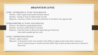
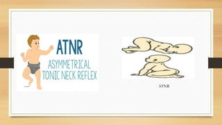
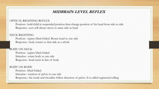
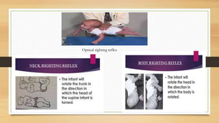
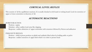
Reflexes are important to understand for all medical professional it is an assessment tool for patients with neurological conditions.
a god knowledge of primitive reflexes can be effective for pediatric health care as well. it helps us in identifying any developmental delay in children.
2. INTRODUCTION
REFLEX is an involuntary (automatic) response to a stimulus which depends on the
integrity of the reflex pathway i.e. the reflex arc
REFLEX ARC
It forms the functional unit of the nervous system and consists of receptor, an
afferent neuron, an efferent neuron and effector organ
3. A stimulus is a detectable change in the environment. A receptor detect the environmental change and
sends the information to an integrating centre. The path travelled by the signal between the receptor and
the integrating centre is called the afferent pathway (afferent means to carry on). The output of the
integrating centre travels along a path known as the efferent pathway (efferent means to bear away from)
to an effector ( a device that constitutes the overall response of the system)
CLASSIFICATION
MONOSYNAPTIC POLYSYNAPTIC
Reflexes in which only one synapse Reflexes in which more than one synapses
is present between the afferent and are present between afferent and efferent
efferent neurons neurons
Example: all stretch reflexes (biceps, Example: withdrawal reflex, gross flexor
triceps, knee jerks etc) reflex, superficial reflex
4.
5.
6. Why do we check reflexes?
• Reflex tests provide an objective sign indicating abnormality and some
indication as to the level of abnormality.
• It helps to assess the integrity of nerve circuits involved.
• To check the integrity of spinal cord, for spinal cord injuries, neuromuscular
diseases.
7. INDICATIONS FOR CHECKING REFLEXES:
• Developmental asymmetries
• Alteration and/or compensation in movement patterns.
• Unstable and/ or poorly adaptive movement patterns.
• Involuntary movements
• Unsteady or altered gait
• Altered sensitivity
• Weakness
• Spinal pain / injury
• Disregard or neglect of a body part
• Lack of awareness of injury or pain
• Side to side asymmetries
8. SUPERFICIAL REFLEXES
Superficial reflexes are elicited by stimulation of certain parts of
the skin or mucous membrane and the end result would be
contraction of one or more muscle
They are polysynaptic reflexes and are lost during corticospinal
tract lesion
Corneal reflexes conjunctival reflex pharyngeal reflex scapular
reflex abdominal reflex cremastric reflex anal reflex
9. CORNEAL REFLEX
The corneal reflex, also known as the blink reflex, is an involuntary blinking of the
eyelids elicited by stimulation of the cornea (such as by touching or by a foreign body),
though could result from any peripheral stimulus. Stimulation should elicit both a direct
and consensual response (response of the opposite eye). The reflex occurs at a rapid rate
of 0.1 seconds. The purpose of this reflex is to protect the eyes from foreign bodies and
bright lights (the latter known as the optical reflex)
The reflex is mediated by:
5th cranial nerve (trigeminal nerve) sensing the stimulus on the cornea only (afferent
fiber).
the temporal and zygomatic branches of the 7th cranial nerve (facial nerve) initiating the
motor response (efferent fiber)
Damage to the ophthalmic branch of the 5th cranial nerve results in absent corneal
reflex when the affected eye is stimulated.
10.
11. ABDOMINAL REFLEX
An abdominal reflex is a superficial neurological reflex stimulated by stroking of
the abdomen around the umbilicus. It can be helpful in determining the level of a
CNS lesion. Being a superficial reflex, it is polysynaptic
12.
13. The cremasteric reflex is a superficial (i.e., close to the skin's surface) reflex observed in human males.
This reflex is elicited by lightly stroking or poking the superior and medial (inner) part of the thigh—regardless of
the direction of stroke. The normal response is an immediate contraction of the cremaster muscle that pulls up
the testis ipsilaterally (on the same side of the body). The reflex utilizes sensory and motor fibers from two
different nerves. When the inner thigh is stroked, sensory fibers of the ilioinguinal nerve are stimulated. These
activate the motor fibers of the genital branch of the genitofemoral nerve which causes the cremaster muscle to
contract and elevate the testis
CREMASTERIC REFLEX
14. DEEP TENDON REFLEXES
In a normal person, when a muscle tendon is tapped briskly, the muscle immediately contracts due to a
two-neuron reflex arc involving the spinal or brainstem segment that innervates the muscle. The afferent
neuron whose cell body lies in a dorsal root ganglion innervates the muscle or Golgi tendon organ
associated with the muscles; the efferent neuron is an alpha motoneuron in the anterior horn of the cord.
The cerebral cortex and a number of brainstem nuclei exert influence over the sensory input of the
muscle spindles by means of the gamma motoneurons that are located in the anterior horn; these
neurons supply a set of muscle fibers that control the length of the muscle spindle itself
Hyporeflexia is an absent or diminished response to tapping. It usually indicates a disease that involves one
or more of the components of the two-neuron reflex arc itself.
Hyperreflexia refers to hyperactive or repeating (clonic) reflexes. These usually indicate an interruption of
corticospinal and other descending pathways that influence the reflex arc due to a suprasegmental lesion,
that is, a lesion above the level of the spinal reflex pathways.
15.
16. BICEPS REFLEX
Biceps reflex is a reflex test that examines the function of the C5 reflex
arc and the C6 reflex arc. The test is performed by using a tendon
hammer to quickly depress the biceps brachii tendon as it passes through
the cubital fossa. Specifically, the test activates the stretch receptors
inside the biceps brachii muscle which communicates mainly with the C5
spinal nerve and partially with the C6 spinal nerve to induce a reflex
contraction of the biceps muscle and jerk of the forearm.
A strong contraction indicates a 'brisk' reflex, and a weak or absent reflex
is known as 'diminished'. Brisk or absent reflexes are used as clues to the
location of neurological disease. Typically brisk reflexes are found in
lesions of upper motor neurons, and absent or reduced reflexes are
found in lower motor neuron lesions.
A change to the biceps reflex indicates pathology at the level of
musculocutaneous nerve, segment C5/6 or at some point above it in the
spinal cord or brain.
17.
18. The triceps reflex, a deep tendon reflex, is a reflex as it elicits
involuntary contraction of the triceps brachii muscle. It is initiated by
the Cervical (of the neck region) spinal nerve 7 nerve root (the small
segment of the nerve that emerges from the spinal cord). The reflex
is tested as part of the neurological examination to assess the sensory
and motor pathways within the C7 and C8 spinal nerves
The test can be performed by tapping the triceps tendon with the
sharp end of a reflex hammer while the forearm is hanging loose
at a right angle to the arm. A sudden contraction of the triceps
muscle causes extension, and indicates a normal reflex.[
TRICEPS REFLEX
19.
20. The brachioradialis reflex (also known as supinator
reflex) is observed during a neurological exam by striking
the brachioradialis tendon (at its insertion at the base of the
wrist into the radial styloid process (radial side of wrist
around 4 inches proximal to base of thumb)) directly with a
reflex hammer when the patient's arm is relaxing. This
reflex is carried by the radial nerve (spinal level: C5, C6)
The reflex should cause slight pronation or supination and
slight elbow flexion. Contrary to popular belief, this reflex
should not cause wrist extension and/or radial deviation,
because the brachioradialis does not cross the wrist.
BRACHIORADIALIS REFLEX
21.
22. The patellar reflex or knee-jerk (myotatic) (monosynaptic) (in
American English knee reflex) is a stretch reflex which tests the
L2, L3, and L4 segments of the spinal cord.
Striking of the patellar tendon with a reflex hammer just below
the patella stretches the muscle spindle in the quadriceps muscle.
This produces a signal which travels back to the spinal cord and
synapses (without interneurons) at the level of L3 in the spinal
cord, completely independent of higher centres. From there, an
alpha motor neuron conducts an efferent impulse back to the
quadriceps femoris muscle, triggering contraction. This
contraction, coordinated with the relaxation of the antagonistic
flexor hamstring muscle causes the leg to kick. This is a reflex of
proprioception which helps maintain posture and balance,
allowing to keep one's balance with little effort or conscious
thought.
PATELLAR REFLEX
23.
24. The ankle jerk reflex, also known as the Achilles reflex, occurs when the Achilles tendon is
tapped while the foot is dorsiflexed. It is a type of stretch reflex that tests the function of the
gastrocnemius muscle and the nerve that supplies it. A positive result would be the jerking of the
foot towards its plantar surface. Being a deep tendon reflex, it is monosynaptic. It is also a stretch
reflex. These are monosynaptic spinal segmental reflexes. When they are intact, integrity of the
following is confirmed: cutaneous innervation, motor supply, and cortical input to the
corresponding spinal segment.
This reflex is mediated by the S1 spinal segment of the spinal cord.
Ankle of the patient is relaxed. It is helpful to support the ball of the foot at least somewhat to put
some tension in the Achilles tendon, but don’t completely dorsiflex the ankle. A small strike is
given on the Achilles tendon using a rubber hammer to elicit the response. If the practitioner is
not able to elicit a response, a Jendrassik maneuver can be tried by having the patient cup their
fingers on each hand and try to pull the hands apart. A positive response is marked by a brisk
plantarflexion of the foot. The response is also graded into Grade 0-4 according to the reflex
grading system.
ACHILLES REFLEX
25.
26. PRIMITIVE REFLEXES
Primitive reflex patterns seen in early stages of development which disappear later on or with advanced age.
These reflexes are essential for normal progressive motor development. There are some children who may
skip some movements but these are not abnormal
However not overcoming these primitive reflex patterns at the right time should be definitely considered as
abnormal.
Initially lower centers such as spinal cord control these movements but later on higher centers like midbrain
and cortex take control over them and dominate the lower ones thus integrating them for various voluntary
functional task
These primitive reflexes are classified according to the level at which they are controlled:-
• Spinal cord
• Brainstem
• Midbrain
• Cortex
27. Spinal level reflexes
• FLEXOR WITHDRAWAL:-
Position:- supine head in neutral position and legs extended
Stimulus:- sole of foot
Response:- uncontrolled flexion of stimulated extremity
This is present since birth and disappear by 2 months
• EXTENSOR TRUST:-
Position :- supine head neutral one leg extended and other flexed
Stimulus:- sole of flexed leg is giving stroking
Response:- immediate extension adduction and internal rotation of flexed leg
with plantar flexion of foot
Present at birth and integrated at 4 months
28. CROSS EXTENSOR
Position:- supine head neutral one leg extended and one flexed
Stimulus:- medial aspect of extended leg
Response:- immediate extension adduction and internal rotation of flexed leg with foot plantar flexion
PLANTAR AND PALMAR GRASP
Stimulus:- press some object on palm from ulnar side
Response:- grasping of the object on the ulnar side
Stimulus:- press object on the plantar side of toes laterally
Response:- clawing and clutching
SUCKING AND ROOTING
Stimulus:- finger touching or going into child’s mouth
Response:- turning of head of child in the direction of stimulus and appears
as though the child is sucking the object
29. BRAINSTEM LEVEL
ATNR ( ASYMMETRICAL TONIC NECK REFLEX)
Position:- child is supine head neutral and limbs relaxed
Stimulus:- turning of head of child towards one side
Response:- extension of limbs on face side and flexion of the limb on the opposite side
STNR(SYMMETRICAL TONIC NECK REFLEX)
Position:- the child id in quadrupedal position
Stimulus:- the head is either flexed or extended
Response:- when the head is in flexion the upper limb gets flexed and
lower limb extended and vice versa
TONIC LABYRINTHINE REFLEX
Position:- supine or prone
Response:- as the position itself is the stimulus if the child is in supine position then there is increase in
tone of extensor group of muscle and when child is kept in prone position then there is increase in
flexor tone
33. MIDBRAIN LEVEL REFLEX
OPTICAL RIGHTING REFLEX
Position:- hold child in suspended position then change position of his head from side to side
Response:- eyes will always move to same side as head
NECK RIGHTING
Position:- supine blind folded. Rotate head to one side
Response:- body rotates to that side as a whole
BODY ON NECK
Position:- supine blind folded
Stimulus:- rotate body to one side
Response:- head turns in line of body
BODY ON BODY
Position:- blind folded
Stimulus:- rotation of pelvis to one side
Response:- the trunk and shoulder follow direction of pelvis. It is called segmental rolling
35. CORTICAL LEVEL REFLEX
This consists of all the equilibrium reactions. It is usually checked on tilt board or rocking board. Look for extension of
upper or lower extremities in direction of tilt
AUTOMATIC REACTIONS
MORO’S REACTION
Position:- supine
Stimulus:- either sudden loud noise like clapping
Response:- sudden abduction of upper extremities with extension followed by flexion and adduction
PARACHUTE REFLEX
Position :- child in prone position on plinth and suddenly lifted either by holding ankle or pelvis
Response:- sudden extension of upper limb which is in order to protect head